. 2024 Oct 2;15(10):725.
doi: 10.1038/s41419-024-07102-w.

Dronedarone hydrochloride (DH) induces pancreatic cancer cell death by triggering mtDNA-mediated pyroptosis

Affiliations

Dronedarone hydrochloride (DH) induces pancreatic cancer cell death by triggering mtDNA-mediated pyroptosis

Ming-Qiao Li et al. Cell Death Dis. .

Abstract

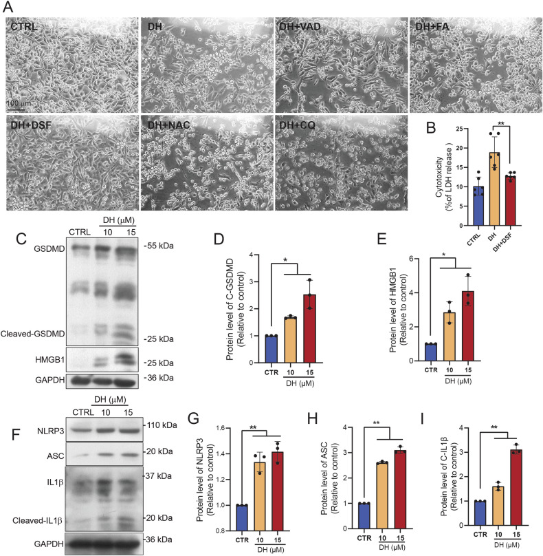
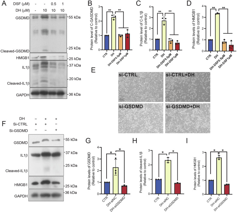
Pancreatic cancer is one of the leading causes of cancer-associated mortality, with a poor treatment approach. Previous study has shown that inducing pyroptosis in pancreatic ductal adenocarcinoma (PDAC) slows the growth of PDACs, implying that pyroptosis inducers are potentially effective for PDAC therapy. Here, we found that Dronedarone hydrochloride (DH), an antiarrhythmic drug, induces pyroptosis in pancreatic cancer cells and inhibits PDAC development in mice. In PANC-1 cells, DH caused cell death in a dosage- and time-dependent manner, with only pyroptosis inhibitors and GSDMD silencing rescuing the cell death, indicating that DH triggered GSDMD-dependent pyroptosis. Further work revealed that DH increased mitochondrial stresses and caused mitochondrial DNA (mtDNA) leakage, activating the cytosolic STING-cGAS and pyroptosis pathways. Finally, we assessed the anti-cancer effects of DH in a pancreatic cancer mouse model and found that DH treatment suppressed pancreatic tumor development in vivo. Collectively, our investigation demonstrates that DH triggers pyroptosis in PDAC and proposes its potential effects on anti-PDAC growth.


Conflict of interest statement

The authors declare no competing interests.

Figures

Fig. 1
Fig. 1. DH induced pancreatic cancer cell death.
A Morphology observation of PANC-1 after treatment of DH at different concentrations. Scale bar, 100 μm. B Released LDH activity in PANC-1 cells after treatment of DH at different concentrations. n = 4. C Morphology observation of PANC-1 after treatment of DH (20 μM) at indicated time point. Scale bar, 100 μm. D Released LDH activity in PANC-1 cells after treatment of DH (20 μM) at indicated time point. n = 8. Data was shown as mean ± SD. Mutiple t-test are followed by Bonferoni correction was used. *p < 0.05; ***p < 0.001.
Fig. 2
Fig. 2. DH triggered inflammasome-related pyroptosis.
A Morphology observation of PANC-1 after treatment of vehicle, DH (15 μM) or DH combined with different inhibitors. Z-VAD-FMK (VAD), pan-caspase inhibitors; Z-FA-FMK (FA), cathepsin B/L inhibitor; disulfiram (DSF), pyroptosis inhibitor; N-acetyl-Lcysteine (NAC), ROS inhibitor; chloroquine (CQ), autophagy inhibitor; Scale bar, 100 μm. B Released LDH activity in PANC-1 cells after treatment of DH or DH combined with DSF. n = 6. C Representative Western Blot images of pyroptosis-related proteins expression in PANC-1 cells after DH treatment. D, E Quantification of Cleaved-GSDMD, HMGB1 protein expressions in Fig. 2C. n = 3. F Representative Western Blot images of inflammasome-related proteins expression in PANC-1 cells after DH treatment. GI Quantification of NLRP3, ASC and cleaved-IL1β protein expressions in Fig. 2F. n = 3. Data was shown as mean ± SD. Mutiple t-test are followed by Bonferoni correction was used. *p < 0.05; **p < 0.01.
Fig. 3
Fig. 3. Inhibition of GSDMD blocked DH-induced cell death.
A Representative images of Western Blot images of pyroptosis-related proteins in PANC-1 cells after DH or DH combined DSF treatment. BD Quantification of cleaved-GSDMD, HMGB1, and cleaved-IL1β protein expressions in Fig. 3A. n = 3. E Morphology observation of PANC-1 after treatment of vehicle or DH. GSDMD was knocked down or not in cells before DH treatment. Scale bar, 100 μm. F Representative images of Western Blot images of pyroptosis-related proteins in cells with different treatment. GI Quantification of cleaved-GSDMD, HMGB1, and cleaved-IL1β protein expressions in Fig. 3F. n = 3. Data was shown as mean ± SD. Mutiple t-test are followed by Bonferoni correction was used. *p < 0.05; **p < 0.01.
Fig. 4
Fig. 4. DH disturbed mitochondria homeostasis.
A Representative images of DCFH-FA staining result in PANC-1 cells with or without DH treatment. White or Green, DCFH positive stained area. Scale bar, 200 μm. B Quantification of DCFH-FA positive staining area in images in Fig. 4A. n = 3. C Representative images of MitoSox staining results in PANC-1 cells with or without DH treatment. Scale bar, 50 μm. Red, MitoSox positive stained area. D Quantification of MitoSox positive staining area in images in Fig. 4C. n = 3. E Representative immunofluorescent images of Tom20 staining results in PANC-1 cells with or without DH treatment. Scale bar, 25 μm. Red, Tom20; Blue, DAPI. Data was shown as mean ± SD. Unpaired t-test was used. **p < 0.01; ***p < 0.001.
Fig. 5
Fig. 5. DH-induced mitochondrial DNA (mtDNA)-mediated pyroptosis.
A Morphology observation of PANC-1 after treatment of vehicle, DH or DH combined ddC. Scale bar, 100 μm. B Released LDH activity in PANC-1 cells after treatment of DH or DH combined with ddC. n = 6. C Representative images of Western Blot images of pyroptosis-related proteins in PANC-1 cells after different treatment. DF Quantification of cleaved-GSDMD, HMGB1, and cleaved-IL1β protein expressions in Fig. 5C. n = 3. G Representative images of MitoSox staining results in PANC-1 cells after different treatments. Scale bar, 100 μm. Red, MitoSOX; Blue, DAPI. H Quantification of MitoSOX positive staining area in images in figure (G). n = 5. Data was shown as mean ± SD. Mutiple t-test are followed by Bonferoni correction was used. *p < 0.05; **p < 0.01; ***p < 0.001.
Fig. 6
Fig. 6. DH induced mtDNA release and triggered cGAS-STING pathway.
A Representative images of PANC1 cells after stained with dsDNA and MitoTracker. Green, MitoTracker; Red, dsDNA. Scale bar, 100 μm. B Determination of extracellular DNA value in the culture medium of PANC-1 cells after DH treatment. n = 3. C Representative images of Western Blot images of cGAS-STING pathway-related proteins in PANC-1 cells after different treatment. D, E Quantification of STING, p-IRF3 protein expressions in Fig. 6B. n = 3. F Determination of extracellular DNA value in the culture medium of PANC-1 cells after DH or DH combined ddC treatment. n = 6. G Representative images of Western Blot images of cGAS-STING pathway-related proteins in PANC-1 cells after DH or DH combined ddC treatment. H, I Quantification of STING, p-IRF3 protein expressions in Fig. 6F. n = 3. Data was shown as mean ± SD. Mutiple t-test are followed by Bonferoni correction was used. *p < 0.05.
Fig. 7
Fig. 7. DH treatment inhibit pancreatic tumor growth in vivo.
A Relative tumor growth rate after DH treatment. n = 9,10. B Comparison of tumor weight after DH treatment. n = 8. C Representative images of tumors from mice with DH treatment or not. n = 8. D Representative images of Western Blot images of pyroptosis-related proteins in tumors from mice with or without DH treatment. EH Quantification of cleaved-GSDMD, HMGB1, cleaved-caspase 1, and cleaved-IL1β protein expressions in (D). I A diagram illustrating working machinery of DH on stimulation of pyroptosis. Data was shown as mean ± SD. Mutiple t-test are followed by Bonferoni correction was used. *p < 0.05; **p < 0.01; ***p < 0.001.

References

    1. Siegel RL, Miller KD, Jemal A. Cancer statistics, 2016. CA Cancer J Clin. 2016;66:7–30. - PubMed
    1. Cui J, Zhou Z, Yang H, Jiao F, Li N, Gao Y, et al. MST1 Suppresses Pancreatic Cancer Progression via ROS-Induced Pyroptosis. Mol Cancer Res. 2019;17:1316–25. - PubMed
    1. Shi J, Zhao Y, Wang K, Shi X, Wang Y, Huang H, et al. Cleavage of GSDMD by inflammatory caspases determines pyroptotic cell death. Nature. 2015;526:660–5. - PubMed
    1. Lu L, Zhang Y, Tan X, Merkher Y, Leonov S, Zhu L, et al. Emerging mechanisms of pyroptosis and its therapeutic strategy in cancer. Cell Death Discov. 2022;8:338 10.1038/s41420-022-01101-6. - PMC - PubMed
    1. Wang L, Qin X, Liang J, Ge P. Induction of Pyroptosis: A Promising Strategy for Cancer Treatment. Front Oncol. 2021;11:635774. - PMC - PubMed

MeSH terms