. 2024 Oct 2;15(1):8542.
doi: 10.1038/s41467-024-52872-8.

Structural basis of thiamine transport and drug recognition by SLC19A3

Affiliations

Structural basis of thiamine transport and drug recognition by SLC19A3

Florian Gabriel et al. Nat Commun. .

Abstract

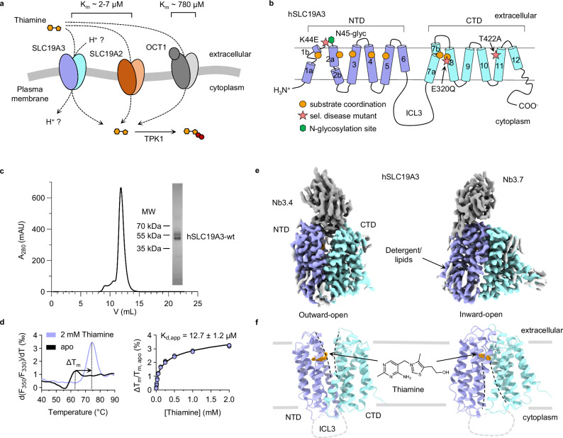
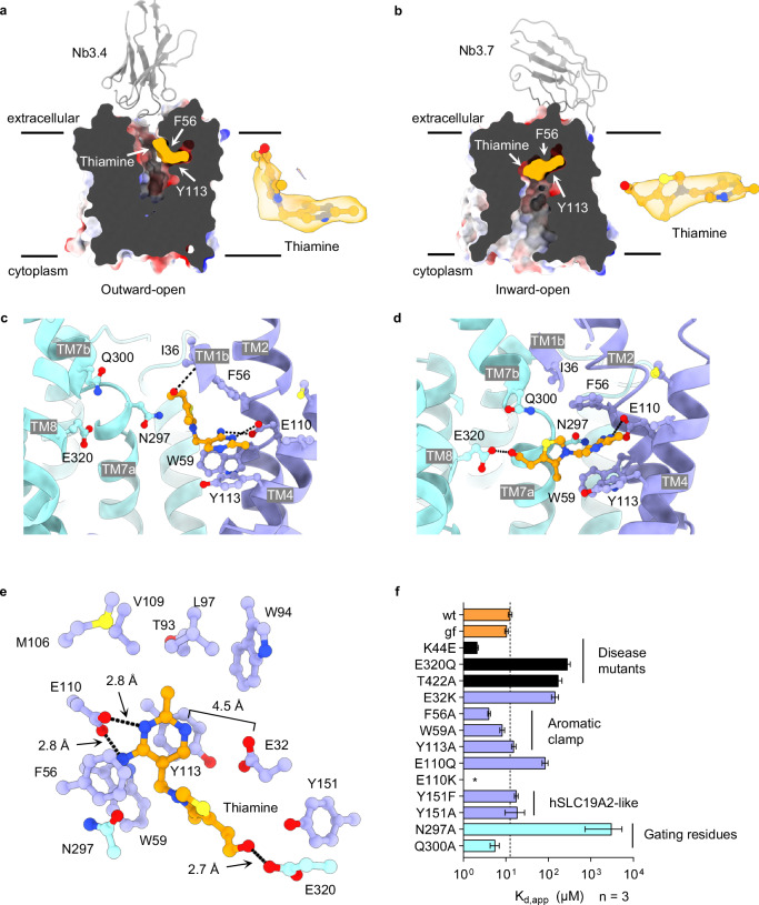
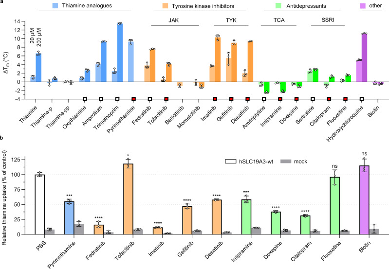
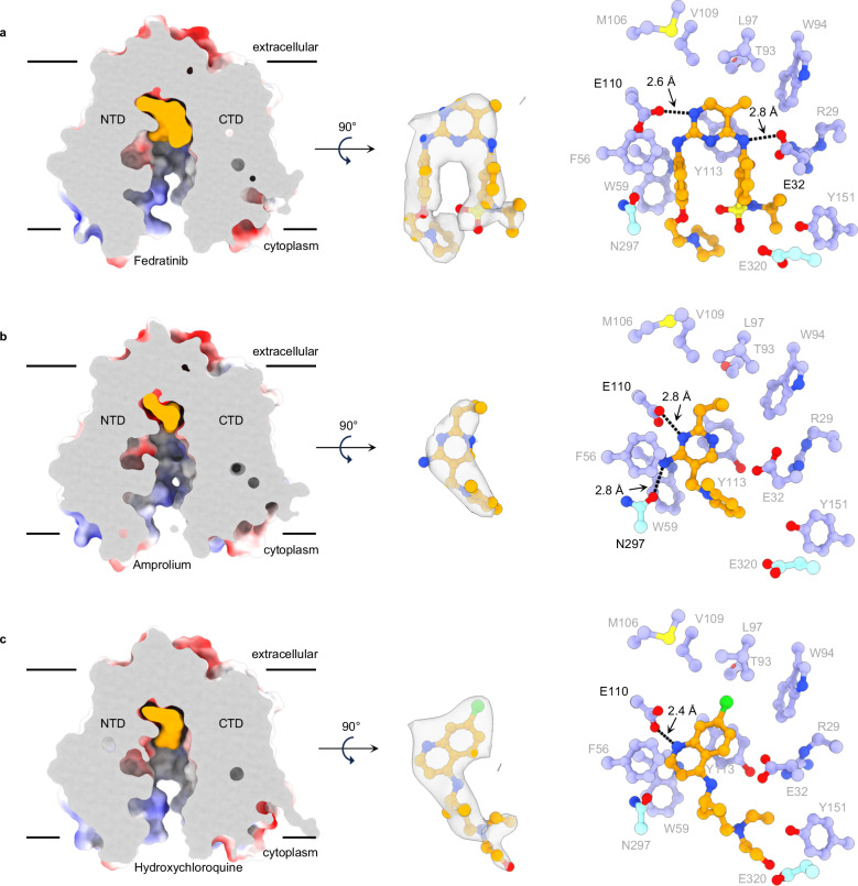
Thiamine (vitamin B1) functions as an essential coenzyme in cells. Humans and other mammals cannot synthesise this vitamin de novo and thus have to take it up from their diet. Eventually, every cell needs to import thiamine across its plasma membrane, which is mainly mediated by the two specific thiamine transporters SLC19A2 and SLC19A3. Loss of function mutations in either of these transporters lead to detrimental, life-threatening metabolic disorders. SLC19A3 is furthermore a major site of drug interactions. Many medications, including antidepressants, antibiotics and chemotherapeutics are known to inhibit this transporter, with potentially fatal consequences for patients. Despite a thorough functional characterisation over the past two decades, the structural basis of its transport mechanism and drug interactions has remained elusive. Here, we report seven cryo-electron microscopy (cryo-EM) structures of the human thiamine transporter SLC19A3 in complex with various ligands. Conformation-specific nanobodies enable us to capture different states of SLC19A3's transport cycle, revealing the molecular details of thiamine recognition and transport. We identify seven previously unknown drug interactions of SLC19A3 and present structures of the transporter in complex with the inhibitors fedratinib, amprolium and hydroxychloroquine. These data allow us to develop an understanding of the transport mechanism and ligand recognition of SLC19A3.


Conflict of interest statement

The authors declare no competing interests.

Figures

Fig. 1
Fig. 1. Structure and function of hSLC19A3.
a Humans and other mammals have at least three distinct membrane transporters that can mediate thiamine uptake: SLC19A3, SLC19A2 and OCT1 (SLC22A1). SLC19A3 is essential for the uptake of thiamine across the intestinal and blood-brain barrier under physiological conditions. In the cytoplasm, thiamine is phosphorylated by the enzyme TPK1 to form the biologically active coenzyme thiamine pyrophosphate. b Cartoon representation of hSLC19A3, based on the determined cryo-EM structures. The transporter follows the canonical MFS fold, with twelve transmembrane helices (TMs) folding in two symmetrically related six-helix bundle domains (NTD and CTD). Substrate coordination is mediated mostly by residues of TM1-5 of the NTD and by TM7 and TM8 of the CTD (orange circles). The positions of three disease mutants which were studied in this work are indicated by pink stars. c Representative size-exclusion chromatography (SEC) trace of purified hSLC19A3-wt. The protein elutes as a monomer of about 50 kDa. A fraction of the protein is glycosylated and appears as an extra band at ~60 kDa in SDS-PAGE (inset). The purification of hSLC19A3-wt has been performed >10 times with close to identical outcomes. d Thermal shift assay of hSLC19A3 using nanoDSF. The left panel shows the first derivative of the melting curve, measured as ratio of the fluorescence recorded at 350 nm (F350) and at 330 nm (F330), in the absence and presence of thiamine. Thiamine induces a strong stabilisation of the transporter (ΔTm = 10.9 ± 0.3 °C by 2 mM thiamine). The right panel illustrates the concentration dependent thermal shifts (n = 3) with the resulting apparent dissociation constant Kd,app for thiamine (mean ± s.d.). e Cryo-EM maps of thiamine-bound hSLC19A3 in its outward-open (left) and inward-open (right) conformation. The NTD (purple) and CTD (cyan) of the transporter, as well as the fiducial nanobodies (grey), are resolved. ChimeraX contour levels: outward-open: 0.876; inward-open: 0.148. f Structure models of the respective conformational states. The density of thiamine is shown in orange. ChimeraX contour levels: outward-open: 0.547; inward-open: 0.148. The N-terminus, C-terminus and ICL3 are expected to be structurally disordered and could consequently not be resolved.
Fig. 2
Fig. 2. Thiamine binding site of hSLC19A3.
a Localisation of the thiamine binding site of hSLC19A3 in the outward- and (b), inward-open conformation of the transporter. The transporter is shown in electrostatic surface representation. Density for thiamine (orange) is shown separately (ChimeraX contour levels: 0.547 and 0.148, Q-scores: 0.45 and 0.57). c Residues involved in thiamine coordination in the outward- and (d), inward-open conformation of hLSC19A3, shown in the membrane plane. e Extracellular view of hSLC19A3 highlighting the thiamine-coordinating residues in the substrate binding site of the transporter. Residues of the NTD are coloured in purple, residues of the CTD in cyan, and thiamine in orange. Black dashes indicate hydrogen bonds (cut-off at 3.0 Å) f Thiamine binding affinities (apparent dissociation constants Kd,app) of single point mutants of hSLC19A3. The corresponding titration curves were measured in three independent experiments (n = 3, see Supplementary Fig. 4). The bar plot shows the mean Kd,app and its standard deviation derived through a global fit of the measured data to Eq. (1), as described in the methods. For the purification of the respective mutants, see Supplementary Fig. 20.
Fig. 3
Fig. 3. Drug interactions of hSLC19A3.
a Thermal shift screen of known and potential thiamine uptake inhibitors (TUIs). Thermal shift assays were performed in three independent experiments (n = 3) in the presence of 20 μM and 200 μM of the respective compound and the stabilisation effect compared to the apo stability of hSLC19A3. The melting temperature was determined as the inflection point of the thermal unfolding curve of the protein (cf. Fig. 1d). Wherever possible, the ratio of the fluorescence at 350 nm and 330 nm (F350/F330) was selected as a readout for the thermal unfolding curve. The bar plot shows the mean ± s.d. of the measured thermal shifts. Due to intrinsic fluorescence of the following compounds, the fluorescence trace at 350 nm (F350) only was used as a readout: oxythiamine, trimethoprim, pyrimethamine, tofacitinib, imatinib, gefitinib, dasatinib, sertraline, fluoxetine and hydroxychloroquine. The two compounds, pyrimethamine and baricitinib, interfere at 200 μM too strongly with the fluorescence detection to provide an unambiguous readout and were omitted in this dataset. The squares indicate identified TUIs. White squares indicate already known TUIs, and red squares highlight hSLC19A3-inderactions identified in this study. b Cellular thiamine-uptake and its inhibition by 200 µM of selected drugs. Shown is the relative uptake of deuterated thiamine (thiamine-d3, normalised to PBS-only control) by Expi293FTM cells overexpressing hSLC19A3-wt (indicated in colour), compared with mock transfected cells (indicated in grey). Three independent measurements were performed (n = 3). The data are shown as mean ± s.d. (ns: p ≥ 0.5, *p ≤ 0.05, **p ≤ 0.01, ***p ≤ 0.001, ****p ≤ 0.0001, Student’s t test, two-tailed).
Fig. 4
Fig. 4. Cryo-EM structures of hSLC19A3 in complex with high-affinity inhibitors.
a–c Bound inhibitors (illustrated in orange) were observed to occupy the same substrate binding site and engage with similar residues as thiamine. Shown on the left are cross sections of the electrostatic surface representations of the ligand-bound structures, with the ligand coloured in orange. The densities for the different compounds are shown in the centre of the respective panels (ChimeraX contour levels/Q-scores for fedratinib: 0.7/0.47, amprolium: 0.15/0.53, hydroxychloroquine: 0.47/0.75); density within 2.5 Å of the atom coordinates is shown. On the right, the coordination of the compounds in the substrate binding site is depicted. Residues of the NTD are shown in purple, the ones of the CTD in cyan and the compound in orange. Black dashes indicate hydrogen bonds (cut-off at 3.0 Å).

References

    1. Bettendorff L. Thiamin. Present Knowledge in Nutrition. 261–279 (John Wiley & Sons, Inc., 2012).
    1. Timm, D. E., Liu, J., Baker, L. J. & Harris, R. A. Crystal structure of thiamin pyrophosphokinase. J. Mol. Biol.310, 195–204 (2001). - PubMed
    1. Baker, L. J., Dorocke, J. A., Harris, R. A. & Timm, D. E. The crystal structure of yeast thiamin pyrophosphokinase. Structure9, 539–546 (2001). - PubMed
    1. Tylicki, A., Lotowski, Z., Siemieniuk, M. & Ratkiewicz, A. Thiamine and selected thiamine antivitamins — biological activity and methods of synthesis. Biosci. Rep.38, 1–23 (2018). - PMC - PubMed
    1. Zhao, R. & Goldman, I. D. Folate and thiamine transporters mediated by facilitative carriers (SLC19A1-3 and SLC46A1) and folate receptors. Mol. Asp. Med.34, 373–385 (2013). - PMC - PubMed

Publication types

LinkOut - more resources