. 2024 Oct 2;15(1):8537.
doi: 10.1038/s41467-024-52844-y.

Cell fixation improves performance of in situ crosslinking mass spectrometry while preserving cellular ultrastructure

Affiliations

Cell fixation improves performance of in situ crosslinking mass spectrometry while preserving cellular ultrastructure

Andrew R M Michael et al. Nat Commun. .

Abstract

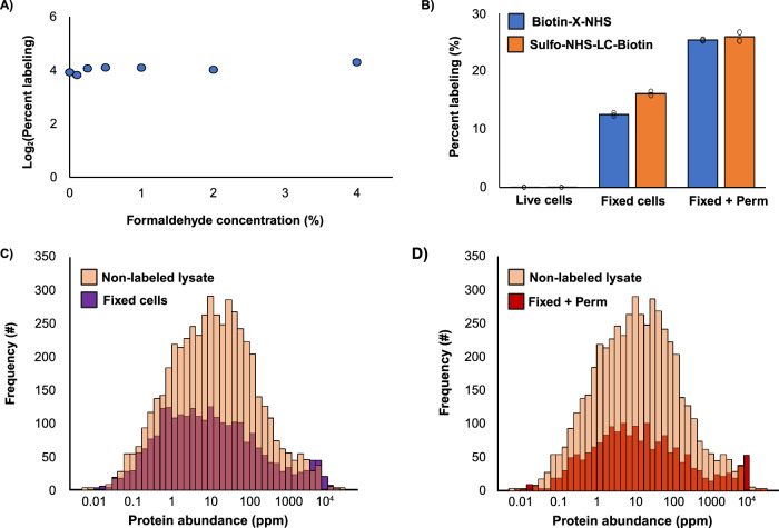
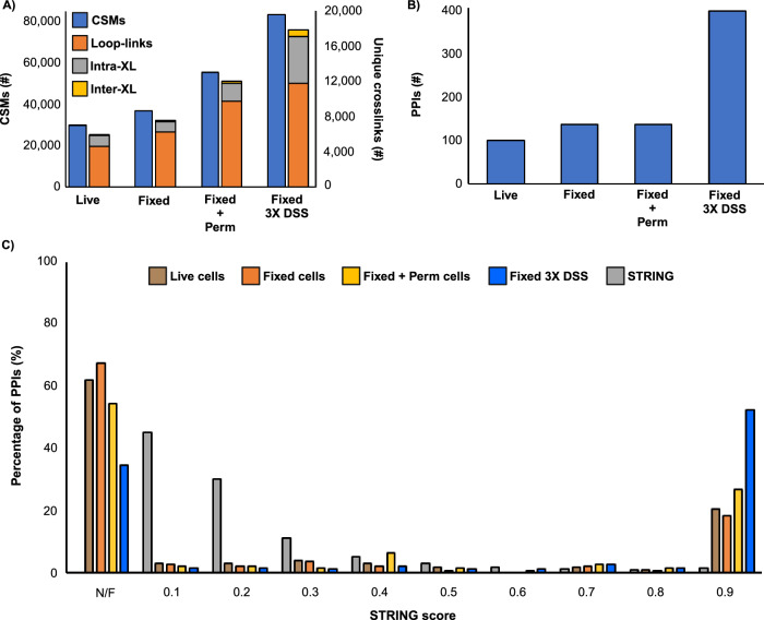
Crosslinking mass spectrometry (XL-MS) has the potential to map the interactome of the cell with high resolution and depth of coverage. However, current in vivo XL-MS methods are hampered by crosslinkers that demonstrate low cell permeability and require long reaction times. Consequently, interactome sampling is not high and long incubation times can distort the cell, bringing into question the validity any protein interactions identified by the method. We address these issues with a fast formaldehyde-based fixation method applied prior to the introduction of secondary crosslinkers. Using human A549 cells and a range of reagents, we show that 4% formaldehyde fixation with membrane permeabilization preserves cellular ultrastructure and simultaneously improves reaction conditions for in situ XL-MS. Protein labeling yields can be increased even for nominally membrane-permeable reagents, and surprisingly, high-concentration formaldehyde does not compete with conventional amine-reactive crosslinking reagents. Prefixation with permeabilization uncouples cellular dynamics from crosslinker dynamics, enhancing control over crosslinking yield and permitting the use of any chemical crosslinker.


Conflict of interest statement

The authors declare no competing interests.

Figures

Fig. 1
Fig. 1. Conventional in situ XL-MS distorts cell structure.
A Fluorescent imaging of actin cytoskeleton (Green) and DNA (blue) in A549 cells for formaldehyde-preserved cells, live cells treated with 1 mM DSS (2% DMSO), live cells treated with 2% DMSO, and formaldehyde-preserved cells treated with 1 mM DSS (2% DMSO). B Cells counted for presence (orange)/absence (blue) of cellular disruptions during respective treatments (n ≥ 100 cells per treatment). See methods for indicators of disruption. Distorted cells in orange, nondistorted cells in blue. Results consistent with 6 similar experiments.
Fig. 2
Fig. 2. Pre-stabilizing cells with formaldehyde does not impact protein labeling.
A Average percent proteome labeling using N-(propionyloxy)succinimide across E. coli and human A549 cells with increasing concentrations of formaldehyde (n = 2 biological replicates). B Comparison of percent proteome labeling with biotin-X-NHS (blue) and sulfo-NHS-LC-biotin (orange) in A549 cells for live, fixed, and fixed + permeabilized states (n = 2 biological replicates). C Histogram of identified protein abundance from human non-labeled lysate (light orange), fixed only (purple), and D fixed + permeabilized (red), labeled with biotin-X-NHS. Protein abundancies retrieved from PaxDb.
Fig. 3
Fig. 3. Workflow for formaldehyde pre-stabilization followed by in situ XL-MS.
Cells are fixed with 4% formaldehyde for 10 minutes. After fixation, excess formaldehyde is washed away prior to the introduction of the crosslinker. After secondary crosslinking, cells are collected, lysed, and formaldehyde linkages are reversed by boiling. Extracted protein is then cleaned up via a single-pot, solid-phase-enhanced sample-preparation (SP3) protocol and digested overnight with trypsin. Peptides then undergo high-pH fractionation for LC-MS/MS data acquisition. Data were then processed using pLink 2. Created with Biorender.com.
Fig. 4
Fig. 4. Effect of fixation and permeabilization on in situ XL-MS.
A Number of Number of Crosslink Spectral Matches (CSMs, blue) and unique crosslinks (orange: loop-links, grey: intraprotein, and yellow: interprotein) identified from in situ crosslinking with DSS, in either live cells, fixed cells, fixed and permeabilized cells, or fixed cells with a 3X DSS treatment. B Number of Protein-Protein Interactions (PPIs) identified from in situ crosslinking with DSS, in either live cells, fixed cells, fixed and permeabilized cells, and fixed cells with a 3X DSS treatment. C STRING score distribution for STRING PPIs (grey), live cells (brown), fixed cells (orange), fixed + permeabilized cells (yellow), and fixed cells with a 3X DSS treatment (blue); PPIs not found in STRING database labeled as not-found (N/F).
Fig. 5
Fig. 5. In situ crosslinking of fixed and permeabilized A549 cells with PhoX.
A Number of Crosslink Spectral Matches (CSMs, blue) and unique crosslinks (orange: loop-links, grey: intraprotein, and yellow: interprotein) identified from in situ crosslinking with 1X or 3X PhoX, in fixed and permeabilized cells. B STRING score distribution for STRING Protein-Protein Interactions (PPIs, grey), 1X in situ PhoX crosslinked cells (orange), and 3X in situ PhoX crosslinked cells (blue); PPIs not present in STRING database labeled as not-found (N/F). C PPI network plot of all detected interactions from 3X in situ PhoX crosslinking. D 3X PhoX crosslinks mapped to the McM—DNA replisome (mapped to PDB 7PLO) and E Ku70/Ku80 (mapped to PDB 1JEQ). Crosslinks below 20 Å are colored yellow, and crosslinks between 20 Å and 35 Å are coloured blue. F Histogram of Cα-Cα distances of 3X PhoX crosslinks mapped to all known structures.

References

    1. Figeys, D., McBroom, L. D. & Moran, M. F. Mass spectrometry for the study of protein-protein interactions. Methods24, 230–239 (2001). - PubMed
    1. Lakey, J. H. & Raggett, E. M. Measuring protein-protein interactions. Curr. Opin. Struct. Biol.8, 119–123 (1998). - PubMed
    1. Ho, Y. et al. Systematic identification of protein complexes in Saccharomyces cerevisiae by mass spectrometry. Nature415, 180–183 (2002). - PubMed
    1. Piersimoni, L., Kastritis, P. L., Arlt, C. & Sinz, A. Cross-Linking Mass Spectrometry for Investigating Protein Conformations and Protein-Protein Interactions-A Method for All Seasons. Chem. Rev.122, 7500–7531 (2022). - PubMed
    1. Rafiei, A. et al. Doublecortin engages the microtubule lattice through a cooperative binding mode involving its C-terminal domain. Elife11, e66975 (2022). - PMC - PubMed

Publication types

LinkOut - more resources