. 2024 Oct 2;15(1):8530.
doi: 10.1038/s41467-024-52810-8.

Interleukin-11 causes alveolar type 2 cell dysfunction and prevents alveolar regeneration

Affiliations

Interleukin-11 causes alveolar type 2 cell dysfunction and prevents alveolar regeneration

Benjamin Ng et al. Nat Commun. .

Abstract

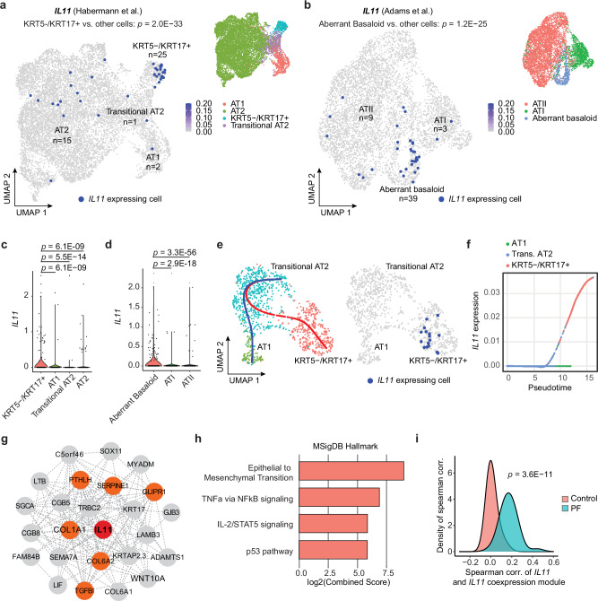
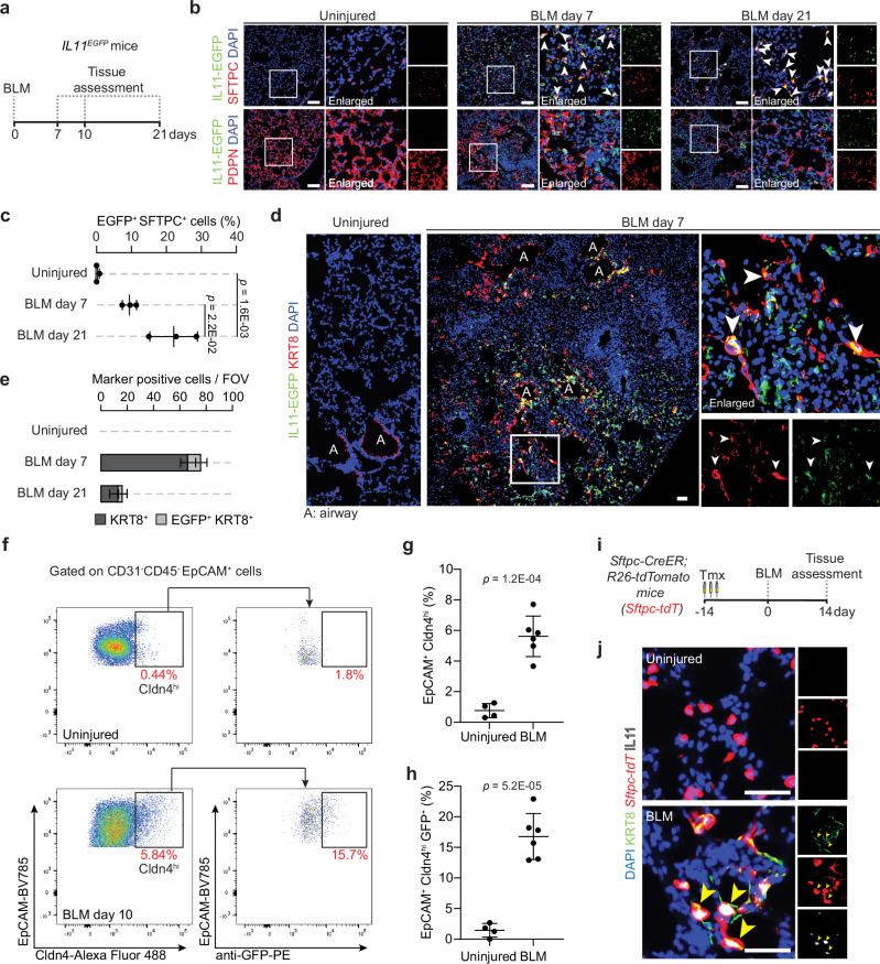
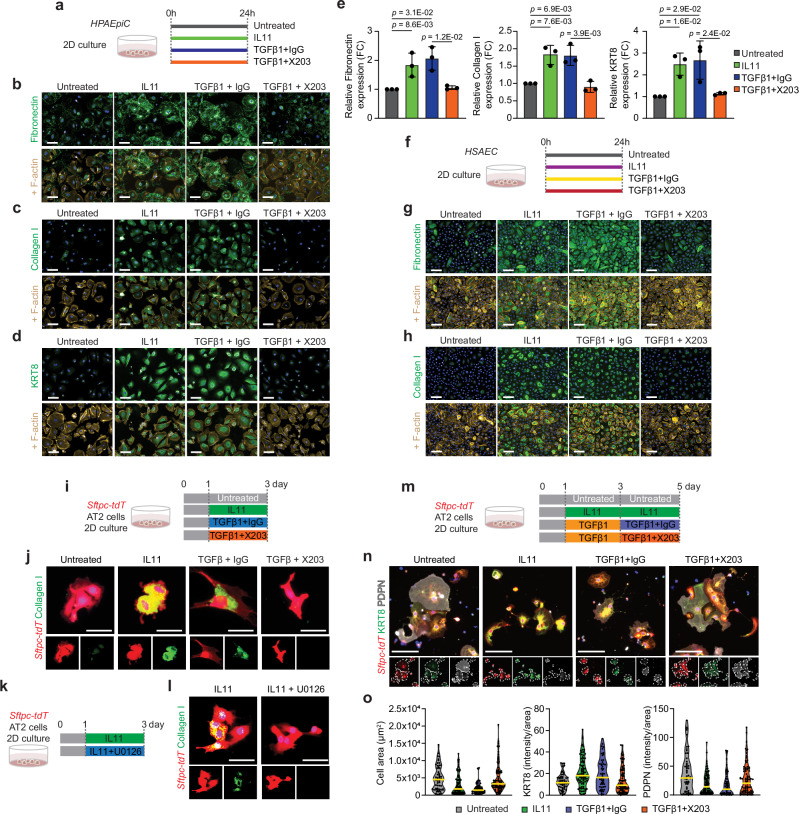
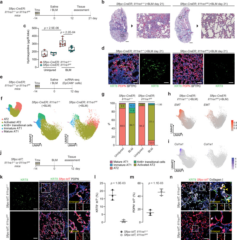
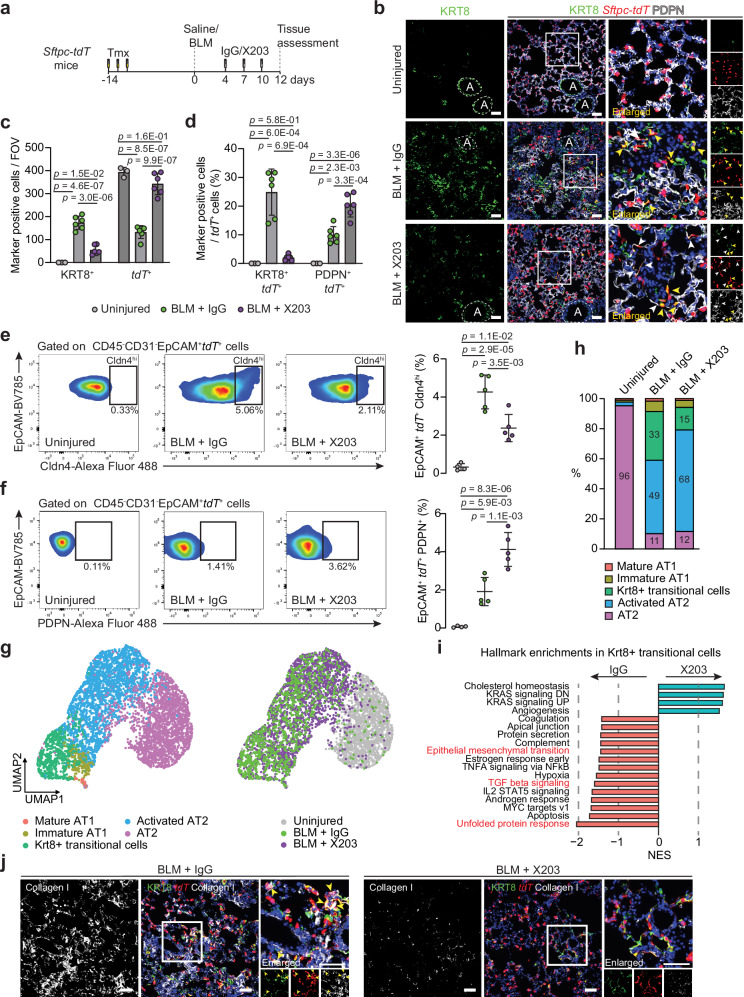
In lung disease, persistence of KRT8-expressing aberrant basaloid cells in the alveolar epithelium is associated with impaired tissue regeneration and pathological tissue remodeling. We analyzed single cell RNA sequencing datasets of human interstitial lung disease and found the profibrotic Interleukin-11 (IL11) cytokine to be highly and specifically expressed in aberrant KRT8+ basaloid cells. IL11 is similarly expressed by KRT8+ alveolar epithelial cells lining fibrotic lesions in a mouse model of interstitial lung disease. Stimulation of alveolar epithelial cells with IL11 causes epithelial-to-mesenchymal transition and promotes a KRT8-high state, which stalls the beneficial differentiation of alveolar type 2 (AT2)-to-AT1 cells. Inhibition of IL11-signaling in AT2 cells in vivo prevents the accumulation of KRT8+ cells, enhances AT1 cell differentiation and blocks fibrogenesis, which is replicated by anti-IL11 therapy. These data show that IL11 inhibits reparative AT2-to-AT1 differentiation in the damaged lung to limit endogenous alveolar regeneration, resulting in fibrotic lung disease.


Conflict of interest statement

S.A.C. is a co-inventor of the patent applications (WO/2017/103108) and (WO/2018/109170). S.A.C., W.-W.L., and B.N. are co-inventors of the patent application (WO/2019/073057). S.A.C. is a co-founder and shareholder of Enleofen Bio PTE LTD, a company that develops anti-IL11 therapeutics, which were acquired for further development by Boehringer Ingelheim. The remaining authors declare no competing interests.

Figures

Fig. 1
Fig. 1. IL11 is specifically expressed by aberrant KRT5-/KRT17+ epithelial cells in human pulmonary fibrosis.
Uniform manifold approximation and projection (UMAP) visualization of IL11 expressing single cells (colored in dark blue) in various alveolar epithelial cell populations (colored by cell types) in scRNA-seq data from Control and pulmonary fibrosis (PF) samples in a Habermann et al. (GSE135893) and b Adams et al. (GSE136831) datasets. The number of IL11-expressing cells in each cell cluster is indicated. P values determined by one-tailed hypergeometric test for enrichment in KRT5-/KRT17+ or aberrant basaloid versus other cell types. Violin plot indicating the expression of IL11 in various alveolar epithelial cell populations in c Habermann et al. dataset and d Adams et al. datasets. P values were determined by a two-tailed Mann–Whitney test between KRT5-/KRT17+ or aberrant basaloid versus other cell types. e UMAP visualization of IL11 expressing single cells colored in dark blue (right panel) and colored dots indicate cell type clustering (left panel). The blue line indicates the differentiation trajectory of transitional AT2 to AT1 cells; the red line indicates the differentiation trajectory from transitional AT2 to KRT5-/KRT17+. Data were composed of cells from PF samples in the Habermann et al. dataset. f Expression of IL11 in the pseudotime trajectory from transitional AT2 to KRT5-/KRT17+ versus from transitional AT2 to AT1 cells in the Habermann et al. dataset. g Network of genes in the IL11 co-expression module in the transitional AT2 to KRT5-/KRT17+ cell trajectory in combined Habermann et al. and Adams et al. datasets. IL11 is colored in red, and genes related to epithelial to mesenchymal transition are colored in orange. h Pathway enrichment of genes in the IL11 co-expression module using MSigDB Hallmark database. i Density plot displaying the distribution of Spearman correlation between the gene expression of IL11 and IL11 co-expression module in Control (salmon color) and PF (turquoise color) transitional AT2 and KRT5-/KRT17+ cells in Habermann dataset. P value was determined by the two-tailed Kolmogorov–Smirnov test.
Fig. 2
Fig. 2. IL11 is expressed by transitional alveolar epithelial cells after bleomycin-induced lung injury in mice.
a Schematic showing the induction of lung fibrosis via oropharyngeal injection of bleomycin (BLM) in IL11EGFP reporter mice. b, c Representative images and quantification of immunostaining for GFP and SFTPC or PDPN in injured regions of the lungs 7 or 21 days post-BLM challenge; n = 3 mice/group. d Representative images of immunostaining for GFP and KRT8 in the injured regions of the lung 7 days post-BLM injury. White arrowheads indicate marker-positive IL11EGFP+ cells. e Quantification of alveolar KRT8+ and IL11EGFP+ KRT8+ cells per field of view (FOV); n = 3 mice/group. fh Flow cytometry analysis and quantification of g EpCAM+ Cldn4hi cells and h EpCAM+ Cldn4hi GFP+ cells; n = 4 control and 6 injured mice/group. i Schematic showing the period of tamoxifen (Tmx) administration and induction of lung fibrosis in Sftpc-CreER; R26-tdTomato (Sftpc-tdT) mice. j Representative images of immunostaining for KRT8 and IL11 in the lungs of Sftpc-tdT mice post-BLM injury. DAPI for nuclei. Yellow arrowheads indicate IL11+ KRT8+ tdT+ cells. Scale bars: 50 µm. Data were representative of three independent experiments (b, d, j). Data (c, d, g, h) were mean ± s.d. P values were determined by one-way ANOVA with Tukey’s multiple comparison test (c) or two-tailed Student’s t-test (g, h).
Fig. 3
Fig. 3. IL11 induces KRT8 expression and profibrotic features in alveolar and distal airway epithelial cells and stalls AT2-to-AT1 cell differentiation in vitro.
a Experimental design for the 2D culture of primary human pulmonary alveolar epithelial cells (HPAEpiC) treated with IL11 (5 ng/ml) or TGFβ1 (5 ng/ml) in the presence of anti-IL11 (X203) or IgG control antibodies (2 µg/ml). be Immunostaining and quantification of Fibronectin, Collagen I or KRT8 expression in HPAEpiC. n = 3/group. FC fold change. f Experimental design for the 2D culture of primary human small airway epithelial cells (HSAEC) treated with IL11 (5 ng/ml) or TGFβ1 (5 ng/ml) with anti-IL11 (X203) or IgG control antibodies (2 µg/ml). g, h Immunostaining of Fibronectin or Collagen I expression in HSAEC. n = 2/group. Quantification data were shown in Supplementary Fig. 11c. i Experimental design and j immunostaining of Collagen I expression in Sftpc-tdT+ AT2 cells treated with IL11 (5 ng/ml), TGFβ1 (5 ng/ml), X203 or IgG control antibodies (2 µg/ml). k Experimental design and l immunostaining images of Collagen I in Sftpc-tdT+ AT2 cells treated with IL11 (5 ng/ml) and MEK inhibitor U1026 (10 µM). Quantification of Collagen I expression are shown in Supplementary Fig. 11g, h. m Experimental design for the in vitro differentiation of Sftpc-tdT+ AT2 cells to AT1 cells in the presence of IL11 (5 ng/ml), TGFβ1 (5 ng/ml), X203 or IgG control antibodies (2 µg/ml). n Immunostaining of KRT8 and PDPN in Sftpc-tdT+ cells at day 5. Individual cells are highlighted within dotted lines in split channels. o Violin plots of cell size and immunostaining quantification of KRT8 or PDPN expression in tdT+ cells at day 5; One representative dataset of two independent experiments are shown (n > 50 cells/group). Data were representative of three independent experiments in (bd) or two independent experiments in (g, h, j, l, n). Scale bars: 100 µm (bd, g, h), 25 µm (j, l), 50 µm (o). Data in (e) are mean ± s.d. and P values determined by one-way ANOVA with Tukey’s multiple comparison test.
Fig. 4
Fig. 4. IL11 signaling in AT2 cells disrupts AT2-to-AT1 differentiation after injury and is required for lung fibrosis.
a Schematic showing the induction of lung injury in Sftpc-CreER; Il11ra1fl/fl or Il11ra1+/+ mice via oropharyngeal injection of BLM and the time points of tissue assessment. b Representative images of Masson’s trichrome staining and c lung hydroxyproline content of right caudal lobes of Sftpc-CreER; Il11ra1fl/fl or Il11ra1+/+ mice 21 days post-BLM injury; n = 4 control and 9 injured mice/genotype. d Immunostaining of KRT8 and PDPN in the lungs of BLM-treated Sftpc-CreER; Il11ra1fl/fl or Il11ra1+/+ mice. e Schematic showing the induction of lung injury in Sftpc-CreER; Il11ra1fl/fl or Il11ra1+/+ mice and at day 12, lung single cells were enriched for EpCAM+ epithelial cells and assessed using scRNA-seq. f UMAP embedding of alveolar epithelial cells from scRNA-seq analysis showing distinct cell states and g distribution of alveolar epithelial cell states from Sftpc-CreER; Il11ra1fl/fl or Il11ra1+/+ mice 12 days post-BLM injury. h UMAP visualization of single cells colored by gene expression signature score for EMT pathway from the MSigDB Hallmark gene sets and i UMAP visualization of Col1a1 expressing single cells (purple dots) from AT2-lineage cells from Sftpc-CreER; Il11ra1fl/fl or Il11ra1+/+ mice post-BLM challenge. j Schematic showing the induction of lung injury in Sftpc-tdT; Il11ra1fl/fl or Il11ra1+/+ mice. Lung tissues were assessed 12 days post-BLM challenge. k Immunostaining of KRT8 and PDPN in the lungs of BLM-treated Sftpc-tdT; Il11ra1 fl/fl or Il11ra1+/+ mice and the proportion of l KRT8+ tdT+ or m PDPN+ tdT+ relative to total tdT+ cells, n = 3 mice/group. n Images of immunostaining for KRT8 and Collagen I in lungs from BLM-treated Sftpc-tdT; Il11ra1 fl/fl or Il11ra1+/+ mice. Data were representative of three independent experiments (d, n). Data were median ± IQR and whiskers represent minimum to maximum values (c) and mean ± s.d. (l, m). P values were determined by one-way ANOVA with Tukey’s multiple comparison test (c), two-tailed Student’s t-test (l, m). Scale bars: 1000 µm (b), 100 µm (d, k, n).
Fig. 5
Fig. 5. Pharmacological inhibition of IL11 prevents the accumulation of profibrotic transitional epithelial cells and enhances AT2-to-AT1 cell differentiation during lung injury in vivo.
a Schematic showing the administration time points of BLM and X203 or IgG antibodies in Sftpc-tdT mice. Lung tissues were assessed 12 days post-BLM challenge. b Images of immunostaining for KRT8 and PDPN in lung sections from BLM-injured Sftpc-tdT mice treated with X203 or IgG antibodies. Yellow arrowheads indicate KRT8+ tdT+ cells. White arrowheads indicate flattened PDPN+ tdT+ cells. Recognizable airway regions are demarcated by white dotted lines. c Numbers of KRT8+, tdT+ per field of view (FOV) and d the proportions of KRT8+ tdT+ cells or PDPN+ tdT+ cells divided by the number of tdT+ cells in injured lung regions (n = 3 control and 6 per BLM+antibody treatment group). e Flow cytometry analysis and quantification of lung. h EpCAM+ tdT+ Cldn4hi cells and f EpCAM+ tdT+ PDPN+ cells (n = 4 control and 5 per BLM+antibody treatment group). g UMAP embedding of alveolar epithelial cells from scRNA-seq analysis showing distinct cell states and treatment groups and h distribution of alveolar epithelial cell states from uninjured and BLM-injured mice (12 days post-BLM) treated with X203 or IgG antibodies. i Normalized enrichment scores (NES) of pathways significantly enriched in Krt8+ transitional cells from the MSigDB Hallmark gene sets. j Representative images of immunostaining for KRT8 and Collagen I in lung sections of BLM-injured mice treated with X203 or IgG antibodies. Yellow arrowheads indicate Collagen I-expressing KRT8+ tdT+ cells. Data were representative of at least three independent experiments (b, j). Scale bars: 100 µm (b, j). Data were mean ± s. d. and P values were determined by one-way ANOVA with Tukey’s multiple comparison test (cf).

References

    1. Barkauskas, C. E. et al. Type 2 alveolar cells are stem cells in adult lung. J. Clin. Invest.123, 3025–3036 (2013). - PMC - PubMed
    1. Desai, T. J., Brownfield, D. G. & Krasnow, M. A. Alveolar progenitor and stem cells in lung development, renewal and cancer. Nature507, 190–194 (2014). - PMC - PubMed
    1. Basil, M. C. et al. The cellular and physiological basis for lung repair and regeneration: past, present, and future. Cell Stem Cell26, 482–502 (2020). - PMC - PubMed
    1. Zepp, J. A. & Morrisey, E. E. Cellular crosstalk in the development and regeneration of the respiratory system. Nat. Rev. Mol. Cell Biol.20, 551–566 (2019). - PMC - PubMed
    1. Xu, Y. et al. Single-cell RNA sequencing identifies diverse roles of epithelial cells in idiopathic pulmonary fibrosis. JCI Insight1, e90558 (2016). - PMC - PubMed

Publication types

MeSH terms

Associated data