. 2024 Oct 3;73(12):238.
doi: 10.1007/s00262-024-03824-0.

The relevance of tumor target expression levels on IgA-mediated cytotoxicity in cancer immunotherapy

Affiliations

The relevance of tumor target expression levels on IgA-mediated cytotoxicity in cancer immunotherapy

Chilam Chan et al. Cancer Immunol Immunother. .

Abstract

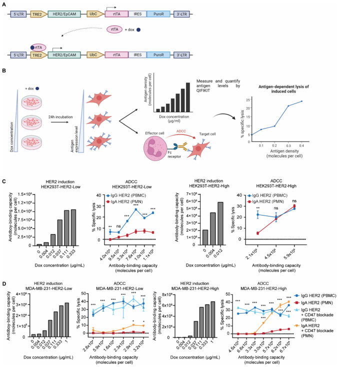
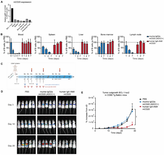
Recent advances in cancer immunotherapy, particularly the success of immune checkpoint inhibitors, have reignited interest in targeted monoclonal antibodies for immunotherapy. Antibody therapies aim to minimize on-target, off-tumor toxicity by targeting antigens overexpressed on tumor cells but not on healthy cells. Despite considerable efforts, some therapeutic antibodies have been linked to dose-limiting side effects. Our hypothesis suggests that the efficacy of IgG leads to a lower target expression threshold for tumor cell killing, contributing to these side effects. Earlier, therapeutic IgG antibodies were reformatted into the IgA isotype. Unlike IgG, which primarily engages Fc gamma receptors (FcγR) to induce antibody-dependent cellular cytotoxicity (ADCC) by NK cells and antibody-dependent cellular phagocytosis (ADCP) by monocytes/macrophages, IgA antibodies activate neutrophils through the Fc alpha receptor I (CD89, FcαRI). In previous studies, it appeared that IgA may require a higher target expression threshold for effective killing, and we aimed to investigate this in our current study. Moreover, we investigated how blocking the myeloid checkpoint CD47/SIRPα axis affect the target expression threshold. Using a tetracycline-inducible expression system, we regulated target expression in different cell lines. Our findings from ADCC assays indicate that IgA-mediated PMN ADCC requires a higher antigen expression level than IgG-mediated PBMC ADCC. Furthermore, blocking CD47 enhanced IgA-mediated ADCC, lowering the antigen threshold. Validated in two in vivo models, our results show that IgA significantly reduces tumor growth in high-antigen-expressing tumors without affecting low-antigen-expressing healthy tissues. This suggests IgA-based immunotherapy could potentially minimize on-target, off-tumor side effects, improving treatment efficacy and patient safety.

Keywords: Antigen expression levels; CD47/SIRPα; IgA; Immunotherapy; Neutrophils.


Conflict of interest statement

The authors declare no competing interests.

Figures

Fig. 1
Fig. 1
Influence of HER2 and CD47 expression levels on IgG and IgA-mediated ADCC capacity in breast cancer cell lines. A HER2 expression levels depicted as number of molecules per cell on breast cancer cell lines: MDA-MB-468, MDA-MB-175, MDA-MB-453, and SK-BR-3. Expression levels were quantified by QIFIKIT using 10 µg/mL mIgG1 anti-human HER2 antibody. Data shown as mean ± SD from three independent experiments. B PBMC- and PMN-mediated ADCC of the breast cancer cell lines, using IgG1 HER2 and IgA HER2, respectively. Significance was determined as compared to MDA-MB-468. Assay was performed in technical triplicates and repeated independently with three donors. Specific lysis is shown as mean ± SEM for one representative experiment. ns > 0.05, ***p < 0.001, by two-way ANOVA followed by Tukey’s multiple comparisons test. C CD47 expression levels depicted as number of molecules per cell on MDA-MB-468, MDA-MB-175, MDA-MB-453, and SK-BR-3. Expression levels were quantified by QIFIKIT using 10 µg/mL mIgG1 anti-human CD47 antibody. Data shown as mean ± SD from three independent experiments. D ADCC assay against breast cancer cell lines mediated by IgG1 HER2, IgA HER2 and IgA HER2 in combination with CD47 blockade. CD47 was pre-blocked using SIRPα fusion protein at 10 µg/mL. In all ADCC assays, PBMCs were used as effector cells in IgG ADCCs (E:T ratio 100:1); while, PMNs (E:T ratio 40:1) were used for IgA ADCCs. Antibody concentrations ranged between 0 and 10 µg/mL. Assay was performed in technical replicates (n = 3) and repeated with three independent donors in separate assays. Specific lysis is shown as mean ± SEM for one representative experiment from one donor. ns > 0.05, **p < 0.01, ***p < 0.001, by two-way ANOVA followed by Tukey’s multiple comparisons test
Fig. 2
Fig. 2
ADCC capacity related to dox-inducible HER2 expression in HEK293T-HER2 and MDA-MB-231-HER2 cells. A Schematic representation of the Tet-On inducible system. A ubiquitin C promoter (UbC) controls the expression of rtTA (reverse tetracycline transactivator) and puromycin resistance (PuroR). In the presence of doxycycline (dox), rtTA undergoes conformational changes and binds to TRE2 (tetracycline response element 2), leading to the translation of the target gene. B Overview of the experimental setup: Cells are cultured with varying dox concentrations for 24-h, resulting in titrated levels of antigen expression. These cells are subsequently tested in ADCC experiments, with concurrent quantification of antigen levels using QIFIKIT. Antigen expression levels are correlated to antibody-mediated killing. The graphs presented here are illustrative and do not represent actual data. C IgG and IgA-mediated ADCC assays against two HEK293T-HER2 clones with low and high HER2 expression levels. The ADCC assays for both clones were performed using effector cells from a single donor and repeated across three different donors (biological triplicates). One representative assay of each clone is shown. D ADCC assay against MDA-MB-231-HER2 (low and high expression) cells, using IgG HER2, IgA HER2, and IgA HER2 in combination with CD47 blockade. Effector cells were isolated from a healthy donor. For MDA-MB-231-HER2 with low expression, assays were performed in technical triplicates and independently repeated with 2 donors; while, those with high expression were done in technical triplicates and independently repeated with 3 donors. A representative graph for each clone is shown. In all ADCC assays, dox in concentration indicated in the graphs were used to induce HER2 expression. HER2 levels were quantified with 10 µg/mL HER2 antibody in a QIFIKIT assay. Lysis was assessed in a 51Cr release assay using PBMCs (E:T 100:1) for IgG-mediated and PMNs (40:1) for IgA-mediated cytotoxicity. Cytotoxicity was evaluated with 10 µg/mL antibody, with optional CD47 blocking by 10 µg/mL SIRPα fusion protein. The mean ± SEM of specific lysis is shown. Statistical significance as compared to IgA HER2: ns > 0.05, *p < 0.05, **p < 0.01, ***p < 0.001, determined by two-way ANOVA followed by Tukey’s multiple comparisons test. Illustrations were created using Biorender
Fig. 3
Fig. 3
Antibody-mediated ADCC capacity on dox-inducible HeLa-EpCAM cells. EpCAM expression was induced using doxycycline concentrations ranging from 0 to 1 µg/mL and quantified through QIFIKIT with a primary EpCAM antibody concentration of 10 µg/mL. ADCC assays utilized PBMCs (E:T 100:1) for IgG and PMNs (40:1) for IgA, with antibody concentrations of 10 µg/mL. CD47 was blocked using 10 µg/mL SIRPα fusion protein. Effector cells were freshly isolated from a healthy donor. Experiment was repeated in at least three different clones. Significance is shown as compared to IgA-mediated lysis. Specific lysis is shown as mean ± SEM. ns > 0.05, ***p < 0.001, by two-way ANOVA followed by Tukey’s multiple comparisons test
Fig. 4
Fig. 4
Anti-tumoral effect of IgG HER2 and IgA HER2 in A431-luc2 tumor-bearing mice. A HER2 expression levels of A431-luc2 and A431-luc2-HER2 in comparison with SK-BR-3, measured via flow cytometry. The mean ± SD of geometric MFI is presented. *p < 0.05, ***p < 0.001, determined using one-way ANOVA with Bonferroni’s multiple comparisons test. B IgG HER2 and IgA HER2 lysis against A431-luc2 and A431-luc2-HER2 cells were evaluated using PBMCs (E:T 100:1) for IgG and PMNs (40:1) for IgA. Antibody concentrations ranged from 0 to 10 µg/mL. CD47 was pre-blocked with 10 µg/mL SIRPα fusion protein. The assay was performed in technical triplicates using effector cells from a single healthy donor and was repeated three times with independent donors (biological triplicates).Individual values and the mean ± SEM of specific lysis from one representative experiment are presented. ns > 0.05, *p < 0.05 ***p < 0.001, as determined by two-way ANOVA followed by Tukey’s multiple comparisons test. C Bioluminescence imaging (BLI) measurements depicting the tumor growth of A431-luc2 tumor in (CD89 Tg) SCID mice. Detailed treatment regimen is outlined in the methods section. Tumor sizes are presented as mean ± SEM. *p < 0.05, **p < 0.01, ***p < 0.001, as determined by two-way ANOVA followed by Tukey’s multiple comparisons test. D Tumor outgrowth of A431-luc2-HER2 tumors in (CD89 Tg) SCID mice assessed through BLI. The detailed treatment regimen is provided in the methods section. BLI measurements are shown as BLI signal intensity, measured in photons per second (e/-s), and are displayed as the mean ± SEM. Statistical significance (ns > 0.05, ***p < 0.001) was determined using two-way ANOVA followed by Tukey’s multiple comparisons test. For both in vivo experiments, CD89 Tg SCID mice were used in the groups receiving IgA; while, wild-type SCID mice were used for the other groups
Fig. 5
Fig. 5
IgA-mediated selective targeting of lymphoma cells while preserving endogenous B cells A The mCD20 expression level of BCL-1-luc2 cells was compared to endogenous B cells in Balb/c mice, measured via flow cytometry. B The depletion of endogenous B cells was monitored following a single dose of either PBS, 5 mg/kg murine afucosylated IgG2a mCD20, or 5 mg/kg human IgA-Alb8 mCD20 in CD89 Tg Balb/c mice for up to 7 days. The percentage of B cells, compared to the PBS vehicle control, was measured in blood, spleen, liver, bone marrow, and lymph nodes, and the results are presented as mean ± SD. C A schematic overview of the syngeneic BCL-1 lymphoma model in CD89 Tg Balb/c mice. In brief, a total of 5 × 105 BCL-1-luc2 cells and a dose of PEG-G-CSF were injected on day 0, followed by another PEG-G-CSF dose on day 7. Treatment with PBS, 6 mg/kg murine afucosylated IgG2a mCD20, or 6 mg/kg human IgA-Alb8 mCD20 began on day 3. IgA was administered on days 3, 5, 7, 10, 12, and 14; while, IgG was given on days 3 and 10. The dosage frequency differs because of the isotype's different half-lives. This dosing regimen is optimized to maintain similar serum titers. Tumor growth was monitored by bioluminescence imaging twice a week. D Representative BLI images of the different treatment groups on day 3, 14 and 28. E BCL-1 tumor outgrowth is depicted as a percentage increase in BLI signal from day 3. The percentages are presented as mean ± SEM. **p < 0.01, determined by two-way ANOVA followed by Tukey’s multiple comparisons test

Similar articles

Cited by

References

    1. Zeller T, Lutz S, Münnich IA, Windisch R, Hilger P, Herold T, Tahiri N, Banck JC, Weigert O, Moosmann A et al (2022) Dual checkpoint blockade of CD47 and LILRB1 enhances CD20 antibody-dependent phagocytosis of lymphoma cells by macrophages. Front Immunol. 10.3389/fimmu.2022.929339 - PMC - PubMed
    1. Fan X, Krieg S, Kuo CJ, Wiegand SJ, Rabinovitch M, Druzin ML, Brenner RM, Giudice LC, Nayak NR (2008) VEGF blockade inhibits angiogenesis and reepithelialization of endometrium. FASEB J. 10.1096/fj.08-111401 - PMC - PubMed
    1. de Taeye SW, Bentlage AEH, Mebius MM, Meesters JI, Lissenberg-Thunnissen S, Falck D, Sénard T, Salehi N, Wuhrer M, Schuurman J et al (2020) FcγR binding and ADCC activity of human IgG allotypes. Front Immunol. 10.3389/fimmu.2020.00740 - PMC - PubMed
    1. Zinn S, Vazquez-Lombardi R, Zimmermann C, Sapra P, Jermutus L, Christ D (2023) Advances in antibody-based therapy in oncology. Nat Cancer 4:165–180 - PubMed
    1. Goydel RS, Rader C (2021) Antibody-based cancer therapy. Oncogene 40:3655–3664 - PMC - PubMed

Substances

LinkOut - more resources