. 2024 Nov;16(11):2918-2945.
doi: 10.1038/s44321-024-00143-w. Epub 2024 Oct 2.

A new class of capsid-targeting inhibitors that specifically block HIV-1 nuclear import

Affiliations

A new class of capsid-targeting inhibitors that specifically block HIV-1 nuclear import

Aude Boulay et al. EMBO Mol Med. 2024 Nov.

Abstract

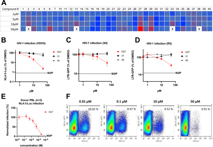
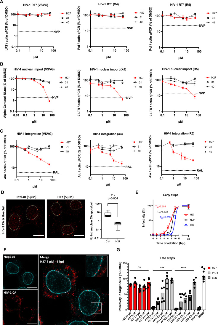
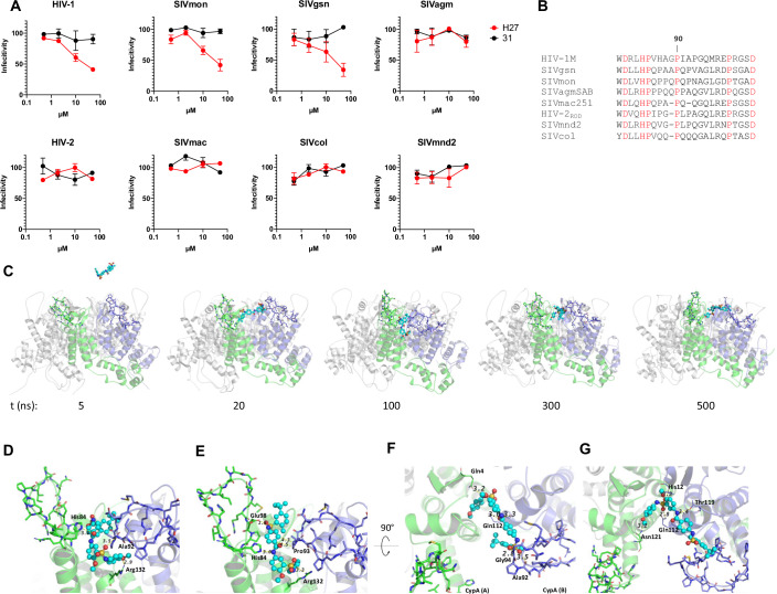
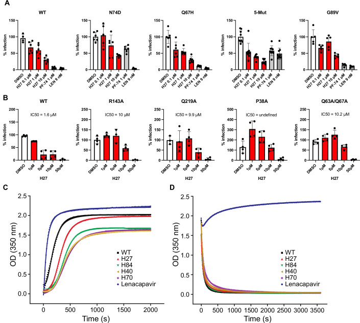
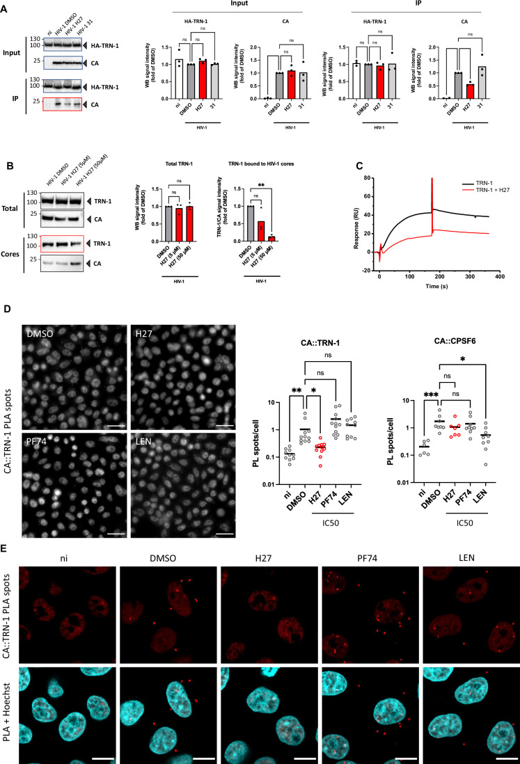
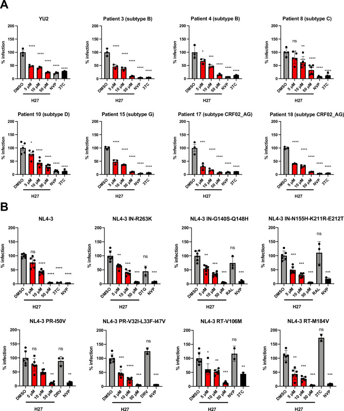
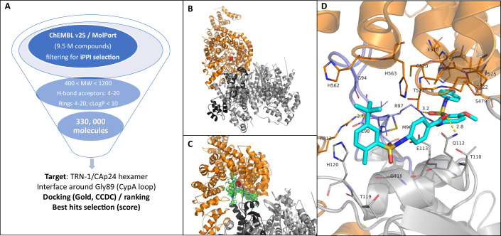
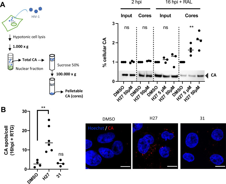
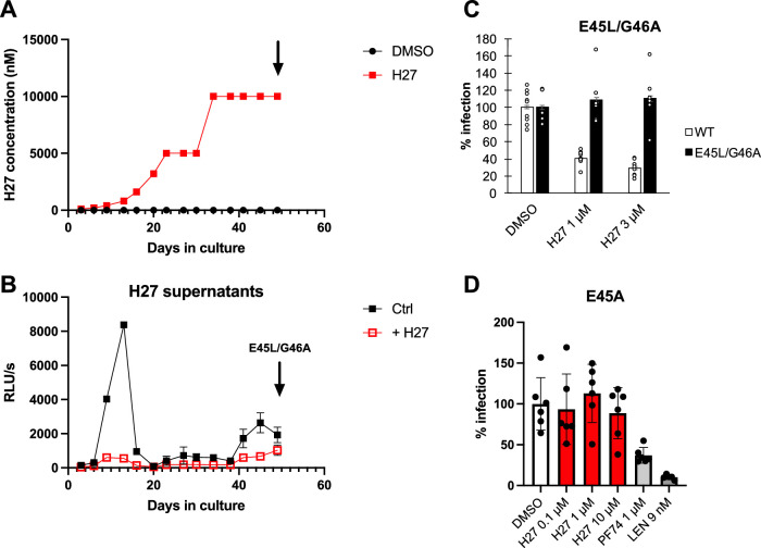
HIV-1 capsids cross nuclear pore complexes (NPCs) by engaging with the nuclear import machinery. To identify compounds that inhibit HIV-1 nuclear import, we screened drugs in silico on a three-dimensional model of a CA hexamer bound by Transportin-1 (TRN-1). Among hits, compound H27 inhibited HIV-1 with a low micromolar IC50. Unlike other CA-targeting compounds, H27 did not alter CA assembly or disassembly, inhibited nuclear import specifically, and retained antiviral activity against PF74- and Lenacapavir-resistant mutants. The differential sensitivity of divergent primate lentiviral capsids, capsid stability and H27 escape mutants, together with structural analyses, suggest that H27 makes multiple low affinity contacts with assembled capsid. Interaction experiments indicate that H27 may act by preventing CA from engaging with components of the NPC machinery such as TRN-1. H27 exhibited good metabolic stability in vivo and was efficient against different subtypes and circulating recombinant forms from treatment-naïve patients as well as strains resistant to the four main classes of antiretroviral drugs. This work identifies compounds that demonstrate a novel mechanism of action by specifically blocking HIV-1 nuclear import.

Keywords: Antiretroviral Therapy; Capsid; Drug Discovery; HIV-1; Nuclear Import.


Conflict of interest statement

Disclosure and competing interests statement The authors declare no competing interests.

Figures

Figure 1
Figure 1. H27 inhibits HIV-1 infection at low micromolar concentrations.
(A) Heat map of the HIV-1 infectivity screen in HeLa-R5 cells of compounds selected by virtual screening. HeLa-R5-Luc cells were treated with compounds at the indicated concentrations for 2 h, then infected with VSV-G pseudotyped HIV-1-CMV-Nluc. Infectivity (NLuc) and viability (Luc) were assessed at 24 h post-infection (hpi). Results show mean NLuc/Luc values relative to DMSO control, and are representative of two independent screens. X = complete cell mortality. (BD) Infectivity assays were performed in HeLa-R5 (B) or JLTR-5G (C, D) cells pre-treated with compounds at the indicated concentrations for 2 h, then infected with VSV-G pseudotyped LAI virus or in JLTR-5G with X4-tropic NL4-3 or R5-tropic BaL virus at 1 ng p24/1000 cells for 48 h. Results show the mean ± SD from three independent experiments. The Y-axes are on a logarithmic scale. (E, F) Primary PBL from healthy donors were pre-treated with H27 at the indicated concentrations for 2 h, then infected with NL4-3-Luc (E) or NL4-3-IRES-eGFP (F), and infectivity was assessed by plate luminometry at 48 hpi and flow cytometry at 72 hpi, respectively. In (E) curves show mean values from three different donors ± SEM. In (F) flow cytometry plots are representative of three independent experiments on three different donors. Source data are available online for this figure.
Figure 2
Figure 2. H27 specifically blocks HIV-1 nuclear import.
(AC) HeLa-R5 cells were pre-treated with compounds at the indicated concentrations, then infected with VSV-G pseudotyped LAI, and JLTR-5G cells were infected with X4-tropic NL4-3 or R5-tropic BaL virus, at 1 ng p24/1000 cells for 24 h. Compounds 31 and 40 were included as negative controls since they do not affect HIV-1 infection. Results are the mean of four independent experiments ± SEM normalised for the corresponding DMSO control. The reverse transcriptase inhibitor NVP (5 µM) or the integration inhibitor RAL (10 µM) were included as a negative control in all experiments. (A) Reverse transcription (RT°) efficiency was assessed by qPCR amplification of late-reverse transcripts (LRT) or Pol gene and normalised for Actin values. (B) HIV-1 nuclear import in HeLa-R5 cells was assessed by Alpha Centauri protein complementation between a small NLuc fragment tagged C-ter of integrase (αHIVNLuc) and a large NLuc fragment fused to a NLS (CenNLSNLuc) (Fernandez et al, 2021). In JLTR-5G, HIV-1 nuclear import was assessed by qPCR amplification of 2-LTR circles and normalised for Actin values. (C) HIV-1 integration was assessed by qPCR and normalised for actin. (D) HIV-1 nuclear import was assessed by determining the localisation of CA at 6–8 hpi. Cells infected with LAI-VSV-G were fixed 6–8 hpi and labelled with anti-CA antibody and Hoechst. Confocal images acquired on an LSM800 microscope were quantified using Imaris on >300 cells from 4 independent experiments. Results are represented as a box and whisker plot with minimum to maximum bounds. Boxes extend from the 25th to 75th percentiles, while the line is plotted at the median. Statistical significance was assessed by unpaired t-test. Representative images with nuclei outlined in yellow are provided. Scale bar = 10 µm. (E) Time of addition assay in HeLa-R5 cells infected with VSV-G pseudotyped NL4-3-Luc and treated with compound 27 (H27, 5 µM), NVP (5 µM) or RAL (10 µM) at the indicated times pre/post-infection (hpi). The T50 infectivity values indicate the times at which 50% of the viral complexes became resistant to the inhibitors. Results are the mean of two independent experiments performed in duplicate ± SEM. (F) HeLa-R5 cells were treated with H27 (3 µM) and then infected with LAI-VSV-G. Cells were fixed at 6 hpi, labelled with anti-CA and -Nup214 antibodies and stained with Hoechst. Images were acquired on an LSM880 Airyscan microscope with 63 × objective. Images are representative of three independent experiments (see Appendix Fig. S5 for full conditions). Scale bar = 10 µm. (G) To test the effect of H27 on HIV-1 assembly, HEK-293T cells were transfected with an NL4-3 molecular clone and treated with H27 at the indicated concentrations after 4 h. Supernatants were collected at 48 hpt and clarified by centrifugation. HeLa-R5 cells were then infected with diluted supernatants, and β-galactosidase luminescence was read at 48 hpi. Results show the mean from two independent experiments normalised for the DMSO control. Statistical significance was assessed using ordinary One-way ANOVA (Brown–Forsythe test). ****p ≤ 0.0001, ***p ≤ 0.001, ns non-significant. Source data are available online for this figure.
Figure 3
Figure 3. Determinants for sensitivity to H27 across divergent lentiviral capsids.
(A) CHO cells were infected with chimeric SIV constructs containing CA from diverse primate lentiviruses. Infection was assessed by GFP quantification, and results are represented as % of infectivity in 0.5% DMSO for each virus. Results show the mean of two independent experiments ± SEM. (B) Sequence alignments of the CypA-binding region of HIV and SIV CA used in Fig. 3A. Fully conserved amino acids are highlighted in red and gaps are indicated as “−“. Full alignments of the CA are available in Appendix Fig. S8. (C) Snapshots from MD simulation with H27 (cyan ball & sticks) and CA hexamer (the two monomers involved in H27 binding are depicted in green and blue cartoon representation). H27 introduced into the proximity of CA selected a binding site located between two monomers near PR loops (residues depicted in sticks) after 20 ns and remained associated for the duration of the simulation (trapped in between two monomers). (DG) Close-up views of the H27 binding site after (D) 100, (E) 150, (F) 300 and (G) 500 ns of simulation. Only hydrogen bonds formed between H27 and residues from the PR loop are shown for clarity. Additional Van der Waals contacts are observed with L83, H84, A92, P93, G94, P123, I124, P125). Source data are available online for this figure.
Figure 4
Figure 4. Sensitivity of CA mutants to H27 and effect on CA assembly and disassembly.
(A) HeLa-R5 cells were treated with H27 at 0.1, 1 or 10 µM, PF74 (1 µM) or LEN (9 nM) for 2 h, then infected with NL4-3 wild-type (WT), N74D, G89V or R9-GFP-VSV-G WT, Q67H and 5Mut (Q67H, K70R, H87P, T107N and L111I) at concentrations that yielded RLU values of ~2000 to normalise infections across mutants. β-galactosidase activity was measured at 48 hpi. Results are the mean of three independent experiments ± SD. Results obtained with NL4-3 and R9 viruses were similar and, therefore, merged in a single panel (left). (B) The effect of H27 on capsid stability R9 mutants was assessed in HeLa-R5 cells in the presence of increasing concentrations of H27 or compound 31 as control. Infectivity was assessed at 48 hpi by β-galactosidase assay. Results from two independent experiments were normalised for DMSO mean values in each dataset ± SD. IC50 values were calculated by non-linear regression with variable slope (four parameters) using Prism 8. (C) Rates of HIV-1 CA assembly. Plots show the time dependence of OD350 after the introduction of HIV-1 CA into high-salt assembly conditions in the presence of the indicated compounds. (D) Rates of HIV-1 CA disassembly. Plots show the time dependence of OD350 after dilution of CA-assemblies from high-salt assembly conditions into a low-salt buffer in the presence of the indicated compounds. In each panel, a representative assay from three to four replicates is shown. The raw data were plotted as points, and the lines are the curve of best fit. Data from curve fitting and error analysis are given in Table EV3. Source data are available online for this figure.
Figure 5
Figure 5. H27 inhibits the binding of HIV-1 cores to TRN-1.
(A) Co-immunoprecipitation assay. HEK- 293T cells were transfected with pcDNA3.1 or pcDNA3.1-HA-TRN-1. At 24 h post-transfection (hpt), cells were treated with 50 µM H27 or compound 31 for 2 h, then infected with LAI-VSV-G for 6 h. Immunoprecipitation was performed using HA beads following hypotonic mechanical lysis. The bar graphs show the mean and scatter of the data from three independent experiments. Statistical significance was assessed by one-way ANOVA. (B) Capsid pull-down assay. HeLa-R5 cells were infected with LAI-VSV-G for 16 h in the presence of RAL to prevent de novo synthesis of Gag, and with 5 or 50 µM H27. Intact cores were isolated by ultracentrifugation on a 50% sucrose cushion, and TRN-1 co-sedimentation with capsids was assessed by Western blotting. The bar graphs show the mean and scatter of the data from three independent experiments. Statistical significance was assessed by one-way ANOVA. (C) Effect of H27 (500 µM) on the binding response of TRN-1 (1 µM) on CA-hexamer covalently immobilised on SPR sensor ship (4800 RU). (D, E) Proximity ligation assay. HeLa-R5 cells were infected with LAI-VSV-G at 1 ng p24/1000 cells in the presence of 4.5 µM H27, 1 µM PF74 or 9 nM LEN. Cells were fixed in 4% PFA at 2 hpi. The interaction between CA and either TRN-1 or CPSF6 in cultured cells was analysed by PLA (Duolink) using mouse anti-CA (AG3.0) and either TRN-1 or CPSF6 rabbit antibodies. Nuclei were stained with Hoechst. (D) Images were acquired using a Leica Thunder Imaging microscope with a 40 × objective. Representative images are shown. Scale bar = 30 µm. Dots indicate the number of PL spots per cell in a given field. Bars indicate mean values from a total of n = 1900–2300 cells per condition from three independent experiments. ***p ≤ 0.001, **p ≤ 0.01, *p ≤ 0.05, ns non-significant (Kruskal–Wallis test with multiple comparison). PL spots were counted using ImageJ software and analysed using PRISM GraphPad. (E) Samples from (D) were imaged using a Zeiss Airyscan microscope with a 63 × objective. Images are representative of three independent experiments. Scale bar = 10 µm. Source data are available online for this figure.
Figure 6
Figure 6. Potency of H27 on HIV-1 isolates from treatment-naïve patients and resistant mutants to NRTIs, NNRTIs, PIs and INSTIs.
(A) Sensitivity of clinical HIV-1 isolates to H27. The infectivity of different HIV-1 subtypes was tested on HeLa-R5 indicator cells in the presence of increasing concentrations of H27, 1 µM NVP or 5 µM 3TC and compared to the R5-tropic YU-2 molecular clone. The following collection dates from PBMC cultures (Appendix Fig. S10) were tested: d11 for patient 3, d32 for patient 4, d39 for patients 10, 15 and 18, and d42 for patients 8 and 17. Results are the mean of two independent experiments. (B) Effect of H27 on ART-resistant mutants. HeLa-R5 cells were pre-treated with the indicated concentrations of H27, then infected with NL4-3 replicative virus bearing mutations associated with clinical resistance to ART. Infection was assessed at 48 hpi by β-galactosidase activity using a plate luminometer. For each virus tested are included antiretroviral drugs to which it is insensitive and sensitive. Individual normalised values from two independent experiments are shown with mean ± SD. Statistical analysis was performed using Brown–Forsythe and Welch ANOVA tests with multiple comparisons to DMSO control. *p < 0.05, **p < 0.005, ***p < 0.0005, ****p < 0.0001, ns non-significant. Source data are available online for this figure.
Figure EV1
Figure EV1. Workflow for discovering new inhibitors of TRN-1 / CA hexamer interface.
(A) Processing of the chEMBL (release 25, 1.9 M molecules) and MolPort (7.6 M of compounds) databases by applying specific criteria to enrich the final library in iPPI (higher molecular weight, lower solubility and high number of hydrogen bond acceptors or rings) resulting in 330,00 final compounds. (B) TRN-1/CA hexamer complex obtained by rigid docking. TRN-1 is depicted in orange cartoon representation and CA in grey; Gly89 from the CypA loop is shown as red spheres. (C) Illustration of the virtual screening using iPPI compounds with the superimposition of 10 first best-ranking hits obtained by docking with the Gold programme (green sticks). (D) Zoom-in view of the interface with residues from TRN-1 or CA interacting with the hit H27 (depicted as cyan sticks).
Figure EV2
Figure EV2. CA accumulation in cells treated with H27.
(A) Fate-of-capsid assay. HeLa-R5 cells were treated with 5 or 50 µM H27 for 2 h, then inoculated with LAI-VSV-G at 1 ng p24/1000 cells for 4 h in serum-free medium to promote cell attachment, and incubated in 10% FCS for a further 2 or 16 h to allow infection to proceed. For the latter time point, cells were treated additionally with RAL (10 µM) to prevent de novo gag synthesis. Cells were collected and lysed in hypotonic buffer, and lysates were either directly analysed by immunoblotting (Input), or were centrifuged through a sucrose cushion to separate pelletable CA (cores) from soluble CA. Results show the immunoblot quantifications from three independent experiments, normalised for the DMSO control. A representative blot is provided below. Statistical significance was assessed by paired non-parametric t-test at 2 hpi (Wilcoxon test) and One-way ANOVA at 16 hpi (Friedman test). **p ≤ 0.01, ns non-significant. (B) HeLa-R5 cells were treated with 50 µM H27 or compound 31, then infected with LAI-VSV-G at 1 ng p24/1000 cells. The effect of compounds on HIV-1 CA accumulation was assessed at 16 hpi with RAL to prevent de novo Gag synthesis. Cells were labelled with anti-CA antibody, stained with Hoechst, and imaged using an LSM880 confocal microscope with 63 x objective. Analysis was performed using the spot detection tool in Imaris 9. The graph indicates the average number of spots per cell for 4 independent experiments and a total of >400 cells. Statistical significance was assessed by ordinary one-way ANOVA with multiple comparison. **p ≤ 0.01, ns non-significant. Representative confocal images show CA and Hoechst staining. Scale bar = 10 µm.
Figure EV3
Figure EV3. Structure activity relationship (SAR).
(A) Chemical structures of the H27 analogues 41 to 85. (B) Conclusions from the structure-activity relationship study.
Figure EV4
Figure EV4. 1H-13C methyl-TROSY NMR mapping of CA-Hex H27 interactions.
(A, B) 1H-13C HMQC methyl-TROSY spectra of 50 µM 13C Ile-δ1 - Met-ε labelled CA-hexamer (blue) and upon addition of 1 mM H27 (red). The Ile region of the spectrum is shown in (A) and the Met region in (B). Introduction of H27 results in only very small chemical shift perturbation <0.1 ppm of the assigned resonances, indicated. (C, D) Crystal structure of a CA-monomer (C) and CA-hexamer (D) taken from, PDB ID: 7ZUD. The protein backbone is shown in cartoon representation, CA-NTD is coloured cyan and CA-CTD is coloured wheat. Ile and Met residues displaying chemical shift perturbation are shown in stick and ball representation mapped onto the CA-monomer and hexamer structures, Ile in red and Met in blue. Residues that do show small chemical shift perturbations are largely dispersed across the structure.
Figure EV5
Figure EV5. Capsid mutants that escape H27.
(A) JLTR-R5 cells were inoculated with NL4-3 replicative virus at MOI 0.1 in the presence of 100 nM H27. Viral replication was assessed by tat transactivation of the LTR-eGFP reporter and H27 was increased by increments of 2–3-fold starting with 0.2 µM on day 1, until 10 µM from day 34 onwards. Control infections were performed in the presence of DMSO and NVP. (B) Virus production and sensitivity to H27 (10 µM) was assessed by inoculating HeLa-R5 cells with 25 µl supernatants from JLTR-R5 cultures, which is ~1:80th of the supernatant volume. β-galactosidase activity indicative of single-cycle infection was assessed at 48 hpi. Results are the mean of 3 independent experiments ± SD. The supernatants at D49 (arrow) exhibited some resistance to H27. TOPO-cloning and sequencing of capsid sequences uncovered a double E45L/G46A escape mutation. (C) Site-directed mutagenesis was performed to introduce the E45L/G46A mutations in pNL4-3. Sensitivity to H27 was tested by comparing it with wild-type virus in HeLa-R5 cells. Results show individual and mean values from three independent experiments ± SEM. (D) Sensitivity of the E45A mutant to H27. HeLa-R5 cells were treated with 0.1, 1 or 10 µM H27, 1 µM PF74 or 9 nM LEN, then infected with the E45A R9 virus. Results show the mean normalised infectivity values ± SD obtained at 48 hpi from three independent experiments.

References

    1. Arhel NJ, Souquere-Besse S, Munier S, Souque P, Guadagnini S, Rutherford S, Prevost MC, Allen TD, Charneau P (2007) HIV-1 DNA Flap formation promotes uncoating of the pre-integration complex at the nuclear pore. EMBO J 26:3025–3037 - PMC - PubMed
    1. Bachmann N, von Siebenthal C, Vongrad V, Turk T, Neumann K, Beerenwinkel N, Bogojeska J, Fellay J, Roth V, Kok YL et al (2019) Determinants of HIV-1 reservoir size and long-term dynamics during suppressive ART. Nat Commun 10:3193 - PMC - PubMed
    1. Basse MJ, Betzi S, Bourgeas R, Bouzidi S, Chetrit B, Hamon V, Morelli X, Roche P (2013) 2P2Idb: a structural database dedicated to orthosteric modulation of protein-protein interactions. Nucleic Acids Res 41:D824–827 - PMC - PubMed
    1. Bester SM, Wei G, Zhao H, Adu-Ampratwum D, Iqbal N, Courouble VV, Francis AC, Annamalai AS, Singh PK, Shkriabai N et al (2020) Structural and mechanistic bases for a potent HIV-1 capsid inhibitor. Science 370:360–364 - PMC - PubMed
    1. Bhargava A, Lahaye X, Manel N (2018) Let me in: control of HIV nuclear entry at the nuclear envelope. Cytokine Growth Factor Rev 40:59–67 - PubMed

LinkOut - more resources