. 2025 Aug 1;20(8):2382-2394.
doi: 10.4103/NRR.NRR-D-23-02040. Epub 2024 May 13.

EZH2-dependent myelination following sciatic nerve injury

Affiliations

EZH2-dependent myelination following sciatic nerve injury

Hui Zhu et al. Neural Regen Res. .

Abstract

JOURNAL/nrgr/04.03/01300535-202508000-00028/figure1/v/2024-09-30T120553Z/r/image-tiff Demyelination and remyelination have been major focal points in the study of peripheral nerve regeneration following peripheral nerve injury. Notably, the gene regulatory network of regenerated myelin differs from that of native myelin. Silencing of enhancer of zeste homolog 2 (EZH2) hinders the differentiation, maturation, and myelination of Schwann cells in vitro. To further determine the role of EZH2 in myelination and recovery post-peripheral nerve injury, conditional knockout mice lacking Ezh2 in Schwann cells (Ezh2fl/fl;Dhh-Cre and Ezh2fl/fl;Mpz-Cre) were generated. Our results show that a significant proportion of axons in the sciatic nerve of Ezh2-depleted mice remain unmyelinated. This highlights the crucial role of Ezh2 in initiating Schwann cell myelination. Furthermore, we observed that 21 days after inducing a sciatic nerve crush injury in these mice, most axons had remyelinated at the injury site in the control nerve, while Ezh2fl/fl;Mpz-Cre mice had significantly fewer remyelinated axons compared with their wild-type littermates. This suggests that the absence of Ezh2 in Schwann cells impairs myelin formation and remyelination. In conclusion, EZH2 has emerged as a pivotal regulatory factor in the process of demyelination and myelin regeneration following peripheral nerve injury. Modulating EZH2 activity during these processes may offer a promising therapeutic target for the treatment of peripheral nerve injuries.


Conflict of interest statement

Conflicts of interest: The authors declare that they have no competing interests.

Figures

Figure 1
Figure 1
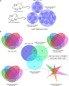
Overview of transcriptomic changes in myelin-related genes after sciatic nerve crush and transection. (A) Dynamic differentially expressed myelin-related gene counts 2-fold at the indicated transitions in a sciatic nerve crush segment and both sciatic nerve stumps post-transection. (B) Venn diagrams showing dynamic overlapping or specific differentially expressed gene counts for myelin function in both models of sciatic nerve crush and transection. Aspartoacylase (ASPA), C-C motif chemokine ligand 2 (Ccl2), C-C motif chemokine ligand 3 like 3 (CCL3L3), interleukin 1 beta (IL1B), matrix metallopeptidase 9 (MMP9), nerve growth factor receptor (NGFR), neuregulin 1 (NRG1), neurotrophic receptor tyrosine kinase 2 (NTRK2), and thyroid hormone receptor beta (THRB) are conserved myelin-related genes differentially expressed in the sciatic nerve crush segment and both sciatic nerve stumps post-transection.
Figure 2
Figure 2
Comparison of differentially expressed genes for demyelination and myelination. (A) Specific and conserved overlapping protein–protein interaction (PPI) networks for demyelination and myelination in a sciatic nerve crush segment and both sciatic nerve stumps post-transection. (B) Ridgeline plots of dynamic differentially expressed demyelination- and myelination-related gene counts 2-fold in the sciatic nerve crush segment and both sciatic nerve stumps post-transection.
Figure 3
Figure 3
Dynamic expression of PRC2 family members and conserved overlapping myelin-related genes and their interactions. (A) Heatmap of dynamic gene expression for PRC2 family members at 0.5, 1, 6, 12, 24 hours, 4 days, and 1, 2, 3, and 4 weeks post-sciatic nerve transection. (B) Heatmap of dynamic gene expression for PRC2 family members in sciatic nerve crush segments at 1, 4, 7, and 14 days post-sciatic nerve crush. (C) Protein–protein interaction (PPI) networks for PRC2 family members and conserved overlapping genes for demyelination and myelination. (D) Heatmap of dynamic gene expression for PRC2 family members in proximal sciatic nerve stumps at 1, 4, 7, and 14 days post-sciatic nerve transection. (E) Heatmap showing dynamic expression of conserved overlapping myelin-related genes in sciatic nerve crush segments at 1, 4, 7, and 14 days post-sciatic nerve crush. (F) Heatmap showing dynamic expression of conserved overlapping myelin-related genes in proximal sciatic nerve stumps at 1, 4, 7, and 14 days post-sciatic nerve transection. (G) Heatmap showing dynamic gene expression of conserved overlapping myelin-related genes in distal sciatic nerve stumps at 0.5, 1, 6, 12, 24 hours, 4 days, and 1, 2, 3, and 4 weeks post-sciatic nerve transection. PRC2: Polycomb repressive complex 2.
Figure 4
Figure 4
In situ hybridization spatial mRNA validation of Ezh2 expression in sciatic nerve of rats. (A) Schematic illustration of sciatic nerve samples obtained from normal, proximal, and distal segments of a rat sciatic nerve transection (SNT) model. Created with Adobe Illustrator CC and Adobe Photoshop CC. (B) Immunofluorescence staining of Schwann cells (SCs) on sciatic nerve or transected nerve probed for Ezh2 mRNA (green) at 4 days post-injury. All SCs were identified using S100B (Cy3, red), neurofilament 200 (NF200; Cy5, grey), and 4′,6-diamidino-2-phenylindole (DAPI; blue). Scale bars: 50 µm for low magnification, 20 µm for high magnification. (C) Percentage of SCs with Ezh2 mRNA ≥ 1 visible spots. (D) Fold change quantification of Ezh2 mRNA per SC. Ratio of number of average spots per SC were calculated for all groups, and then standardized to the normal sciatic nerve group value. SNT may induce expression of Ezh2 in SCs of both sciatic nerve stumps compared with the normal group. Data are expressed as mean ± SE (n = 6). **P < 0.01, vs. normal group (one-way analysis of variance followed by Bonferroni post hoc test).
Figure 5
Figure 5
Ablation of Ezh2 in Schwann cells of mice leads to abnormal growth and myelination deficits in the sciatic nerve. (A) Diagram outlining the generation of Ezh2 conditional knockout (Ezh2fl/fl;Mpz-Cre) mice. (B) Representative images of wild-type (Ezh2fl/fl), heterozygous (Ezh2fl/+;Mpz-Cre), and homozygous conditional knockout mice (Ezh2fl/fl;Mpz-Cre) at 7, 14, 21 days, and 8 weeks after birth. (C) Sciatic nerve without injury in wild-type control and Ezh2fl/fl;Mpz-Cre mice at 8 weeks post-birth stained for S100B (Cy5, grey), EZH2 (Cy3, red), MPZ (Alexa Fluor 488, green), and DAPI (blue). Genetic ablation of Ezh2 in Schwann cells leads to abnormal growth and deficits in the myelination of mouse sciatic nerve. Scale bars: 10 µm. DAPI: 4,6-Diamidino-2-phenylindole; MPZ: myelin protein zero.
Figure 6
Figure 6
Knockout of Ezh2 in Schwann cells leads to hypomyelination of mouse sciatic nerve and suppresses remyelination post-SNC. (A) Transmission electron microscopy analysis of sciatic nerves from both Ezh2 cKO mice (Ezh2fl/fl;Dhh-Cre and Ezh2fl/fl;Mpz-Cre) showed plenty of axons remain unmyelinated in mutant sciatic nerves. (B) Transmission electron microscopy analysis showed most axons remyelinated at the crush site of the control nerve while Ezh2fl/fl;Mpz-Cre mice had significantly fewer remyelinated axons than control nerve at 21 days after SNC. (C) Quantification of myelinated axons in sciatic nerves among both Ezh2 cKO mice and their wild-type littermate control mice at 8 weeks after birth and 21 days after SNC. Data are expressed as mean ± SE (n = 3). *P < 0.05, ***P < 0.001, vs. wildtype control (Ezh2fl/fl) group (one-way analysis of variance followed by Bonferroni post hoc test). (D) The percentage of myelinated axons in sciatic nerves among both Ezh2 cKO mice and wild-type control mice at 8 weeks after birth (P8W) and 21 days after SNC (21 dpi). (E) Sciatic nerve from wild-type control (Ezh2fl/fl) and Ezh2fl/fl;Dhh-Cre P8W mice at 14 days post sciatic nerve crush (14 dpi) stained for S100B (Cy5, grey), EZH2 (Cy3, red), MPZ (Alexa Fluor 488, green), and DAPI (blue). Scale bars: 10 µm. cKO: Conditional knockout; DAPI: 4,6-diamidino-2-phenylindole; MPZ: myelin protein zero; SNC: sciatic nerve crush.
Figure 7
Figure 7
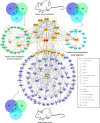
Interaction between PRC2 family members and specific myelin–related genes in a sciatic nerve crush segment and both sciatic nerve stumps post-transection. Protein–protein interaction networks of myelin-related genes showing that SOX2, insulin-like growth factor 1 (IGF1), and signal transducer and activator of transcription 3 (STAT3) were hub genes in the proximal sciatic nerve stump, while MYC was the hub gene in the distal sciatic nerve stump post-transection. Tumor necrosis factor (TNF) was the hub gene in the sciatic nerve crush segment post-crush. PRC2 complex members interacted with specific myelin-related PPI networks in a sciatic nerve crush segment and both sciatic nerve stumps post-transection. EZH2 (red) was the hub gene (blue) in sciatic nerve crush segments via interactions with TNF, interleukin (IL)-1, N-Myc downregulated gene 1 (NDRG1), Erb-B2 receptor tyrosine kinase 4 (ERBB4), chemokine (C-X-C motif) receptor 3 (CXCR3), chemokine (C-X-C motif) receptor 4 (CXCR4), hepatocyte nuclear factor-4 alpha (HNF4A), IL-12B, and IL-12 receptor subunit beta 2 (IL12RB2) (dark yellow). EZH2 (red) interacted with specific myelin-related PPI networks (blue) in proximal sciatic nerve stump post-transection via interferon beta 1 (IFNB1), IGF1, STAT3, and O-linked N-acetylglucosamine transferase (OGT) (green). EZH2 (red) interacted with specific myelin-related PPI networks (cyan) in distal sciatic nerve stump post-transection via C-X-C motif chemokine ligand 2 (CXCL2), colony stimulating factor 3 (CSF3), and MYC (dark yellow). PRC2: Polycomb repressive complex 2.

Similar articles

Cited by

References

    1. An Q, Sun C, Li R, Chen S, Gu X, An S, Wang Z. Calcitonin gene-related peptide regulates spinal microglial activation through the histone H3 lysine 27 trimethylation via enhancer of zeste homolog-2 in rats with neuropathic pain. J Neuroinflammation. 2021;18:117. - PMC - PubMed
    1. Balakrishnan A, Belfiore L, Chu TH, Fleming T, Midha R, Biernaskie J, Schuurmans C. Insights into the role and potential of Schwann cells for peripheral nerve repair from studies of development and injury. Front Mol Neurosci. 2020;13:608442. - PMC - PubMed
    1. Bankhead P, Loughrey MB, Fernández JA, Dombrowski Y, McArt DG, Dunne PD, McQuaid S, Gray RT, Murray LJ, Coleman HG, James JA, Salto-Tellez M, Hamilton PW. QuPath: Open source software for digital pathology image analysis. Sci Rep. 2017;7:16878. - PMC - PubMed
    1. Bardou P, Mariette J, Escudié F, Djemiel C, Klopp C. jvenn: an interactive Venn diagram viewer. BMC Bioinformatics. 2014;15:293. - PMC - PubMed
    1. Bitgood MJ, McMahon AP. Hedgehog and Bmp genes are coexpressed at many diverse sites of cell-cell interaction in the mouse embryo. Dev Biol. 1995;172:126–138. - PubMed

LinkOut - more resources