Case Reports
. 2024 Sep 4;29(17):102501.
doi: 10.1016/j.jaccas.2024.102501.

Revascularization in a Time After CORAL

Affiliations
Case Reports

Revascularization in a Time After CORAL

Anne R Dionne et al. JACC Case Rep. .

Abstract

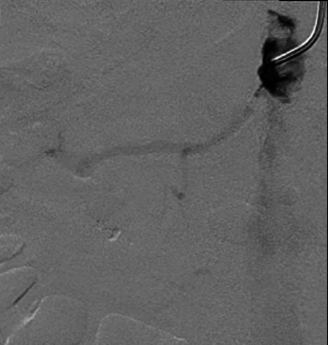
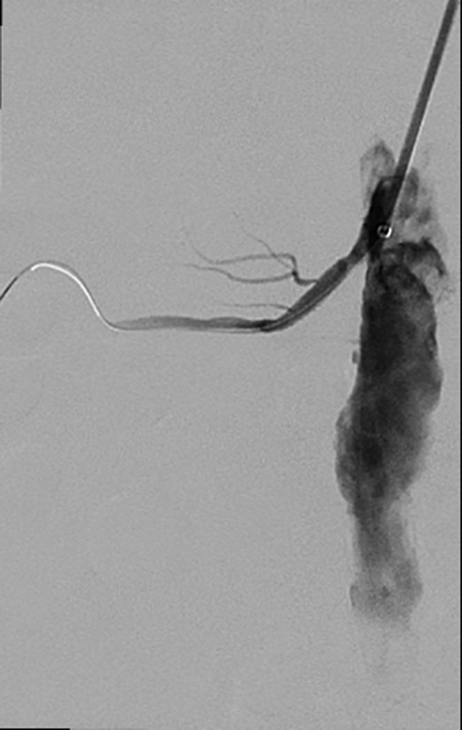
Revascularization of renal artery stenosis became less common following randomized controlled trials that failed to demonstrate benefit in low-risk patients. An 88-year-old patient with recurrent acute pulmonary edema and progressive kidney disease in the setting of high-grade renal artery stenosis, a phenotype excluded from these trials, underwent revascularization.

Keywords: hypertension; stenosis; stents; vascular disease.


Conflict of interest statement

Dr Columbo has received support from the National Heart, Lung and Blood Institute of the National Institutes of Health (K08HL165087). All other authors have reported that they have no relationships relevant to the contents of this paper to disclose.

Figures

None
Graphical abstract
Figure 1
Figure 1
Computed Tomography Angiography of the Abdomen and Pelvis With Contrast Aortic calcification at the origin of the renal arteries, with the right renal artery indicated by the arrow.
Figure 2
Figure 2
Computed Tomography Angiography of the Abdomen and Pelvis With Contrast Calcified plaques along the abdominal aorta with involvement of both renal arteries and associated atrophy of the left kidney.
Figure 3
Figure 3
Pre-Stenting Angiography Directed angiography of the right renal artery demonstrating a severely stenosed, heavily diseased, but patent, artery.
Figure 4
Figure 4
Post-Stenting Angiography Post-stenting image showing a 6 mm × 19 mm noncovered balloon-expandable stent, demonstrating patency of the proximal renal artery.
Figure 5
Figure 5
Post-Stenting Angiography With Distal Flow Post-stenting angiogram demonstrating excellent flow into the distal right renal artery branches.

References

    1. Radermacher J., Chavan A., Bleck J., et al. Use of Doppler ultrasonography to predict the outcome of therapy for renal-artery stenosis. N Engl J Med. 2001;344(6):410–417. doi: 10.1056/NEJM200102083440603. - DOI - PubMed
    1. Hicks C.W., Clark T.W.I., Cooper C.J., et al. Atherosclerotic renovascular disease: a KDIGO (Kidney Disease: Improving Global Outcomes) controversies conference. Am J Kidney Dis. 2022;79(2):289–301. doi: 10.1053/j.ajkd.2021.06.025. - DOI - PMC - PubMed
    1. de Silva R., Loh H., Rigby A.S., et al. Epidemiology, associated factors, and prognostic outcomes of renal artery stenosis in chronic heart failure assessed by magnetic resonance angiography. Am J Cardiol. 2007;100(2):273–279. doi: 10.1016/j.amjcard.2007.02.098. - DOI - PubMed
    1. Conlon P.J., O’Riordan E., Kalra P.A. New insights into the epidemiologic and clinical manifestations of atherosclerotic renovascular disease. Am J Kidney Dis. 2000;35(4):573–587. doi: 10.1016/s0272-6386(00)70002-3. - DOI - PubMed
    1. Rimoldi S.F., Yuzefpolskaya M., Allemann Y., Messerli F. Flash pulmonary edema. Prog Cardiovasc Dis. 2009;52(3):249–259. doi: 10.1016/j.pcad.2009.10.002. - DOI - PubMed

Publication types

LinkOut - more resources