Case Reports
. 2024 Sep 4;29(17):102499.
doi: 10.1016/j.jaccas.2024.102499.

Crinkle-Cut Coronary Arteries in a Patient With Granulomatosis With Polyangiitis

Affiliations
Case Reports

Crinkle-Cut Coronary Arteries in a Patient With Granulomatosis With Polyangiitis

Noor M A Alsammarraie et al. JACC Case Rep. .

Abstract

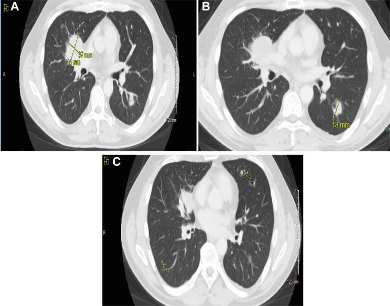
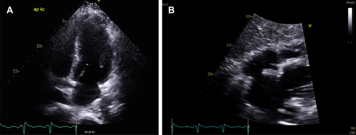
Granulomatosis with polyangiitis (GPA) is a rare type of small to medium vessel necrotizing vasculitis that usually affects vessels of the upper or lower airways and kidneys. Cardiac involvement in GPA is often subclinical and if clinically significant has been rarely reported, even less so as an initial presentation. We describe the case of a 44-year-old man who presented with what appeared to be inferior ST-segment elevation myocardial infarction and was found to have small vessel vasculitis of the coronary arteries with associated myocarditis as a presenting manifestation of GPA, which was ultimately treated with steroids, rituximab, and avacopan.

Keywords: cardiac magnetic resonance; coronary vasculitis; granulomatosis with polyangiitis.


Conflict of interest statement

The authors have reported that they have no relationships relevant to the contents of this paper to disclose.

Figures

None
Graphical abstract
Figure 1
Figure 1
Initial Electrocardiography The tracing shows ST-segment elevation in leads II, III, and aVF, with reciprocal ST-segment depression in lateral leads I, aVL, V5, and V6.
Figure 2
Figure 2
Imaging of Lung Nodules: Computed Tomography Angiography of the Chest (A) An irregular heterogenous mass anteriorly in the right hilum extending into the right upper and middle lobes and measuring 4.5 cm × 3.7 cm. (B) An ill-defined nodule measuring up to 18 mm in the left lower lobe. (C) Arrow on right side of image indicating a tiny 2-mm nodule medially in left lower lobe. Arrow on the left side of image indicating another tiny approximately 2mm rounded nodule in right lower lobe.
Figure 3
Figure 3
Initial Transthoracic Echocardiogram (A) Left ventricular ejection fraction of 45% with mild left ventricular global hypokinesis. (B) Subxiphoid pericardial view showing a normal left ventricular cavity size and a normal left atrium.
Figure 4
Figure 4
Initial Cardiac Magnetic Resonance (A) A 4-chamber view with arrow highlighting area of late gadolinium enhancement (LGE). (B and C) Multiple 2-chamber views with arrows highlighting areas of LGE. (D) A short-axis view with arrow highlighting area of LGE. (E) T1 mapping. (F) Elevated T2 mapping.
Figure 5
Figure 5
Crinkle-Cut Coronary Arteries Cardiac catheterization imaging showing diffuse small vessel coronary vasculitis. (A and B) Coronary angiogram demonstrating the left coronary artery system. The left anterior descending (LAD) artery exhibits a distinctive “crinkle-cut” appearance characterized by irregular, undulating contour suggestive of segmental stenosis and dilation along the vessel wall that are especially prominent distally. The left circumflex (LCx) artery here is visible in the proximal portion and appears to have less involvement in this view. (C) The distal LAD artery and its smaller branches. This image highlights the impact of vasculitis on the microvasculature, where the damage appears more pronounced. (D) The proximal and mid segments of the LAD distorted vessel architecture. (E) Coronary angiogram demonstrating the right coronary artery “crinkle cut” appearance especially in the mid to distal segment.

References

    1. Ntatsaki E., Watts R.A., Scott D.G. Epidemiology of ANCA-associated vasculitis. Rheum Dis Clin North Am. 2010;36(3):447–461. - PubMed
    1. McGeoch L., Carette S., Cuthbertson D., et al. Vasculitis Clinical Research Consortium Cardiac involvement in granulomatosis with polyangiitis. J Rheumatol. 2015;42(7):1209–1212. doi: 10.3899/jrheum.141513. - DOI - PMC - PubMed
    1. Greco A., Marinelli C., Fusconi M., et al. Clinic manifestations in granulomatosis with polyangiitis. Int J Immunopathol Pharmacol. 2016;29(2):151–159. doi: 10.1177/0394632015617063. - DOI - PMC - PubMed
    1. Ruisi M., Ruisi P., Finkielstein D. Cardiac manifestations of Wegener’s granulomatosis: Case report and review of the literature. J Cardiol Cases. 2010;2(2):e99–e102. doi: 10.1016/j.jccase.2010.04.002. - DOI - PMC - PubMed
    1. Roubille C., Henriquez S., Mercuzot C., et al. Impact of cardiovascular risk factors on the occurrence of cardiovascular events in antineutrophil cytoplasmic antibody (ANCA)-associated vasculitides. J Clin Med. 2021 May 25;10(11):2299. doi: 10.3390/jcm10112299. - DOI - PMC - PubMed

Publication types

LinkOut - more resources