. 2024 Sep 18:14:1399761.
doi: 10.3389/fcimb.2024.1399761. eCollection 2024.

SNX27:Retromer:ESCPE-1-mediated early endosomal tubulation impacts cytomegalovirus replication

Affiliations

SNX27:Retromer:ESCPE-1-mediated early endosomal tubulation impacts cytomegalovirus replication

Igor Štimac et al. Front Cell Infect Microbiol. .

Abstract

Introduction: Cytomegaloviruses (CMVs) extensively reorganize the membrane system of the cell and establish a new structure as large as the cell nucleus called the assembly compartment (AC). Our previous studies on murine CMV (MCMV)-infected fibroblasts indicated that the inner part of the AC contains rearranged early endosomes, recycling endosomes, endosomal recycling compartments and trans-Golgi membrane structures that are extensively tubulated, including the expansion and retention of tubular Rab10 elements. An essential process that initiates Rab10-associated tubulation is cargo sorting and retrieval mediated by SNX27, Retromer, and ESCPE-1 (endosomal SNX-BAR sorting complex for promoting exit 1) complexes.

Objective: The aim of this study was to investigate the role of SNX27:Retromer:ESCPE-1 complexes in the biogenesis of pre-AC in MCMV-infected cells and subsequently their role in secondary envelopment and release of infectious virions.

Results: Here we show that SNX27:Retromer:ESCPE1-mediated tubulation is essential for the establishment of a Rab10-decorated subset of membranes within the pre-AC, a function that requires an intact F3 subdomain of the SNX27 FERM domain. Suppression of SNX27-mediated functions resulted in an almost tenfold decrease in the release of infectious virions. However, these effects cannot be directly linked to the contribution of SNX27:Retromer:ESCPE-1-dependent tubulation to the secondary envelopment, as suppression of these components, including the F3-FERM domain, led to a decrease in MCMV protein expression and inhibited the progression of the replication cycle.

Conclusion: This study demonstrates a novel and important function of membrane tubulation within the pre-AC associated with the control of viral protein expression.

Keywords: Cytomegalovirus; ESCPE-1; assembly compartment; beta-herpesvirus secondary envelopment; retromer; sorting nexin 27; tubular endosomes.


Conflict of interest statement

The authors declare that the research was conducted in the absence of any commercial or financial relationships that could be construed as a potential conflict of interest. The author(s) declared that they were an editorial board member of Frontiers, at the time of submission. This had no impact on the peer review process and the final decision.

Figures

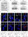
Figure 1
Figure 1
Distribution of SNX27, Vps35 and SNX1 in uninfected and MCMV-infected cells. (A) Balb3T3 fibroblasts were infected with Δm138-MCMV (MOI of 10) or left uninfected. Samples were stained with antibodies against SNX27, Vps35 or SNX1 (red), the pIE1 of MCMV (green), or DAPI (blue), followed by confocal imaging and quantitative analysis. The percentage of cells with pericentriolar accumulation of SNX27, Vps35 and SNX1 in MCMV-infected cells was presented as the mean±SD. Arrows indicate pericentriolar pre-AC. Bars, 10 μm. (B) Western blots of the expression kinetics of SNX27, Vps35 and SNX1 in the course of MCMV infection. Signals were quantified using ImageJ and expressed as a percentage of the initial expression. Shown are the individual results (empty circles) and the average (red bars) of three (SNX27 and SNX1) and four (Vps35) experiments. (C) Colocalization of recombinant SNX27 (EGFP-mSNX27) with endogenous Vps35 and SNX1. NIH3T3 cells expressing EGFP mSNX27 were either uninfected or infected with Δm138-MCMV (MOI of 10, 16 hpi). After fixation and permeabilization, immunofluorescence labeling with primary and appropriate secondary antibodies was performed, and cells were analyzed by confocal microscopy. Arrows indicate pericentriolar pre-AC in MCMV-infected cells; asterisks indicate endosomal partitioning of SNX1 and SNX27, and arrowheads indicate SNX27+/Vps35+/SNX1+ tubules. Mander’s coefficients (M1 and M2) on serial images were calculated to quantify colocalization. Data represents mean ± SD of 15-22 cells (number shown within the column) from three independent experiments. Bars, 10 μm.
Figure 2
Figure 2
siRNA depletion of SNX27, Vps35, SNX1 and SNX1 + 2 prevents pericentriolar accumulation of Rab10 in the early phase of MCMV infection. (A) Balb3T3 fibroblasts were transfected with scrambled siRNA or siRNA for SNX27, Vps35, SNX1, and SNX1 + 2. After 48 h, the expression of depleted proteins was detected by Western blot using β-actin as a loading control. (B) siRNA-treated cells (48 h after transfection) were infected with Δm138-MCMV (MOI of 10), and at 16 hpi the expression of depleted proteins was analyzed by Western blot. The pIE1 expression served as a marker for infection, and β-actin was used as a loading control. (C-F) Infected cells (16 hpi) transfected with either control siRNA or siRNA for SNX27, Vps35, SNX1 and SNX1 + 2 were stained with antibodies for either SNX27 (C) or Vps35 (D) or SNX1 (E) or SNX1 + 2 (F) to visualize these proteins (green fluorescence) combined with antibodies for Rab10 (red fluorescence) and pIE1 (blue). The stained cells were analyzed by confocal imaging, and the percentage of cells expressing pericentriolar Rab10 accumulation was determined by epifluorescence image analysis. The data show individual results from five independent experiments. Ctrl., control level (mean ± SD) from 18 independent experiments. Mean values are shown as red bars, and statistical significance was determined using a two-tailed Student t-test (***p < 0.001). Bars, 10 μm.
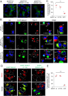
Figure 3
Figure 3
The F3-FERM subdomain of SNX27 is required for pericentriolar Rab10 and SNX1 accumulation in the pre-AC. (A-E) Balb3T3 fibroblasts were transfected with pEGFP-N1-mSNX27, pEGFP-N1-mSNX27 H112A, or pEGFP-N1 SNX27 ΔF3 plasmids. After 30 h, the cells were fixed (A) or infected with Δm138-MCMV (MOI of 10) for a further 16 h before fixation (B-E). Subsequently, the permeabilized cells were labeled with anti-Rab10 or anti-SNX1 antibodies (red), anti-IE1 antibodies (blue, infected cells) or DAPI (blue, uninfected cells) and analyzed by confocal microscopy. (B) Asterisks represent cells with pericentriolar Rab10, and (B, D) arrowheads represent transfected cells. (C, E) The graphs show the percentage of MCMV-infected cells (IE1-positive) with pericentriolar accumulation of Rab10 (C) or SNX1 (E), determined in three independent experiments (empty circles), and the mean values (red bars). Statistical significance was determined using a two-tailed Student t-test (***p < 0.001; **p < 0.01). NT, non-transfected. Bars, 25 μm and 10 μm (B), and 10 μm (D).
Figure 4
Figure 4
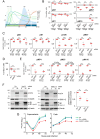
Depletion of SNX27 inhibits progression through the productive infection. Balb3T3 fibroblasts were transfected with scrambled (Scr.) or SNX27 siRNAs. After 48 hours, cells were infected with Δm138-MCMV (MOI of 10) and analyzed at 6, 16, 24, and 40 hpi by immunofluorescence and Western blot. (A) Schematic presentation of the viral replication cycle program. The available antibody reagents against seven MCMV-encoded proteins can distinguish immediate early (ie), three temporal classes of early (e I, e II, and e III), and two temporal classes of late (l I and l II) gene expression. The kinetics of the temporal classes of transcription are shown by dotted lines, and protein expression by full lines using different colour codes. The temporal effect of MCMV infection on cellular DNA replication (cdr) and the timing of viral DNA replication (vdr) are shown in the light-blue area. (B) Percentage of cells expressing viral proteins at 6, 16, and 40 hpi. Triple-stained (viral protein, SNX27, and DAPI) immunofluorescence samples were captured by confocal imaging ( Supplementary Figures S9, S10A ) and quantitatively analyzed using epifluorescence microscopy. Open circles represent data from three independent experiments, and red bars show the average value. Ctrl., control level (mean ± SD) determined in independent experiments (n=25 for pIE1, n=22 for pE1, n=12 for pM57, and n=8 for pM74). (C) Quantification of ie, e I, e II, and e III viral protein expression by Western blot. Viral proteins pIE1, pE1, pm06, and pM57 were analyzed simultaneously (at the same membrane) with SNX27 and actin ( Supplementary Figure S7 ). The signals were quantified, viral protein expression in SNX27-depleted cells normalized to actin expression, and the result was shown as a relative to control (scr-siRNA-treated cells). Results are presented as fold changes compared to scr-siRNA in the respective kinetics (means ± SD) by empty circles, and averages by red bars. (D) EdU labeling of viral DNA replication. Cells transfected with Scr. and SNX27 siRNA were infected and labeled 16-24 hpi with 10 μM EdU, followed by staining with antibodies against SNX27 and pIE1, and visualization of EdU-labeled DNA with the click reaction. Shown is the percentage of EdU-positive cells, whereas the triple-labeled images are shown in Supplementary Figure S10B . The empty circles show the values in an independent experiment, and the red bars show the mean value. Ctrl., control level (mean ± SD) determined in 15 independent experiments. (E) Quantification of l I and l II viral protein expression (pM55, pM74, and pm116) was performed by Western blot as described in (C). The representative images of Western blots are shown in Supplementary Figure S7 . Two forms of pM55 (55 and 130 kDa) were analyzed. (F) Effect of depletion of Vps35 and SNX1+2 on the expression of the late protein pM74. Shown are the representative Western blots of pM74, Vps35, and SNX1+2 expression in Vps35- and SNX1+2-depleted cells at 40 hpi, respectively. The asterisk represents remnants of Vps35 detection overlaying the lower band of pIE1. The pM74 signals were quantified, normalized to actin, and shown as a fold change compared to the scr-siRNA (empty circles). The average values are shown as red bars. (G) Depletion of SNX27 reduces virus production. Single-step growth kinetics in non-transfected (NT), scr-siRNA- or SNX27 siRNA-transfected Balb3T3 cells after infection with Δm138-MCMV (MOI of 10). Supernatants and cells were harvested at indicated times post-infection, and frozen at -80°C. After two rounds of freeze/thaw, the virus was quantitated by the plaque assay on MEFs. Data represents log10 infectious units/mL of sample, and error bars indicate standard errors of the means of six biological replicates. Statistical significance was determined using a two-tailed Student t-test (B-F) or one-way ANOVA analysis (G). The colour of the asterisks (G) denotes the statistical difference of NT (green) and Scr. (blue) from SNX27. ***p < 0.001; **p < 0.01; *p < 0.05.
Figure 5
Figure 5
Expression of pM74 after MCMV infection of SNX27 wt, SNX27 H112A and SNX27 ΔF3 overexpressing cells. (A) Immunofluorescence analysis of transfected and non-transfected cells. Cells were transfected with EGFP SNX27 wt, EGFP SNX27 H112A and EGFP SNX27 ΔF3 and 48 hours later infected with Δm138-MCMV at an MOI of 10. 48 hours after infection, the cells were stained for the proteins pM74 (red) and pIE1 (blue). The cell boundaries are indicated by fine dashed lines. Arrowheads indicate transfected cells and arrows indicate ring-shaped pM74 expression. Bars, 10 μm. (B) The percentage of SNX27-transfected and MCMV-infected cells expressing pM74 was determined in three independent experiments (empty circles). The red bars represent the mean value. Statistical significance was determined using a two-tailed Student t-test (*** p < 0.001). NT, non-transfected.
Figure 6
Figure 6
A model showing the role of the SNX27:Retromer:ESCPE-1 complex in the formation of AC in the early phase of MCMV infection. (A) In uninfected cells, the SNX27:Retromer:ESCPE-1 pathway involves cargo molecule recognition and recruitment of the Vps35 Retromer component by different parts of the SNX27 PDZ domain, followed by recognition of the SNX1/2-SNX5/6 (ESCPE-1) heterodimer by the SNX27-F3-FERM subdomain (1). ESCPE-1 can also recognize additional cargo (not shown). This process allows mobilization of actin by WASH1 (not shown), initiation of tubulation at the endosomal membrane, and transient binding of Rab10, which is required for tubule growth (2) and their fission into transport carriers (3) that release SNX27:Retromer:ESCPE-1 components (4) and Rab10 (5). The transport carriers migrate to the subset of ERC membranes to further sort and recycle the cargo to the PM, which is known as the slow recycling pathway (6). (B) The initial process of EE tubule formation (1) is functional in MCMV-infected cells. SNX27:Retromer:ESCPE-1 components and Rab10 are recruited to membranes. However, the mechanism limiting tubule growth is blocked and the development of transport carriers is inhibited (3), leading to the formation of elongated tubules (2), which retain cargo and SNX27:Retromer:ESCPE-1 components (4), resulting in reduced supply to the ERC and subsequently a reduced ERC recycling rate (5). Elongated tubules can concentrate CMV envelope proteins and serve as membranes for secondary envelopment. In addition, elongated tubules with retained SNX27:Retromer:ESCPE-1 and viral glycoproteins can serve as signalosomes by retaining or recruiting signaling proteins and establishing a signaling cascade that influences the entire MCMV replication cycle.

References

    1. Angulo A., Ghazal P., Messerle M. (2000). The major immediate-early gene ie3 of mouse cytomegalovirus is essential for viral growth. J. Virol. 74, 11129–11136. doi: 10.1128/JVI.74.23.11129-11136.2000 - DOI - PMC - PubMed
    1. Babbey C. M., Ahktar N., Wang E., Chen C. C. H., Grant B. D., Dunn K. W. (2006). Rab10 regulates membrane transport through early endosomes of polarized Madin-Darby Canine Kidney cells. Mol. Biol. Cell 17, 3156–3175. doi: 10.1091/mbc.E05-08-0799 - DOI - PMC - PubMed
    1. Beltran P. M. J., Mathias R. A., Cristea I. M., Beltran P. M. J., Mathias R. A., Cristea I. M. (2016). A portrait of the human organelle proteome in space and time during cytomegalovirus infection. Cell Syst. 3, 361–373. doi: 10.1016/j.cels.2016.08.012 - DOI - PMC - PubMed
    1. Bergant Marušič M., Ozbun M. A., Campos S. K., Myers M. P., Banks L. (2012). Human papillomavirus L2 facilitates viral escape from late endosomes via sorting nexin 17. Traffic 13, 455–467. doi: 10.1111/J.1600-0854.2011.01320.X - DOI - PMC - PubMed
    1. Birzer A., Kraner M. E., Heilingloh C. S., Mühl-Zürbes P., Hofmann J., Steinkasserer A., et al. . (2020). Mass spectrometric characterization of HSV-1 L-particles from human dendritic cells and BHK21 cells and analysis of their functional role. Front. Microbiol. 11. doi: 10.3389/fmicb.2020.01997 - DOI - PMC - PubMed

MeSH terms

Substances

LinkOut - more resources