

Case Reports
doi: 10.1016/j.jaccas.2024.102524.
Cardiac Computed Tomography for Cardioembolic Stroke Evaluation in a Patient With a Mechanical Aortic Valve
Affiliations
- PMID: 39359973
- PMCID: PMC11442204
- DOI: 10.1016/j.jaccas.2024.102524
Item in Clipboard
Case Reports
Cardiac Computed Tomography for Cardioembolic Stroke Evaluation in a Patient With a Mechanical Aortic Valve
JACC Case Rep.
.
Abstract
We present a case of cardioembolic stroke in a patient with a history of mechanical aortic valve who was compliant with anticoagulation medication. Cardiac computed tomography was used as an alternative, noninvasive means of evaluation for the cardioembolic source of stroke and identified subvalvular mobile pannus of the mechanical aortic valve.
Keywords: cardiac computed tomography; cardioembolic stroke; mechanical valve.
Conflict of interest statement
The authors have reported that they have no relationships relevant to the contents of this paper to disclose.
Figures
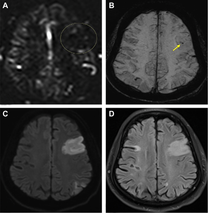

Noncontrast Magnetic Resonance Imaging of the Head Showing Evidence of Acute Infarcts in Multiple Vascular Territories Concerning for Cardioembolic Etiology of Stroke (A) Perfusion: an asymmetrically decreased area of cortical perfusion in the left frontal lobe (circle) that correlates with diffusion weighted imaging. (B) Susceptibility weighted image: within the frontal lobe infarct territory are areas of susceptibility blooming (arrow), suggesting the presence of thrombus or slow flow in the distal middle cerebral artery. (C) Diffusion weighted image: large area of diffusion restriction in the left frontal lobe and smaller patchy area seen in the parietal lobe indicating acute multifocal infarct. (D) Fluid-attenuated inversion recovery (FLAIR): Hyperintense signal in the left frontal lobes, correlating with the diffusion weighted images. The left parietal territory is not evident on this image.
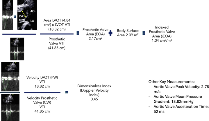
Key Measurements of the Prosthetic Valve Function Obtained Using Transthoracic Echocardiography (Top) Velocity time integral (VTI) measurements obtained using transthoracic echocardiography with continuous-wave Doppler of both the left ventricular outflow tract and prosthetic aortic valve used to calculate the indexed prosthetic valve area. (Bottom) VTI measurements of the left ventricular outflow tract and prosthetic aortic valve obtained using continuous-wave Doppler with transthoracic echocardiography used to calculate the dimensionless index.
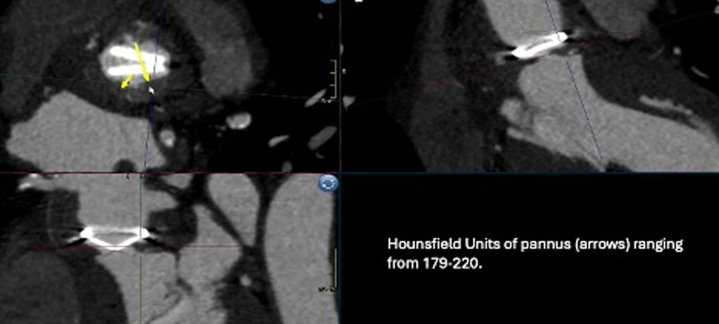
Cardiac Computed Tomography Multiplanar Reconstruction of the Subvalvular Pannus Multiple cardiac computed tomography images showing evidence of subvalvular pannus (crosshairs). Hounsfield units of the measured pannus (arrows) seen in bottom left of image. Please refer to the videos for further images and videos of the pannus. AO = aorta; CW = continuous-wave Doppler; EOA = effective orifice area; LA = left atrium; LVOT = left ventricular outflow tract; PW = pulsed-wave Doppler.
References
-
- de Bruijn S.F.T.M., Agema W.R.P., Lammers G.J., et al. Transesophageal echocardiography is superior to transthoracic echocardiography in management of patients of any age with transient ischemic attack or stroke. Stroke. 2006;37(10):2531–2534. - PubMed
-
- Easton J.D., Saver J.L., Albers G.W., et al. Definition and evaluation of transient ischemic attack: a scientific statement for healthcare professionals from the American Heart Association/American Stroke Association Stroke Council; Council on Cardiovascular Surgery and Anesthesia; Council on Cardiovascular Radiology and Intervention; Council on Cardiovascular Nursing; and the Interdisciplinary Council on Peripheral Vascular Disease. Stroke. 2009;40(6):2276–2293. - PubMed
-
- Otto C.M., Nishimura R.A., Bonow R.O., et al. 2020 ACC/AHA guideline for the management of patients with valvular heart disease: a report of the American College of Cardiology/American Heart Association Joint Committee on clinical practice guidelines. J Am Coll Cardiol. 2021;77(4):e25–e197. - PubMed
-
- Bouzas-Mosquera A., Álvarez-García N. Orientation of bileaflet mechanical aortic valve prostheses for optimal evaluation by transthoracic echocardiography. J Thorac Cardiovasc Surg. 2015;150(2):428–430. - PubMed
-
- Apfaltrer G., Lavra F., De Cecco C.N., et al. Predictive value of cardiac CTA, cardiac MRI, and transthoracic echocardiography for cardioembolic stroke recurrence. AJR Am J Roentgenol. 2021;217(2):336–346. - PubMed
Publication types
LinkOut - more resources
Full Text Sources
