Review
. 2024 Sep 19;15(41):16887-16907.
doi: 10.1039/d4sc02625j. Online ahead of print.

Two-dimensional nanomaterials based on rare earth elements for biomedical applications

Affiliations
Review

Two-dimensional nanomaterials based on rare earth elements for biomedical applications

Mingjun Bai et al. Chem Sci. .

Abstract

As a kind of star materials, two-dimensional (2D) nanomaterials have attracted tremendous attention for their unique structures, excellent performance and wide applications. In recent years, layered rare earth-based or doped nanomaterials have become a new important member of the 2D nanomaterial family and have attracted significant interest, especially layered rare earth hydroxides (LREHs) and layered rare earth-doped perovskites with anion-exchangeability and exfoliative properties. In this review, we systematically summarize the synthesis, exfoliation, fabrication and biomedical applications of 2D rare earth nanomaterials. Upon exfoliation, the LREHs and layered rare earth-doped perovskites can be dimensionally reduced to ultrathin nanosheets which feature high anisotropy and flexibility. Subsequent fabrication, especially superlattice assembly, enables rare earth nanomaterials with diverse compositions and structures, which further optimizes or even creates new properties and thus expands the application fields. The latest progress in biomedical applications of the 2D rare earth-based or doped nanomaterials and composites is also reviewed in detail, especially drug delivery and magnetic resonance imaging (MRI). Moreover, at the end of this review, we provide an outlook on the opportunities and challenges of the 2D rare earth-based or doped nanomaterials. We believe this review will promote increasing interest in 2D rare earth materials and provide more insight into the artificial design of other nanomaterials based on rare earth elements for functional applications.


Conflict of interest statement

There are no conflicts to declare.

Figures
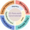
Fig. 1
Fig. 1. Schematic illustration of the properties and biomedical applications of rare earth nanomaterials.
Fig. 2
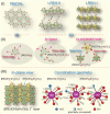
Fig. 2. Structure illustrations of LREHs. (I) RE(OH)3, LREH-II and LREH-I structures. (II) Connectivity of the polyhedral unit for the three structures. (III) In-plane view of the anion-exchangeable LREH-I structure and representative coordination geometries of RE cations. Adapted from ref. . Copyright 2023, Elsevier Inc.
Fig. 3
Fig. 3. Scanning electron microscopy (SEM) and transmission electron microscopy (TEM) images of synthesized LREHs. (a) As-prepared LEuH through the homogeneous precipitation method. Adapted from ref. . Copyright 2008, John Wiley & Sons, Inc. (b) As-prepared LYH through the homogeneous precipitation method. Adapted from ref. . Copyright 2008, American Chemical Society. As-prepared (c) LGdH, (d) LDyH, (e) LErH through the homogeneous precipitation method. Adapted from ref. . Copyright 2009, American Chemical Society. (f) As-prepared LYH through the hydrothermal method with NH4NO3 as a mineralizer. Adapted from ref. . Copyright 2012, Elsevier Inc. (g and h) As-prepared LYH through the hydrothermal method with SDS as a surfactant. Adapted from ref. . Copyright 2017, the Royal Society of Chemistry.
Fig. 4
Fig. 4. AFM images of LREH nanosheets. (a) LNdH nanosheet through ultrasonication of the NO3 intercalated LNdH powder in deionized water. Adapted from ref. . Copyright 2009, John Wiley & Sons, Inc. (b) LGdH nanosheet through ultrasonication of the Cl intercalated LGdH powder in deionized water. Adapted from ref. . Copyright 2009, the Royal Society of Chemistry. (c) LEuH nanosheet through shaking of the DS intercalated LEuH powder in formamide. Adapted from ref. . Copyright 2010, John Wiley & Sons, Inc. (d) LTmH nanosheet through magnetic stirring of the DS intercalated LTmH powder in formamide. Adapted from ref. . Copyright 2017, the Royal Society of Chemistry. (e) LYH nanosheet through magnetic stirring of the anion-exchanged NO3 intercalated LYH powder in toluene. Adapted from ref. . Copyright 2015, Springer Nature. (f) LGdH nanosheet through ultrasonication of the DS intercalated LGdH powder in formamide. Adapted from ref. . Copyright 2021, the Royal Society of Chemistry.
Fig. 5
Fig. 5. Morphologies of ultrathin rare earth hydroxide or oxide platelets. (a) SEM, (b) TEM and (c) AFM images of Y/Yb/Er hydroxides. Adapted from ref. . Copyright 2018, the Royal Society of Chemistry. (d) SEM image, (e) luminescence intensity ratio (LIR) values and (f) relative sensitivity (Sr) values of each mode of the Y2O3:Bi3+,Sm3+. Adapted from ref. . Copyright 2021, American Chemical Society.
Fig. 6
Fig. 6. AFM images of rare earth-doped perovskite nanosheets. (a) La0.90Eu0.05Nb2O7 nanosheets. Adapted from ref. . Copyright 2007, American Chemical Society. (b) Eu0.56Ta2O7 nanosheets. Adapted from ref. . Copyright 2008, American Chemical Society. (c) Gd1.4Eu0.6Ti3O10 nanosheets. Adapted from ref. . Copyright 2008, American Chemical Society. (d) Ln2Ti3O10:Tm3+/Er3+ nanosheets. Adapted from ref. . Copyright 2021, Elsevier Inc. (e) [K1.5(Tb0.8Sm0.2)]Ta3O10 nanosheets. Adapted from ref. . Copyright 2022, the Royal Society of Chemistry. (f) GdMgWO6:60%Eu3+ nanosheets. Adapted from ref. . Copyright 2019, Elsevier Inc.
Fig. 7
Fig. 7. TEM and AFM images of 2D MOF based on rare earth elements. (a) TEM and (b) AFM images of 2D MOFs with Tb as nodes and porphyrin as the ligand (Tb-TCPP). Adapted from ref. . Copyright 2020, John Wiley & Sons, Inc. (c) TEM and (d) AFM images of MB/Yb-TCPP-SO4, in which MB/Yb-TCPP-SO4 represents methylene blue (MB) modified porphyrin-based 2D Yb-TCPP MOF. Adapted from ref. . Copyright 2021, the Royal Society of Chemistry. (e) TEM and (f) AFM images of rare earth-containing MOF nanosheets with 2,2′-thiodiacetic acid as a surfactant. Adapted from ref. . Copyright 2017, Springer Nature. (g) SEM and (h) AFM images of the 2D Tb/Eu-bop MOF nanosheets, in which Tb/Eu-bop represents 2D MOFs with Tb/Eu as nodes and 5-boronoisophthalic acid as the ligand. Adapted from ref. . Copyright 2022, Elsevier Inc.
Fig. 8
Fig. 8. Multilayer rare earth hydroxide films. (a) Scheme of the fabrication process and (b) photographs of multilayer LGdH/PSS films. Adapted from ref. . Copyright 2010, John Wiley & Sons, Inc. (c) SEM images and schematic illustration of Gd(OH)2.5Cl0.5·0.9H2O:0.05Eu film and Gd2O3:0.05Eu film. Reproduced from ref. . Copyright 2009, John Wiley & Sons, Inc.
Fig. 9
Fig. 9. Rare earth perovskite superlattice films through the LBL fabrication method. (a) XRD patterns and (b) photoluminescence emission spectra of (PEI/La0.90Dy0.05Nb2O7)10 films. Adapted from ref. . Copyright 2012, Elsevier Inc. (c) CIE diagram and upconversion emission spectra of (Ln2Ti3O10:Er3+,Yb3+/Ln2Ti3O10:Tm3+,Yb3+)60 film. Adapted from ref. . Copyright 2022, Elsevier Inc.
Fig. 10
Fig. 10. LREH superlattice films through the LBL fabrication method. (a) Scheme of the fabrication process of LREHs/semiconducting oxide superlattice films. Adapted from ref. . Copyright 2021, the Royal Society of Chemistry. (b) Scheme of the fabrication process and (c) photographs of (Gd2O3:Re/SiO2) films under daylight and 254 nm UV irradiation, in which Gd2O3:Re represents Gd2O3 doped with different rare earth ions. Adapted from ref. . Copyright 2012, John Wiley & Sons, Inc.
Fig. 11
Fig. 11. Rare earth-doped perovskite lamellar flocculation. (a) TEM image and (b) possible model of the energy diagram of Eu3+ flocculated Ti0.92O2 nanosheet (ex-Ti0.92O2/Eu) composite. Adapted from ref. . Copyright 2004, American Institute of Physics. (c) TEM image of (Ho0.096Yb0.23Y0.164)Ca1.76Nb3O10 composite. Adapted from ref. . Copyright 2014, American Chemical Society. (d) Fabrication process and growth inhibition of Ag+ intercalated Tm3+/Er3+ co-doped Ln2Ti3O10 composite. Adapted from ref. . Copyright 2022, Elsevier Inc.
Fig. 12
Fig. 12. (a) XRD patterns, (b) TEM image and (c) SEM and elemental mapping images of the GdEu/TiO membrane, in which GdEu and TiO represent Eu3+ doped LGdH nanosheets and Ti0.87O20.52− nanosheets, respectively. Adapted from ref. . Copyright 2022, John Wiley & Sons, Inc.
Fig. 13
Fig. 13. Release kinetics of (a) diclofenac, ibuprofen, and naproxen from LGdH. Adapted from ref. Copyright 2018, the Royal Society of Chemistry. (b) Release kinetics of aspirin from LTbH, in which ASA-LTbH-1 : 1 and ASA-LTbH-1 : 2 represent ASA intercalated LTbH at a molar ratio of ASA to NaOH of 1 : 1 or 1 : 2, respectively. Reproduced from ref. . Copyright 2017, John Wiley & Sons, Inc. (c) Scheme of drug loading and release process and (d) release kinetics of ASA from Y/Tb/Gd ternary layered rare earth hydroxide nanocones, in which YTG-DS, YTG-ASA and YTG-CL represent DS, ASA and Cl intercalated Y/Tb/Gd ternary layered rare earth hydroxide nanocones, respectively. Reproduced from ref. . Copyright 2023, John Wiley & Sons, Inc. (e) Drug loading and (f) release capacity of the Gd-based porphyrin paddlewheel framework (PPF-Gd). Adapted from ref. . Copyright 2020, Oxford University Press.
Fig. 14
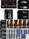
Fig. 14. MRI properties of Gd3+-containing 2D nanomaterials. (a) T1-weighted and T2-weighted phantom images of Gd/Y hydroxide nanosheets. Reproduced from ref. . Copyright 2020, Multidisciplinary Digital Publishing Institute. (b) In vivo T2-weighted MRI of mouse body with intravenous injection of LGdH-FS-PEGP, in which LGdH-FS-PEGP represents poly(ethylene glycol)-phospholipid-modified LGdH. Adapted from ref. . Copyright 2009, John Wiley & Sons, Inc. (c) In vitro and in vivo T1-weighted MRI effect of LGdH/Ti3C2. Adapted from ref. . Copyright 2023, the Royal Society of Chemistry. In vivo T1-weighted MR coronal images of a nude mouse bearing A375 tumor before and after (d) intravenous injection and (e) subcutaneous injection of PPF-Gd nanosheets, where PPF-Gd represents Gd-porphyrin MOF nanosheets. Adapted from ref. . Copyright 2020, Oxford University Press. (f) In vivo MR and (g) CT imaging of nude mice bearing HeLa tumors at different time points after intravenous injection of SN38&ICG/Gd&Yb-LDH. (h) In vivo fluorescence imaging and drug bio-distribution for nude mice bearing HeLa tumors at different time points after intravenous injection of PBS, SN38&ICG/Gd&Yb-LDH and SN38&ICG, respectively, together with ex vivo imaging of ICG in the tumour and five different organs collected from mice sacrificed at 24 h, in which SN38&ICG/Gd&Yb-LDH represents a chemotherapeutic drug (SN38) and indocyanine green (ICG) co-modified Gd3+ and Yb3+ co-doped LDH. Adapted from ref. . Copyright 2018, the Royal Society of Chemistry.
Fig. 15
Fig. 15. Tumor therapy, antibacterial activity and living cell sensing properties of 2D rare earth nanomaterials. (a) Images of CT-26 tumor tissues collected on day 15 after being treated with saline, PLGdH, Phy@PLGdH, saline + RT, Gd-NCPs + RT, PLGdH + RT, Phy@PLGdH + RT (group 1–7), in which PLGdH represents PEG-modified LGdH, Phy@PLGdH represents physcion modified PLGdH, Gd-NCPs represents spherical Gd-based nanoscale coordinate polymers and RT represents radiation therapy. Adapted from ref. . Copyright 2022, John Wiley & Sons, Inc. Photographs of (b) tumors from different groups after 14 days treatment and (c) representative tumors in mice from different groups, in which group I to IV represent phosphate-buffered saline (PBS), gadolinium-based porphyrin paddlewheel framework (PPF-Gd), doxorubicin (DOX) and DOX modified PPF-Gd (PPF-Gd/DOX) treatment, respectively. Reproduced from ref. . Copyright 2020, Oxford University Press. (d) Heating curves of the LGdH/Ti3C2 hybrid under NIR laser irradiation. Adapted from ref. . Copyright 2023, the Royal Society of Chemistry. (e) Representative digital photographs of excised tumor invidious treating groups of tumor-bearing mice after 14 days' treatment. (g) The relative tumor volume of tumor-bearing mice treated under different conditions. Adapted from ref. . Copyright 2020, John Wiley & Sons, Inc. (f) Antibacterial properties and (g) antibiofilm properties of Tm3+/Er3+ co-doped K2La2Ti3O10 nanomaterials before and after Ag+ intercalation. Adapted from ref. . Copyright 2022, Elsevier Inc. (h) Confocal images of intracellular visualization for adenosines after treatment with the dye (tetramethylrhodamine (TAMRA) or fluorescein (FAM))-labelled MOF–La complex after an incubation time of 2 to 6 h and quantification of adenosines by two-color fluorescence. Adapted from ref. . Copyright 2017, Springer Nature.

Similar articles

References

    1. Novoselov K. S. Geim A. K. Morozov S. V. Jiang D. Zhang Y. Dubonos S. V. Grigorieva I. V. Firsov A. A. Science. 2004;306:666–669. doi: 10.1126/science.1102896. - DOI - PubMed
    1. Liu H. Neal A. T. Zhu Z. Luo Z. Xu X. Tománek D. Ye P. D. ACS Nano. 2014;8:4033–4041. doi: 10.1021/nn501226z. - DOI - PubMed
    1. Shi Z. Zhang Z. Kutana A. Yakobson B. I. ACS Nano. 2015;9:9802–9809. doi: 10.1021/acsnano.5b02753. - DOI - PubMed
    1. Watanabe K. Taniguchi T. Kanda H. Nat. Mater. 2004;3:404–409. doi: 10.1038/nmat1134. - DOI - PubMed
    1. Zhang Y. Pei Q. Wang C. Appl. Phys. Lett. 2012;101:081909. doi: 10.1063/1.4747719. - DOI - PMC - PubMed

LinkOut - more resources