. 2024 Nov;20(11):7788-7804.
doi: 10.1002/alz.14246. Epub 2024 Oct 3.

A novel peptide-based tau aggregation inhibitor as a potential therapeutic for Alzheimer's disease and other tauopathies

Affiliations

A novel peptide-based tau aggregation inhibitor as a potential therapeutic for Alzheimer's disease and other tauopathies

Anthony Aggidis et al. Alzheimers Dement. 2024 Nov.

Abstract

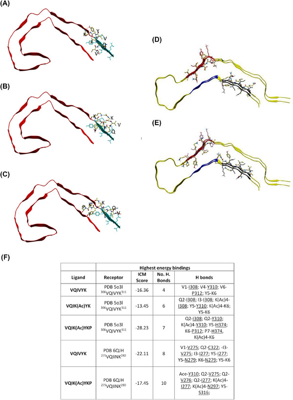
Introduction: As aggregation underpins Tau toxicity, aggregation inhibitor peptides may have disease-modifying potential. They are therefore currently being designed and target either the 306VQIVYK311 aggregation-promoting hotspot found in all Tau isoforms or the 275VQIINK280 aggregation-promoting hotspot found in 4R isoforms. However, for any Tau aggregation inhibitor to potentially be clinically relevant for other tauopathies, it should target both hotspots to suppress aggregation of Tau isoforms, be stable, cross the blood-brain barrier, and rescue aggregation-dependent Tau phenotypes in vivo.

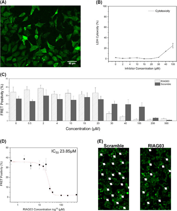
Methods: We developed a retro-inverso, stable D-amino peptide, RI-AG03 [Ac-rrrrrrrrGpkyk(ac)iqvGr-NH2], based on the 306VQIVYK311 hotspots which exhibit these disease-relevant attributes.

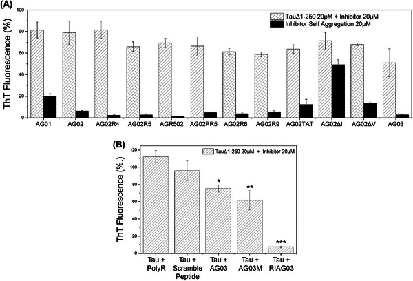
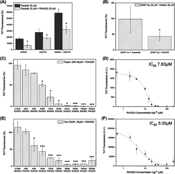
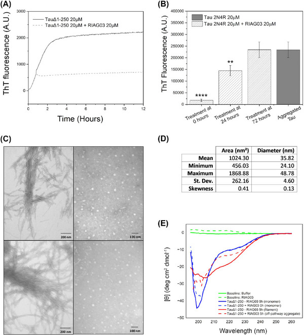
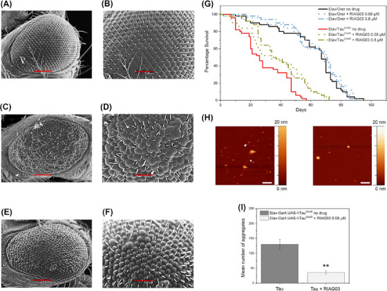
Results: Unlike other aggregation inhibitors, RI-AG03 effectively suppresses aggregation of multiple Tau species containing both hotspots in vitro and in vivo, is non-toxic, and suppresses aggregation-dependent neurodegenerative and behavioral phenotypes.

Discussion: RI-AG03 therefore meets many clinically relevant requirements for an anti-aggregation Tau therapeutic and should be explored further for its disease-modifying potential for Tauopathies.

Highlights: Our manuscript describes the development of a novel peptide inhibitor of Tau aggregation, a retro-inverso, stable D-amino peptide called RI-AG03 that displays many clinically relevant attributes. We show its efficacy in preventing Tau aggregation in both in vitro and in vivo experimental models while being non-toxic to cells. RI-AG03 also rescues a biosensor cell line that stably expresses Tau repeat domains with the P301S mutation fused to Cer/Clo and rescues aggregation-dependent phenotypes in vivo, suppressing neurodegeneration and extending lifespan. Collectively our data describe several properties and attributes of RI-AG03 that make it a promising disease-modifying candidate to explore for reducing pathogenic Tau aggregation in Tauopathies such as Alzheimer's disease. Given the real interest in reducing Tau aggregation and the potential clinical benefit of using such agents in clinical practice, RI-AG03 should be investigated further for the treatment of Tauopathies after validation in mammalian models. Tau aggregation inhibitors are the obvious first choice as Tau-based therapies as much of Tau-mediated toxicity is aggregation dependent. Indeed, there are many research efforts focusing on this therapeutic strategy with aggregation inhibitors being designed against one of the two aggregation-promoting hotspots of the Tau protein. To our knowledge, RI-AG03 is the only peptide aggregation inhibitor that inhibits aggregation of Tau by targeting both aggregation-promoting hotspot motifs simultaneously. As such, we believe that our study will have a significant impact on drug discovery efforts in this arena.

Keywords: Alzheimer's disease; Drosophila; VQIINK; VQIVYK; aggregation; amino‐acid; dementia; drug development; in silico; in vitro; in vivo; inhibitor; melanogaster; peptide; tau; tauopathies; therapeutic.


Conflict of interest statement

The authors declare that they have no conflict of interest. Author disclosures are available in the supporting information.

Figures

FIGURE 1
FIGURE 1
VQIK(Ac)YKP peptides dock to VQIVYK. Experimental peptides docked in parallel to the superior portion of PDB 5o3l 2N4R PHF (A–C) demonstrate preferential binding to 306VQIVYK311, whereas experimental peptides docked to PDB 6QJH 2N4R Tau snake filament (D–E) bind to both 306VQIVYK311 and 275VQIINK280. (A) Ac‐VQIVYK‐NH2; (B) AC‐VQIK(Ac)YK‐NH2; (C) Ac‐VQIK(Ac)YKP‐NH2, notice the peptide extending to interact with the parallel β‐sheet of 306VQIVYK311. (D) Ac‐VQIVYK‐NH2 binding in parallel to the filament; (E) Ac‐VQIK(Ac)YKP‐NH2 binding in anti‐parallel to the filament at 306VQIVYK311 position, and in parallel at the 275VQIINK280 position. (F) Summary table of the highest computationally calculated energy values describing the docked compounds to PDB‐5o3l and PDB‐5QJH (emphasis on VQIINK). Lower scores indicate more powerful interactions. ICM score of <–32 indicates a strong binding.
FIGURE 2
FIGURE 2
AG03 utilizes the VQIK(Ac)YKP recognition sequence. Aggregation end‐point measurements using ThT fluorescence after 24 h from 20 µM TauΔ1‐250 and/or 20 µM peptide inhibitor in the presence of 30 mM Tris buffer, 1 mM DTT, 15 µM ThT, 5 µM heparin (pH 7.4). (A) Aggregation of TauΔ1‐250 with different peptide inhibitors in the presence of heparin (white, hatched) and attempted self‐assembly of inhibitors without the presence of Tau (black). Key: AG01 [RG‐VQIINK‐GR], AG02 [RG‐VQIVYK‐GR], AG02R4 [RRG‐VQIVYK‐GRR], AG02R5 [RG‐VQIVYK‐GRRRR], AGR502 [RRRRG‐VQIVYK‐GR], AG02PR5 [RG‐VQIVYKP‐GRRRR], AG02R9 [RG‐VQIVYK‐GRRRRRRRR], AG02TAT [RG‐VQIVYKGRYGRKKRRQRRR], AG02ΔI [RG‐VQK(Ac)VYK‐GR], AG02ΔV [RG‐VQIK(Ac)YK‐GR], AG03 [RG‐VQIK(Ac)YKP‐GRRRRRRRR]. (B) Aggregation of TauΔ1‐250 in the presence of heparin with either octa‐arginine, scrambled AG03 peptide, AG03, N‐methylated AG03, or retro‐inverso AG03. Experiments were conducted in triplicate and error bars were reported as standard deviation. Statistical analysis: p > 0.05, *p ≤ 0.05, **p ≤ 0.01, ***p ≤ 0.001.
FIGURE 3
FIGURE 3
Lead peptide RI‐AG03 dose‐response and target specificity. (A) Aggregation end‐point measurements using ThT fluorescence after 24 h from 20 µM VQIINK, 20 µM VQIVYK, or a combination of both, in the presence of 30 mM Tris buffer, 1 mM DTT, 15 µM ThT, 5 µM heparin (pH 7.4), and in the presence (hatched gray bars) or absence (solid gray bars) of 20 µM RI‐AG03. (B) Aggregation end‐point measurements using ThT fluorescence after 216 h (9 days) from Tau2N4R in the presence of PBS buffer, 1 mM DTT, 20 µM ThT, 10 µM heparin (pH 7.4), and 20 µM RI‐AG03 or 20 µM Scramble peptide. (C) Concentration‐dependent inhibition of by TauΔ1‐250 by RI‐AG03. Aggregation end‐point measurements using ThT fluorescence after 24 h from 20 µM TauΔ1‐250 in the presence of 30 mM Tris buffer, 1 mM DTT, 15 µM ThT, 5 µM heparin (pH 7.4), and RI‐AG03 in a concentration range of 0.5–200 µM. (D) log10 scatter graph of TauΔ1‐250 inhibition by RI‐AG03 employing a curve fitting algorithm to calculate the IC50 (the concentration of RI‐AG03 required for 50% inhibition of aggregation) at 7.83 µM. (E) Concentration‐dependent inhibition of by Tau2N4R by RI‐AG03. Aggregation end‐point measurements using ThT fluorescence after 216 h (9 days) of Tau2N4R in the presence of PBS buffer, 1 mM DTT, 20 µM ThT, 10 µM heparin (pH 7.4), and RI‐AG03 in a concentration range of 0.5–200 µM. (F) log10 scatter graph of Tau2N4R inhibition by RI‐AG03 employing a curve fitting algorithm to calculate the IC50 at 5 µM. Experiments were conducted in triplicate and error bars were reported as standard deviation. One factor repeated measures ANOVA + Tukey post hoc statistical analysis: p > 0.05, *p ≤ 0.05, **p ≤ 0.01, ***p ≤ 0.001.
FIGURE 4
FIGURE 4
Lead peptide RI‐AG03 suppresses Tau β‐sheet content and forms of off‐pathway structures. (A) Aggregation kinetics measurements using ThT fluorescence from 20 µM TauΔ1‐250 in the presence of 30 mM Tris buffer, 1 mM DTT, 15 µM ThT, and 5 µM heparin (pH 7.4), for 12 h in the absence of RI‐AG03 (solid gray line) or with the addition of 20 µM RI‐AG03 1 h after aggregation was initiated (hatched gray line). (B) Aggregation end‐point measurements using ThT fluorescence after 216 h (9 days) from 20 µM Tau2N4R in the presence of PBS buffer, 1 mM DTT, 20 µM ThT, and 10 µM heparin (pH 7.4). 20 µM RI‐AG03 was added after 0, 24, and 72 h, of Tau aggregation (hatched gray bars), or not at all (solid gray bar representing end‐point aggregation of Tau2N4R at 9 days). Note that Tau2N4R aggregation plateaued at ∼72 h. ThT fluorescence was recorded after 216 h (9 days) for all conditions. One factor repeated measures ANOVA + Tukey post hoc statistical analysis: p > 0.05, *p ≤ 0.05, **p ≤ 0.01, ***p ≤ 0.001. (C) Negative stain TEM images using a Joel JEM‐1010 after the aggregation of 20 µM TauΔ1‐250 for 24 h, in the absence of RI‐AG03 (left), and in the presence of 20 µM RI‐AG03 (right). (D) The area and diameter of 200 large spherical species formed in the presence of 20 µM RI‐AG03 and recorded by TEM were quantified using iTEM software. (E) Circular dichroism data depicting secondary structural changes after 20 µM TauΔ1‐250 aggregation in the presence and absence of 20 µM RI‐AG03, including the baseline buffer spectrum (green solid line), and RI‐AG03 alone in buffer (green dashed line), monomeric TauΔ1‐250 (blue solid line) and TauΔ1‐250 aggregated for 5 h (red solid line), monomeric TauΔ1‐250 in the presence of RI‐AG03 (blue dashed line) and TauΔ1‐250 aggregated for 5 h in the presence of RI‐AG03 (dashed red line).
FIGURE 5
FIGURE 5
Lead peptide RI‐AG03 penetrates cells and reduces the seeding capacity of Tau seeds. (A) Fluorescence microscopy image depicting cellular uptake of 15 µM 5(6)‐carboxyfluorescein tagged RI‐AG03 [FAM‐RI‐AG03] by HEK‐293 cells incubated in DMEM/10% FBS at 37°C, with 5% CO2, after 24 h. Cells were seeded to a 96‐well plate at 20,000 cells/100 µL per well and supplemented with FAM‐RI‐AG03. Cells were visualized on a Nikon Eclipse Ti fluorescent microscope using the FITC filter. (B) LDH cytotoxicity assay using varying concentrations of RI‐AG03 co‐incubated with HEK‐293 cells. LDH Cytotoxicity (%) denotes lysed cells. Cells were seeded to a 96‐well plate at 10,000 cells/100 µL per well. Toxicity begins to increase at 40 µM. Experiments were conducted in triplicate. (C) FRET positivity indicative of Tau aggregation in a HEK‐293 biosensor cell line stably expressing Tau repeat domains with the P301S mutation fused to Cer/Clo. Cells were exposed to preformed Tau2N4R fibril seeds that were preincubated with either RI‐AG03 or Scramble peptide for 16 h before transduction. (D) Seeded aggregation is inhibited by RI‐AG03 at doses between 20 and 100 mM, with the IC50 being 23.85 mM. (E) Fewer green puncta indicative of Tau aggregates are seen in representative fluorescence microscopy images of the biosensor cells after treatment with preformed fibrils incubated with 30 µM RI‐AG03 compared with those incubated with Scramble peptide.
FIGURE 6
FIGURE 6
Lead peptide RI‐AG03 reduces tau aggregation and suppresses phenotypes in a model of tauopathy. (A–F) SEM images of the eyes of drosophila melanogaster at 100 (A, C, E) and 50 (B, D, F) microns from left to right; N = 6 (A, B) healthy GMR‐GAL4 (C, D) GMR/hTau2N4R flies without treatment (E, F) GMR‐hTau2N4R flies treated with RI‐AG03 20 µM. (G) Survival curves of control (Elav/Orer) and Tau overexpressing flies (Elav/hTau2N4R) treated with low (0.08 µM) and high (0.8 µM) doses of the RI‐AG03, respectively, and no treatment. Log‐Rank test n = 100 per genotype/treatment group, p = 0.0007 and 0.0004, respectively. (H and I) Visualization (H) and quantitation (I) of reduction of Tau aggregates after 0.08 µM inhibitor treatment using atomic force microscopy imaging on an insoluble Tau prep obtained from 6‐week transgenic flies expressing hTau2N4R pan‐neurally. Without RIA03 treatment there is evidence of fibrils (arrow) and oligomers (arrowhead). After treatment, no fibrils are seen, only large spherical structures. n = 3, Unpaired t‐test p = 0.0045.

References

    1. Wimo A, Guerchet M, Ali GC, et al. The worldwide costs of dementia 2015 and comparisons with 2010. Alzheimers Dement. 2017;13(1):1‐7. doi:10.1016/j.jalz.2016.07.150 - DOI - PMC - PubMed
    1. Monteiro AR, Barbosa DJ, Remião F, Silva R. Alzheimer's disease: insights and new prospects in disease pathophysiology, biomarkers and disease‐modifying drugs. Biochem Pharmacol. 2023;211:115522. doi:10.1016/j.bcp.2023.115522 - DOI - PubMed
    1. Mayes J, Tinker‐Mill C, Kolosov O, Zhang H, Tabner BJ, Allsop D. β‐amyloid fibrils in Alzheimer disease are not inert when bound to copper ions but can degrade hydrogen peroxide and generate reactive oxygen species. J Biol Chem. 2014;289(17):12052‐12062. doi:10.1074/jbc.M113.525212 - DOI - PMC - PubMed
    1. Kaffy J, Brinet D, Soulier JL, et al. Designed glycopeptidomimetics disrupt protein‐protein interactions mediating amyloid β‐peptide aggregation and restore neuroblastoma cell viability. J Med Chem. 2016;59(5):2025‐2040. doi:10.1021/acs.jmedchem.5b01629 - DOI - PubMed
    1. Cohen SIA, Linse S, Luheshi LM, et al. Proliferation of amyloid‐β42 aggregates occurs through a secondary nucleation mechanism. Proc Natl Acad Sci USA. 2013;110(24):9758‐9763. doi:10.1073/pnas.1218402110 - DOI - PMC - PubMed

LinkOut - more resources