. 2024 Oct;72(10):611-622.
doi: 10.1369/00221554241287267. Epub 2024 Oct 3.

Intestinal Tuft Cells Are Enriched With Protocadherins

Affiliations

Intestinal Tuft Cells Are Enriched With Protocadherins

Rachel Stubler et al. J Histochem Cytochem. 2024 Oct.

Abstract

Intestinal tuft cells are rare cells that regulate diverse functions. They harbor chemosensory receptors and signal to the mucosal immune system in response to external stimuli, though their full function and structure remain unclear. Named for their apical "tuft" of long actin-rich microvilli, tuft cells facilitate chemoreception and other physiological responses. In enterocytes, microvilli are stabilized by intermicrovillar adhesion complexes (IMACs) composed of several proteins, including cadherin-related family member-2 (CDHR2) and cadherin-related family member-5 (CDHR5), Myosin 7b, and Usher syndrome type 1 C (USH1C). We hypothesized that IMACs would be enriched in tuft cells to regulate microvillar organization. Immunostaining of murine intestinal tissue revealed that CDHR2 and CDHR5 colocalize with the tuft cell markers, DCLK1, phospho-EGFR, advillin, and cytokeratin 18. CDHR2 was dispersed throughout murine tuft cells, while CDHR5 was concentrated on the apical surface. USH1C and Myosin 7b were present in tuft cells, but at lower levels. Human single-cell RNA sequencing revealed robust CDHR2 and CDHR5 expression in tuft cells in the small intestine and colon. Immunostaining of human intestinal tissue confirmed CDHR2 and CDHR5 localization to the apical surface of tuft cells. Our findings demonstrate that protocadherins are key components of murine and human intestinal tuft cells.

Keywords: IMAC; epithelium; gut; microvilli.


Conflict of interest statement

Competing InterestsThe author(s) declared no potential conflicts of interest with respect to the research, authorship, and/or publication of this article.

Figures

Figure 1.
Figure 1.
CDHR2 is highly expressed in murine tuft cells. (A) Immunohistochemical staining of CDHR2 in the small intestine, highlighting cells that express CDHR2 throughout the cell body. (B-D) Immunofluorescent staining of small intestine and colon for CDHR2 (magenta), apical membrane marker γ-actin (yellow), tuft cell markers (green): DCLK1 (B), CK-18 (C), and Ac-TUBA (D). Scale bar = 50 µm (A low magnification), 5 µm (A Inset, B-D).
Figure 2.
Figure 2.
CDHR5 localizes to the apical surface of murine tuft cells. (A) Immunohistochemical staining of CDHR5 in the small intestine, highlighting enrichment of CDHR5 in a select cell. (B-D) Immunofluorescent staining of murine small intestine and colon for CDHR5 (magenta) with tuft markers: (B) DCLK1 (green) and apical marker γ-actin (yellow), (C) POU2F3 (green) and γ-actin (yellow), and (D) AVIL (green) and CK-18 (yellow). Scale bar = 50 µm (A low magnification), 5 µm (A Inset, B-D).
Figure 3.
Figure 3.
CDHR2 and CDHR5 are expressed by a high percentage of murine tuft cells. (A) Immunostaining of CDHR2 (magenta), DCLK1 (green), and γ-actin (yellow) with quantification of the percentage of DCLK1-positive cells that co-express CDHR2. (B) Immunostaining of CDHR5 (magenta), DCLK1 (green), and γ-actin (yellow) with quantification of the percentage of DCLK1-positive cells that co-express CDHR5 and γ-actin. n=4 mice/group. Scale bar = 50 µm.
Figure 4.
Figure 4.
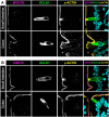
CDHR2 and CDHR5 display differential staining patterns in tuft cells. Immunostaining of murine small intestine and colon showing that CDHR2 (magenta) and CDHR5 (green) are both enriched at the apical surface of tuft cells, marked by P-EGFR (yellow). CDHR2 displays diffuse more cytoplasmic staining compared with CDHR5. Scale bar = 5 µm.
Figure 5.
Figure 5.
Other IMAC components are not enriched in tuft cells compared with neighboring enterocytes. Immunostaining of murine small intestine and colon for (A) MYO7B (magenta) and (B) USH1C (magenta) with DCLK1 (green) and γ-actin (yellow). Scale bar = 5 µm.
Figure 6.
Figure 6.
Tuft cells in murine intestinal organoids are enriched for CDHR2 and CDHR5 and display staining patterns consistent with murine tissue. Immunostaining for: (A) CDHR2 (magenta) with DCLK1 (green) and γ-actin (yellow), (B) CDHR5 (magenta) with DCLK1 (green) and γ-actin (yellow), (C) CDHR2 (magenta) with CK-18 (green) and γ-actin (yellow), (D) CDHR5 (magenta) with AVIL (green) and CK-18 (yellow), (E) CDHR2 (magenta) with P-EGFR (green) and Ac-TUBA (yellow), (F) CDHR5 (magenta) with POU2F3 (green) and γ-actin (yellow), and (G) CDHR2 (magenta) and CDHR5 (green) with P-EGFR (yellow). Scale bar = 10 µm on low magnification and 5 µm on high magnification insets.
Figure 7.
Figure 7.
CDHR2 and CDHR5 localize to the apical surface of tuft cells in the human small intestine and colon. (A) Single cell RNA sequencing of tuft cells in healthy control human small intestine and colon from CellXGene publicly available database. (B-C) Immunostaining of human small intestine and colon for (B) CDHR2 (magenta) and (C) CDHR5 (magenta) with P-EGFR (green) and γ-actin (yellow). Scale bar = 10 µm on low magnification and 2 µm on high magnification insets.

References

    1. Isomäki AM. A new cell type (tuft cell) in the gastrointestinal mucosa of the rat. A transmission and scanning electron microscopic study. Acta Pathol Microbiol Scand A. 1973; 240:1–35. - PubMed
    1. Nabeyama A, Leblond CP. “Caveolated cells” characterized by deep surface invaginations and abundant filaments in mouse gastro-intestinal epithelia. Am J Anat. 1974;140(2):147–65. doi: 10.1002/aja.1001400203. - DOI - PubMed
    1. Tsubouchi S, Leblond CP. Migration and turnover of entero-endocrine and caveolated cells in the epithelium of the descending colon, as shown by radioautography after continuous infusion of 3H-thymidine into mice. Am J Anat. 1979;156(4):431–51. doi: 10.1002/aja.1001560403. - DOI - PubMed
    1. Howitt MR, Cao YG, Gologorsky MB, Li JA, Haber AL, Biton M, Lang J, Michaud M, Regev A, Garrett WS. The taste receptor TAS1R3 regulates small intestinal tuft cell homeostasis. Immunohorizons. 2020;4(1):23–32. doi: 10.4049/immunohorizons.1900099. - DOI - PMC - PubMed
    1. Luo XC, Chen ZH, Xue JB, Zhao DX, Lu C, Li YH, Li S-M, Du Y-W, Liu Q, Wang P, Liu M, Huang L. Infection by the parasitic helminth Trichinella spiralis activates a Tas2r-mediated signaling pathway in intestinal tuft cells. Proc Natl Acad Sci U S A. 2019;116(12):5564–9. doi: 10.1073/pnas.1812901116. - DOI - PMC - PubMed

Publication types