. 2024 Oct 3;134(23):e180160.
doi: 10.1172/JCI180160.

Transcription factor KROX20 marks epithelial stem cell ancestors for hair follicle formation

Affiliations

Transcription factor KROX20 marks epithelial stem cell ancestors for hair follicle formation

Elnaz Ghotbi et al. J Clin Invest. .

Abstract

Epidermal stem cells control homeostasis and regeneration of skin and hair. In the hair follicle (HF) bulge of mammals, populations of slow-cycling stem cells regenerate the HF during cyclical rounds of anagen (growth), catagen (regression), and telogen (quiescence). Multipotent epidermal cells are also present in the HF above the bulge area, contributing to the formation and maintenance of sebaceous gland and upper and middle portions of the HF. Here, we report that the transcription factor KROX20 is enriched in an epidermal stem cell population located in the upper/middle HF. Expression analyses and lineage tracing using inducible Krox20-CreERT showed that Krox20-lineage cells migrate out of this HF region and contribute to the formation of the bulge in the HF, serving as ancestors of bulge stem cells. In vivo depletion of these cells arrests HF morphogenesis. This study identifies a marker for an epidermal stem cell population that is indispensable for hair homeostasis.

Keywords: Development; Mouse stem cells; Stem cells.


Figures
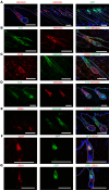
Figure 1
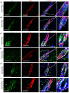
Figure 1. Comparative analysis of Krox20 expression in telogen and anagen HFs in relation to other stem cell niches within the HF.
(A) Immunofluorescence analysis depicting Krox20 expression with KROX20 antibody in telogen (left) and anagen (middle) and by GFP immunofluorescence in anagen HFs of Krox20-GFP mice (right). (BE) Colocalization analysis demonstrating the absence of colocalization between Krox20 expression and bulge stem cell markers, K15 and CD34, in anagen (B and C) and telogen (D and E) HFs. (F and G) Colocalization analysis of GFP expression with upper stem cell niche markers such as SCA1 (F) and LRIG1 (G) in telogen HFs of Krox20-GFP mice. All dashed circles and ovals represent the bulge area. Arrows point to autofluorescence from hair shafts. Asterisks represent the sebaceous glands. n ≥ 3. Scale bars: 100 mm.
Figure 2
Figure 2. Lineage tracing with an inducible Krox20-CreERT confirms the contribution of Krox20-lineage cells to the HF bulge.
(A) Krox20-CreERT; R26-tdTomato mice were induced with tamoxifen at P1. Lineage tracing shows that Krox20-lineage cells are initially restricted to the upper and middle HF but, over time, they expand downward, contributing to the formation of the bulge and hair shaft. (B) Colocalization analysis of Krox20-lineage cells with hair shaft marker AE13. (C) Diagram of an anagen HF and a telogen HF. (D) Quantification of HF regions labeled with tdTomato at various time points in Krox20-CreERT; R26-tdTomato mice when induced at P1. n ≥ 3. Scale bars: 100 μm. Tom, tdTomato. Statistical significance was determined for D by 2-way ANOVA; statistics represent mean ± SEM, *P < 0.05, **P < 0.01, ****P < 0.0001. Non-significant values were not plotted on the graphs due to space constraints.
Figure 3
Figure 3. Krox20 marks an epidermal stem cell niche in the HF excluding the bulge region.
Colocalization analysis showing Krox20-lineage cells in relation to bulge stem cell markers K15 (A and B) and CD34 (CF) within anagen (P36, A and D) and telogen (P60, B; P22, C; P60, E; and P100, F) HFs, induced at P1. Dashed circles represent the bulge area, which is shown at higher magnification in the panels at the end of each row (×400 for AC, E, and F, and ×200 for D). n ≥ 3. Scale bars: 100 μm. TOM, tdTomato.
Figure 4
Figure 4. Krox20-positive cells are indispensable for hair development.
(AC) Krox20-DTA; K14-Cre pups at P1 show normal gross appearance (A), skin histology (B), and K15-positive stem cell distribution (C). (D) Krox20-DTA; K14-Cre pups show no difference compared with littermate controls at P1 but are lighter in skin color and weight by P6. (E) H&E staining at P6 shows that the HFs of Krox20-DTA; K14-Cre mice are shorter and random in orientation. (F) Immunostaining shows the absence of KROX20 protein in the skin of Krox20-DTA; K14-Cre mice. (GL) Immunostaining reveals that HFs in P6 Krox20-DTA; K14-Cre pups have aberrant expression of Ki67 (G), K15 (H), versican (I), DCT (J), AE13 (K), and P-cadherin (P-CAD) (L). n = 5. Scale bars: 100 μm.
Figure 5
Figure 5. Krox20-positive cells are essential for hair regeneration.
(A and B) Ablation of Krox20-positive cells via tamoxifen induction in Krox20-DTA; Krox20-CreERT mice during telogen I (P17) (A) and telogen II (P36) (B) hinders hair regeneration following depilation (n = 3). (C) H&E analysis of the skin in telogen II Krox20-positive cell–depleted mice shows hair growth arrest and HF miniaturization. (D) Quantification of number of normal HFs per millimeter of skin cross section of Krox20 cell–depleted and littermate control mice (n = 2). (EG) Immunofluorescence staining at P56 reveals the absence of KROX20-positive cells (E), and bulge CD34-positive and K15-positive cells (F and G). n = 3. Scale bars: 100 μm. Statistical significance was determined for D by unpaired 2-tailed Student’s t test; statistics represent mean ± SEM, ****P < 0.0001.
Figure 6
Figure 6. Lineages of Krox20- and Sox9-coexpressing cells in the upper HF move down to contribute to the first formation of the bulge.
(A) Ablation of Krox20-positive cells in the K14 lineage results in diminished levels of SOX9 expression within the entire HF at P6. (B) Sox9- and Krox20-coexpressing cells overlap in the upper and middle telogen HF at P17. (C and D) Ablation of Krox20-expressing cells at P17 results in the complete absence of SOX9 (C) and CD34 (D) bulge stem cell markers at P29. (E) Krox20-lineage cells encompass a population of the Sox9-expressing cells in the bulge. Dashed ovals represent the bulge area. Dashed rectangles represent the area coexpressing SOX9 and KROX20 in control skin, or regions devoid of KROX20 and/or SOX9 expression in KROX20-ablated skin. Higher magnifications of the areas represented by the circles or rectangles are shown in the panels at the end of each row (×200 for A, bottom, and D, bottom; and ×400 for A, top, B, C, D, top, and E). Arrows point to the cells coexpressing KROX20 and SOX9. n = 3. Scale bars: 100 μm.
Figure 7
Figure 7. KROX20 protein is critical for epidermal tissue homeostasis.
(AC) Approximately 83% of Krox20fl/fl; K14-Cre mice show pattern hair loss during aging (A) (n = 18); in the balding skin, the majority of HFs are deformed, with some HFs arrested at anagen while others are miniaturized (B). (C) Quantification of number of normal HFs per millimeter of skin cross section of Krox20fl/fl; K14-Cre mice and their littermate controls (n = 7). (D) Immunofluorescence staining of the skin confirmed the reduced expression of epithelial KROX20 protein in Krox20fl/fl; K14-Cre mice. (E and F) The miniaturized HFs in the Krox20fl/fl; K14-Cre mice lack normal bulge structure, characterized by the absence of HF stem cell markers K15 (E) and CD34 (F). n = 3. Dashed circles represent the bulge area. Arrows point to the traces of KROX20 in the HFs. Scale bars: 100 μm. Statistical significance was determined for C by unpaired 2-tailed Student’s t test; statistics represent mean ± SEM, ****P < 0.0001.

References

    1. Horsley V. Skin in the game: stem cells in repair, cancer, and homeostasis. Cell. 2020;181(3):492–494. doi: 10.1016/j.cell.2020.03.019. - DOI - PubMed
    1. Hirsch T, et al. Regeneration of the entire human epidermis using transgenic stem cells. Nature. 2017;551(7680):327–332. doi: 10.1038/nature24487. - DOI - PMC - PubMed
    1. Tumbar T, et al. Defining the epithelial stem cell niche in skin. Science. 2004;303(5656):359–363. doi: 10.1126/science.1092436. - DOI - PMC - PubMed
    1. Morris RJ, et al. Capturing and profiling adult hair follicle stem cells. Nat Biotechnol. 2004;22(4):411–417. doi: 10.1038/nbt950. - DOI - PubMed
    1. Cotsarelis G, et al. Label-retaining cells reside in the bulge area of pilosebaceous unit: implications for follicular stem cells, hair cycle, and skin carcinogenesis. Cell. 1990;61(7):1329–1337. doi: 10.1016/0092-8674(90)90696-C. - DOI - PubMed