. 2025 Jan;292(1):129-152.
doi: 10.1111/febs.17265. Epub 2024 Oct 3.

Substrate preference, RNA binding and active site versatility of Stenotrophomonas maltophilia nuclease SmNuc1, explained by a structural study

Affiliations

Substrate preference, RNA binding and active site versatility of Stenotrophomonas maltophilia nuclease SmNuc1, explained by a structural study

Kristýna Adámková et al. FEBS J. 2025 Jan.

Abstract

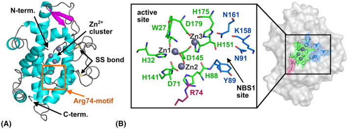
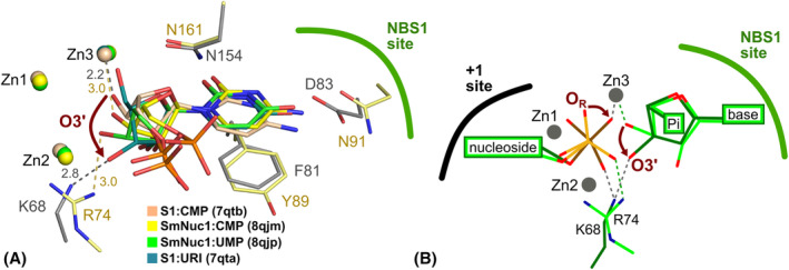
Nucleases of the S1/P1 family have important applications in biotechnology and molecular biology. We have performed structural analyses of SmNuc1 nuclease from Stenotrophomonas maltophilia, including RNA cleavage product binding and mutagenesis in a newly discovered flexible Arg74-motif, involved in substrate binding and product release and likely contributing to the high catalytic rate. The Arg74Gln mutation shifts substrate preference towards RNA. Purine nucleotide binding differs compared to pyrimidines, confirming the plasticity of the active site. The enzyme-product interactions indicate a gradual, stepwise product release. The activity of SmNuc1 towards c-di-GMP in crystal resulted in a distinguished complex with the emerging product 5'-GMP. This enzyme from an opportunistic pathogen relies on specific architecture enabling high performance under broad conditions, attractive for biotechnologies.

Keywords: Stenotrophomonas maltophilia; RNA; S1/P1 nuclease; X‐ray crystallography; c‐di‐GMP cleavage.


Conflict of interest statement

The authors declare no conflict of interest.

Figures

Fig. 1
Fig. 1
Overall structure of SmNuc1. (A) Cartoon representation of SmNuc1 coloured by secondary structure (α‐helices in cyan, β‐strands in magenta and loops in grey). The N‐terminal Trp27 is involved in the coordination of the zinc cluster. The Arg74‐motif is marked by the orange rectangle. (B) The active site of SmNuc1. The active site consists of three zinc ions (grey spheres) which are coordinated by nine residues (C atoms in green), residues of the NBS1 site (C in blue), and the stabilising complementary positive residue Arg74 (C in pink). Localization of the active site on the surface of SmNuc1 is highlighted. Graphics were created using pymol (Schrödinger, LLC, New York, NY, USA) and the SmNuc1:free structure (PDB: 8QJL).
Fig. 2
Fig. 2
Zinc cluster of SmNuc1 and binding of phosphate ion. (A) Zinc cluster of the ligand‐free structure of SmNuc1 (PDB: 8QJL). Zinc ions Zn1, Zn2 and Zn3 (shown as grey spheres) are coordinated by the Trp27 main chain and side chains of eight residues (C atoms in grey). Water molecules W1, W2, W3 and W4 (red spheres) are coordinated by zinc ions. (B) Binding of the phosphate ion (Pi) in the structure of the SmNuc1:GMP complex (PDB: 8QJO). Oxygen ON of Pi replaces the nucleophilic water W1, oxygen OR replaces W2, and oxygen OS replaces W4. W3 is replaced by the oxygen O3′ of the ribose moiety of the bound ligand 5′‐GMP (C in magenta). All contacts are shown as grey dashes and distances are in Å. Graphics were created using pymol (Schrödinger, LLC).
Fig. 3
Fig. 3
Ligand binding in the active site of SmNuc1:AMP (PDB: 8QJN), SmNuc1:GMP (PDB: 8QJO) and SmNuc1:GMP* (PDB: 9EMG) with 2mF o − DF c (blue mesh, [45]) composite omit map around ligands at 1σ level. (A) The active site of the SmNuc1:AMP complex with bound phosphate ion (P atom in orange) and (B) 5′‐AMP (C in cyan). (C) The active site of chain A of AU of the SmNuc1:GMP complex with bound phosphate ion (P in orange) and (D) 5′‐GMP (C in magenta). (E) The active site of chain B of AU of the SmNuc1:GMP complex with bound phosphate ion (P in orange) and (F) 5′‐GMP (C in magenta). (G) The active site of chain B of AU of the SmNuc1:GMP* complex (product of c‐di‐GMP cleavage) with bound waters and 5′‐GMP (C in violet). Graphics were created using pymol (Schrödinger, LLC).
Fig. 4
Fig. 4
Binding modes of purine nucleotides in SmNuc1. (A) Binding of 5′‐AMP (C atoms in cyan) in chain B in the SmNuc1:AMP complex (PDB: 8QJN) and (B) binding of 5′‐GMP (C in magenta) in chain B in the SmNuc1:GMP complex (PDB: 8QJO). All ligand contacts are displayed as dashes and all distances are in Å. For simplicity, contacts of phosphate ions are not shown (these contacts are displayed in Fig. 2B). (C) Superposition of the active sites of the SmNuc1:AMP and SmNuc1:GMP complexes. SmNuc1:GMP is coloured in shades of magenta, and SmNuc1:AMP in shades of cyan. (D) Comparison of the active site of SmNuc1:GMP (C in magenta) and SmNuc1:GMP* (PDB: 9EMG, C in violet). SmNuc1:GMP is coloured in shades of magenta and SmNuc1:GMP* in shades of violet. Note the alternative conformations of Lys158 and Asn161 in SmNuc1:GMP* and the absence of Arg74 in the stabilisation of 5′‐GMP in the active site. Waters W1, W2, W4 and W449 replace oxygens of phosphate and W710 is in the previous position of Arg74. In both cases, oxygen O2′ of 5′‐GMP is stabilised by WO2′ and the phosphate moiety by interaction with WP. Graphics and the alignment, using the automated multi‐step superposition algorithm based on active site residues, were created using pymol (Schrödinger, LLC).
Fig. 5
Fig. 5
Binding modes of 5′‐CMP nucleotides in SmNuc1 structures. (A) Schematic representation of positions occupied by 5′‐CMP (+1 site and − 1 site) in the complex SmNuc1:CMP (PDB: 8QJM). (B) Binding of 5′‐CMP in chain B of AU of the SmNuc1:CMP complex. 5′‐CMP (C atoms in yellow) binds in the −1 site and +1 site mimicking the post‐cleavage state of a dinucleotide. H‐bonds and the interactions are marked by grey dashes. (C) Binding of 5′‐CMP in the +1 site of chain A of the SmNuc1:CMP complex. (D) The comparison of binding of phosphate moiety in the +1 site of chain A and B of the SmNuc1:CMP complex. Phosphate moiety is rotated in the zinc cluster and binds in two binding modes – inverted post‐cleavage state (chain B) and state of the leaving product (chain A). (E) Non‐canonical binding mode of 5′‐CMP in the NBS1 site of chain B of SmNuc1:CMPinh complex (PDB: 8QJQ). Note the position of the ribose moiety in the site normally occupied by the nucleobase. (F) Conformation of the ribose moiety of 5′‐CMP in the structures of the SmNuc1 complexes. In the +1 site the C3′‐endo conformation of the ribose moiety is preferred (chain B, SmNuc1:CMP). In the −1 site the ribose moiety has a C2′‐endo conformation (chain B, SmNuc1:CMP), whereas the C3′‐endo conformation is not possible in this site due to steric constraints. In the non‐canonical binding position of 5′‐CMP (SmNuc1:CMPinh complex), the ribose moiety is in C4′‐exo conformation. Graphics were created using pymol (Schrödinger, LLC). All ligand contacts are displayed as dashes. Distances in (D) are in Å.
Fig. 6
Fig. 6
Binding of 5′‐CMP in the active site of the SmNuc1:CMP complex (PDB: 8QJM). (A) The active site of chain A of AU with 2mF o − DF c composite omit map at 1σ level (blue mesh; [45]) around 5′‐CMP (C atoms in yellow) bound in +1 position with respect to the cleaved phosphodiester bond, and (B) with displayed H‐bonds and O–Zn distances between 5′‐CMP, zinc ion Zn3, waters, and residues of the active site (grey dashes). (C) Active site of chain B with composite omit map 2mF o − DF c at 1σ level (blue mesh) around two molecules of 5′‐CMP (C in yellow) with (D) displayed H‐bonds and O–Zn distances of 5′‐CMP bound to NBS1, and (E) all contact distances of 5′‐CMP bound to +1 site. All distances are in Å. Graphics were created using pymol (Schrödinger, LLC).
Fig. 7
Fig. 7
Binding of 5′‐CMP in the active site of the SmNuc1:CMPinh complex (PDB: 8QJQ). (A) Active site of chain A of AU with 2mF o − DF c composite omit map at 1σ level (blue mesh; [45]) around 5′‐CMP (C atoms in yellow) bound to +1 site with (B) displayed H‐bonds and O–Zn distances between ligand and residues of the active site (grey dashes). (C) Active site of chain B of AU with 2mF o − DF c composite omit map at 1σ level (blue mesh) around Pi (P in orange) and 5′‐CMP (C in yellow) bound in an inhibitory binding mode with (D) displayed H‐bonds and O–Zn distances of Pi and 5′‐CMP bound to the NBS1 site. All distances are in Å. Graphics were created using pymol (Schrödinger, LLC).
Fig. 8
Fig. 8
Binding of 5′‐UMP in the SmNuc1:UMP complex (PDB: 8QJN). (A) Binding of 5′‐UMP in chain B of AU of SmNuc1:UMP (C atoms of 5′‐UMP in green). (B) Binding of 5′‐UMP in chain A blocked by contact with the C terminus of chain A of the symmetry‐related molecule. Residue Arg272sym and the His‐tag residue His275sym (C in dark grey) are from the symmetry‐related molecule with translation vector in fractional coordinates t (x + 1, y, z). Selected interaction contacts are displayed as dashes. Graphics were created using pymol (Schrödinger, LLC).
Fig. 9
Fig. 9
Binding of 5′‐UMP in the active site of the SmNuc1:UMP complex (PDB: 8QJP). (A) Active site of chain A of AU with 2mF o − DF c composite omit map at 1σ level (blue mesh; [45]) around 5′‐UMP (C atoms in green) with (B) displayed H‐bonds and O–Zn distances between 5′‐UMP, zinc ion Zn3, waters, and residues of the active site (grey dashes). (C) Active site of chain B of AU with 2mF o − DF c composite omit map at 1σ level (blue mesh) around 5′‐UMP (C in green) with (D) displayed H‐bonds and O–Zn distances of 5′‐UMP bound to the NBS1 site. All distances are in Å. Graphics were created using pymol (Schrödinger, LLC).
Fig. 10
Fig. 10
Crystal packing of the SmNuc1:UMP complex with blocked active site of chain A and accessible active site of chain B. There are two molecules of SmNuc1 in AU – (chain A and chain B) shown as light grey cartoon. Symmetry‐related molecules (chain Asym and chain Bsym) with translation vector t (x + 1, y, z) (fractional coordinates) are shown as dark grey cartoon. C‐termini of all displayed chains are highlighted in purple. Zinc ions are displayed as pink spheres, 5′‐UMP molecules are shown as sticks (C atoms in green). Graphics were created using pymol (Schrödinger, LLC).
Fig. 11
Fig. 11
The comparison of binding of 5′‐UMP (C atoms in green) in the active site of the SmNuc1:UMP complex (C in light green; PDB: 8QJP) and uridine (URI, C in dark grey) in the active site of the S1:URI complex (C in grey; PDB: 7QTA, [13]). Important contacts are shown as dashes. Graphics were created using pymol (Schrödinger).
Fig. 12
Fig. 12
Crystal contacts in the SmNuc1:free structure involving the Arg74‐motif. The original molecule (x, y, z) is shown as grey cartoon with contact residues as sticks and the (x, y + 1, z) symmetry‐related molecule is shown as yellow cartoon with contact residues as sticks. Zinc ions are shown as spheres and coloured in pink. Contacts are shown as dashes and distances are in Å. Graphics were created using pymol (Schrödinger, LLC).
Fig. 13
Fig. 13
Different states of the Arg74‐motif (Asp71‐Tyr89) in the SmNuc1 structures. (A) Detail of SmNuc1:CMP (PDB: 8QJM) with the closed Arg74‐motif (surface coloured in orange, Asp71, Arg74 and Tyr89 in sticks). (B) Detail of SmNuc1:UMP (PDB: 8QJP) with the Arg74‐motif in the intermediate position (surface coloured in green, Asp71, Arg74 and Tyr89 in sticks). (C) Detail of SmNuc1:free (PDB: 8QJL) with the open Arg74‐motif (blue surface). Arg74 (shown as sticks) is flipped out. (D) Superposition of the SmNuc1:free, SmNuc1:UMP and SmNuc1:CMP structures with different positions of Arg74. Zinc ions are shown as spheres and coloured in pink. Distances in Å show the shift of the guanidium group of Arg74. The alignment was calculated based on Cα atoms, and all graphics were generated using pymol (Schrödinger, LLC).
Fig. 14
Fig. 14
Comparison of the nucleolytic activity of SmNuc1WT (green) with SmNuc1R74K (blue) and SmNuc1R74Q (purple) mutants towards (A) RNA, (B) ssDNA, and (C) dsDNA substrates. All reactions were performed in triplicates at pH 7. The measured activities (including standard deviations) were converted into percentages of SmNuc1WT specific activity towards the indicated substrate type (58 550 ± 2330 U·μg−1 for RNA, 71 000 ± 8910 U·μg−1 for ssDNA, and 2150 ± 170 U·μg−1 for dsDNA). One unit (U) of nuclease activity was defined as change of absorbance of 0.001 at 260 nm in 1 cm path per 1 min [23]. Results were processed and graphs were produced using the graphpad prism8 software version 8.2 (GraphPad Software, Boston, MA, USA).
Fig. 15
Fig. 15
The comparison of binding of the purine nucleotide 5′‐AMP in SmNuc1 (C atoms in cyan) and adenosine nucleotides in structures of S1 nuclease complexes. (A) Alignment of 5′‐AMP from the SmNuc1:AMP structure (C in cyan) with 5′‐AMP (C in grey; PDB: 5FBB, [13]) bound to S1 nuclease in the inhibitory/inverted binding mode and (B) with 5′‐dAMP(S) (C in grey, PDB: 5FBC, [13]) bound in the remodelled NBS1 in an inhibitory position. Zinc cluster (spheres) and the displayed part of the NBS1 site are coloured light blue for SmNuc1:AMP and grey for the S1 nuclease structures. Alignments of the active site residues were calculated using an algorithm implemented in pymol (Schrödinger, LLC), used for generation of graphics.
Fig. 16
Fig. 16
Binding of nucleic acids to SmNuc1 and comparison of the Arg74‐motif within known structures of the S1/P1 family nucleases. (A) SmNuc1 surface coloured by electrostatic potential (−5 to +5 kT·e−1) with predicted binding of the substrate (green arrow) on the SmNuc1 nuclease surface. This model is based on binding of ligand in the SmNuc1:CMP complex (yellow sticks). Zinc ions are displayed as magenta spheres. Residues Lys187, Arg170, Asn107 and Arg106 potentially involved in binding of longer substrates are marked. Electrostatic potential was calculated using the apbs software [46]. (B) The superposition of SmNuc1 nuclease in the complex with 5′‐CMP (PDB: 8QJM) and assembly of S1 nuclease structures (PDB entries 5FBF and 5FBD, [13]) in complexes with 5′‐dCMP ligands. SmNuc1 is shown as blue cartoon with Arg74 and Tyr89 shown as sticks. 5′‐CMP molecules are displayed as sticks (C atoms in blue). S1 nuclease is shown as yellow cartoon with important residues Tyr183 (Half‐Tyr site), Tyr69 (stabilisation of +1 site), Lys68 and Phe81 (substrate/product binding) as yellow sticks. Nucleotides 5′‐dCMP bound to S1 nuclease are shown as sticks (C in orange). (C) Sequence alignment of the SmNuc1 Arg74‐motif within the S1/P1 family. Amino acid sequences of several nucleases from the family were chosen: SmNuc1 from Stenotrophomonas maltophilia (GenBank: WP_005410840.1), S1 from Aspergillus oryzae (UniProt: P24021), P1 from Penicillium citrinum (UniProt: P24289), TBN1 from Solanum lycopersicum (UniProt: Q0KFV0), AtBFN2 from Arabidopsis thaliana (UniProt: Q9C9G4), Lpn1 from Legionella pneumophila (UniProt: Q5ZV70) and LmaC1N from Leishmania major (UniProt: Q8T4M4). The alignment was calculated in Clustal Omega [41] and visualised in espript 3.0 (https://espript.ibcp.fr, [47]). (D) Superposition of the Arg74‐motif (Asp71–Tyr89) of SmNuc1 in the open position (light blue, PDB: 8QJL) with SmNuc1 in the closed position (dark blue, PDB: 8QJM), S1 nuclease (yellow, PDB: 5FB9, [13]), and AtBFN2 nuclease (dark green, PDB: 4CXO, [14]). Zinc ions are shown as grey spheres. The alignments were calculated, and all graphics were created using pymol (Schrödinger, LLC).
Fig. 17
Fig. 17
Sequence alignment of SmNuc1 with selected S1/P1 nucleases. SmNuc1 from Stenotrophomonas maltophilia (GenBank: WP_005410840.1), S1 from Aspergillus oryzae (UniProt: P24021), P1 from Penicillium citrinum (UniProt: P24289), TBN1 from Solanum lycopersicum (UniProt: Q0KFV0), AtBFN2 from Arabidopsis thaliana (UniProt: Q9C9G4), Lpn1 from Legionella pneumophila (UniProt:Q5ZV70), and LmaC1N from Leishmania major (UniProt entry Q8T4M4). The secondary structure elements of SmNuc1 are shown above the alignment. Conserved residues are marked by dark blue background, partially conserved or similar residues are in dark blue bold letters, and globally similar residues are in boxes. The Zn‐binding, NBS1 residues, and the Arg74‐motif are marked. Disulphides of SmNuc1 are marked as SS1 and SS2. The alignment was calculated in clustal omega [32] and visualised in espript 3.0 (https://espript.ibcp.fr, [33]).
Fig. 18
Fig. 18
Product‐leaving states of S1/P1 nucleases. (A) The rotation of O3′ oxygen from the active site zinc cluster. A comparison of RNA cleavage products bound to S1 (PDB: 7QTA and PDB: 7QTB; [16]) and SmNuc1 nuclease (PDB: 8QJM and PDB: 8QJP). O3′ is firstly in contact with Zn3 and then moves out, ending up in contact with a positively charged (stabilising) residue Arg74 (Lys68 in S1 nuclease). The active site residues are shown as sticks (C atoms in light yellow for SmNuc1 nuclease, C atoms in dark grey for S1 nuclease), zinc ions are coloured according to ligands. (B) A schema of the cooperative movement of the phosphate group and O3′ of products in the active site. The O3′ oxygen of the nucleotide in the NBS1 site rotates out from the zinc cluster and the oxygen OR of the nucleotide in +1 position rotates (50°) and takes a shallower binding pose with respect to the zinc cluster. The scheme is based on the after‐cleavage state captured in the structure SmNuc1:CMP (C in light green, P in yellow) and the product‐leaving state in the structure of S1 nuclease in complex with 5′‐dCMP (C in dark green, P in brown, PDB: 5FBF, [13]). All graphics and alignments were done using pymol (Schrödinger, LLC).
Fig. 19
Fig. 19
The first crystal of SmNuc1 nuclease, leading to the structure of SmNuc1:free (PDB: 8QJL). The crystal is shown in visible light, polarised light and UV light (from left to right). Images were captured by an RI1000 (Formulatrix).

Similar articles

References

    1. Ando T (1966) A nuclease specific for heat‐denatured DNA isolated from a product of Aspergillus oryzae . Biochim Biophys Acta 114, 158–168. - PubMed
    1. Mouri T, Hashida W, Shiga I & Teramoto S (1966) Studies on nucleic acid related substances in foodstuff. VII. Nucleic acid degrading enzymes of shiitake (Lentinus edodes). J Ferment Technol 44, 245–258.
    1. Kuninaka A, Otsuka S‐I, Kobayashi Y & Sakaguchi K‐I (1959) Studies on 5′‐phosphodiesterases in microorganisms. Bull Agric Chem Soc Japan 23, 239–243.
    1. Koval T & Dohnalek J (2017) Characteristics and application of S1‐P1 nucleases in biotechnology and medicine. Biotechnol Adv 36, 603–612. - PubMed
    1. Husťakova B, Trundova M, Adamkova K, Koval T, Duskova J & Dohnalek J (2023) A highly active S1‐P1 nuclease from the opportunistic pathogen Stenotrophomonas maltophilia cleaves c‐di‐GMP. FEBS Lett 597, 2103–2118. - PubMed

Associated data

LinkOut - more resources