. 2024 Oct 3;22(10):e3002827.
doi: 10.1371/journal.pbio.3002827. eCollection 2024 Oct.

MMTV RNA packaging requires an extended long-range interaction for productive Gag binding to packaging signals

Affiliations

MMTV RNA packaging requires an extended long-range interaction for productive Gag binding to packaging signals

Suresha G Prabhu et al. PLoS Biol. .

Abstract

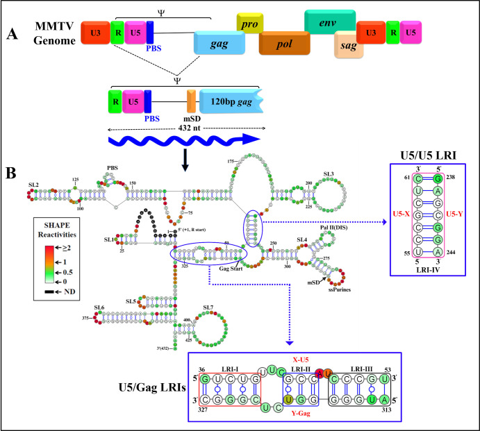
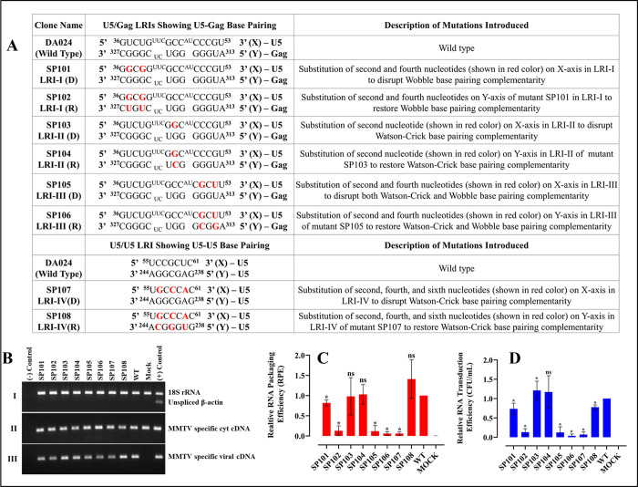
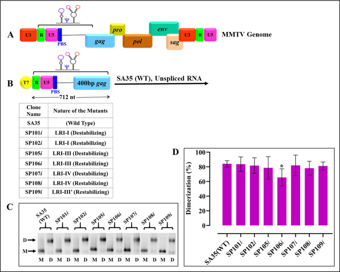
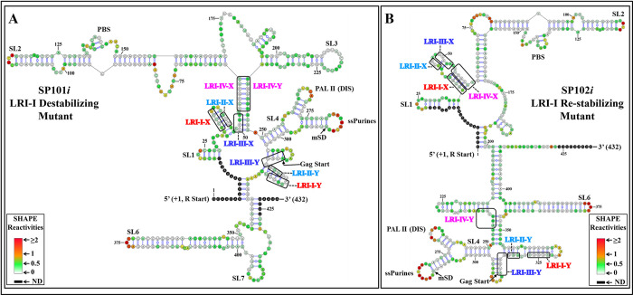
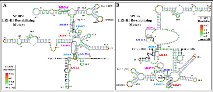
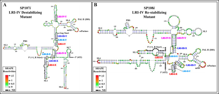
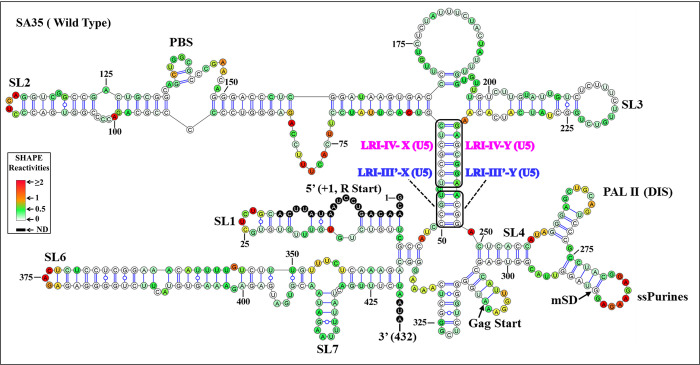
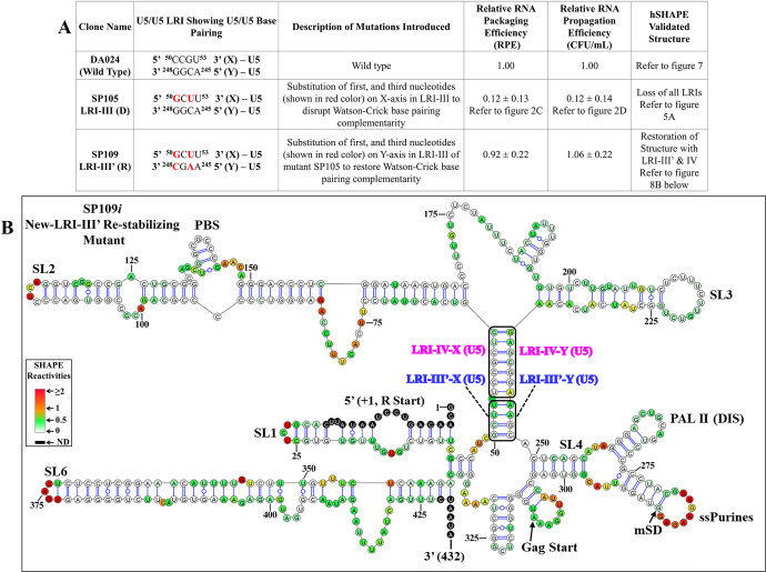
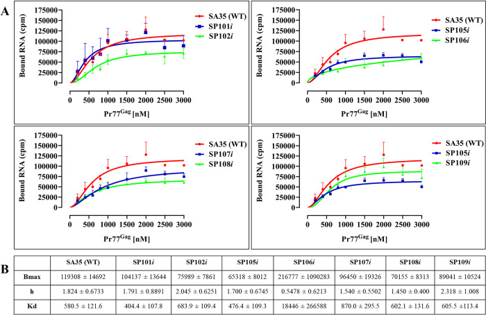
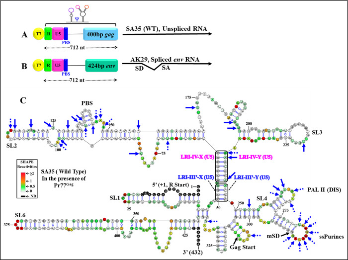
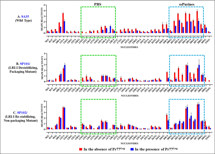
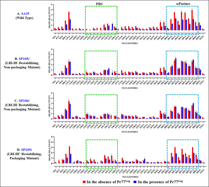
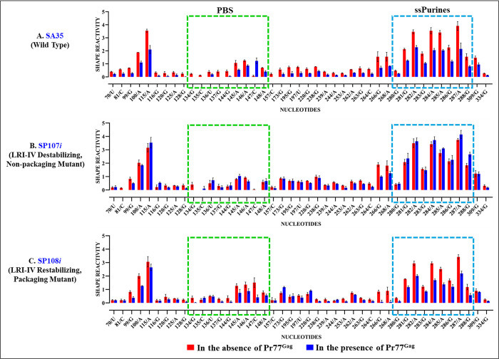
The packaging of genomic RNA (gRNA) into retroviral particles relies on the specific recognition by the Gag precursor of packaging signals (Psi), which maintain a complex secondary structure through long-range interactions (LRIs). However, it remains unclear whether the binding of Gag to Psi alone is enough to promote RNA packaging and what role LRIs play in this process. Using mouse mammary tumor virus (MMTV), we investigated the effects of mutations in 4 proposed LRIs on gRNA structure and function. Our findings revealed the presence of an unsuspected extended LRI, and hSHAPE revealed that maintaining a wild-type-like Psi structure is crucial for efficient packaging. Surprisingly, filter-binding assays demonstrated that most mutants, regardless of their packaging capability, exhibited significant binding to Pr77Gag, suggesting that Gag binding to Psi is insufficient for efficient packaging. Footprinting experiments indicated that efficient RNA packaging is promoted when Pr77Gag binds to 2 specific sites within Psi, whereas binding elsewhere in Psi does not lead to efficient packaging. Taken together, our results suggest that the 3D structure of the Psi/Pr77Gag complex regulates the assembly of viral particles around gRNA, enabling effective discrimination against other viral and cellular RNAs that may also bind Gag efficiently.


Conflict of interest statement

The authors have declared that no competing interests exist.

Figures

Fig 1
Fig 1. Schematic representation of the MMTV genome depicting the 5′ end harboring RNA packaging sequences (Ψ), along with its current RNA secondary structure model.
(A) Genome organization of MMTV and its RNA packaging signal (Ψ) beginning from the R region to 120 nucleotides of Gag, denoted by a blue wavy arrow. (B) hSHAPE-validated RNA secondary structure of MMTV packaging sequences located at the 5′ end, including LRIs-I-IV depicted in blue insets. Nucleotides are color-coded according to hSHAPE reactivity, with features annotated, such as stem-loops (SL1-7), PBS, DIS, single-stranded purines (ssPurines), mSD, and Gag start site. DIS, dimerization initiation site; LRI, long-range interaction; MMTV, mouse mammary tumor virus; mSD, major splice donor; PBS, primer binding site.
Fig 2
Fig 2. Role of LRIs-I-IV in MMTV gRNA packaging and propagation.
(A) List of substitutions in the U5/Gag and U5/U5 LRIs, with mutations highlighted in red. (B) Representative gel images of the controls necessary for validating different aspects of the three-plasmid in vivo packaging and propagation assay: (I) multiplex amplification for nucleocytoplasmic fractionation technique, (II) PCR amplification for cDNAs prepared from cytoplasmic RNA fraction validating stability and nuclear export of transfer vector RNA, and (III) PCR amplification of packaged transfer vector RNA. (C) Packaging efficiency of mutant transfer vector RNAs relative to the wild type (DA024). (D) Relative propagation of MMTV transfer vector RNAs measured as normalized hygromycin resistant CFUs/ml for mutant transfer vectors compared to the wild type (DA024) vector. Mock samples contained only the transfer vector and no packaging construct. Data are presented as mean ± standard deviation from a minimum of 3 independent experiments performed in triplicates for RNA packaging (panel C) and in duplicates for RNA propagation (panel D). Differences compared to the wild-type were considered significant when p < 0.05 according to the nonparametric Mann–Whitney U test. The data underlying this Fig 2C and 2D can be found in S1 Data. CFU, colony-forming unit; gRNA, genomic RNA; LRI, long-range interaction; MMTV, mouse mammary tumor virus.
Fig 3
Fig 3. Dimerization of LRI mutants remains unaffected by the introduced mutations.
(A) Schematic representation of the MMTV genome indicating the location of MMTV gRNA packaging determinants. (B) Illustration of the packageable vector RNA from R to 712 nucleotides expressed from a T7 expression plasmid, with the table detailing the names of the clones and the nature of the mutations. (C) Representative gel images displaying in vitro dimerization of WT (SA35) and LRI mutant RNAs in TBM buffer. M and D labels below the lanes indicate monomer and dimer buffers used for dimerization experiments. Monomeric and dimeric RNA species are denoted by letters M and D, respectively, on the gel’s horizontal margin. Gels have been cropped as indicated by vertical white spaces to show the relevant areas only. (D) Histograms illustrating the dimerization efficiencies of mutant RNAs compared to WT (SA35) calculated through densitometric analysis of bands from 3 independent experiments. Dimerization efficiency was determined by dividing the intensity of the dimeric RNA band by the intensity of the band from the total RNA (i.e., sum of dimer and monomer bands). No statistically significant differences (p-values > 0.05) were observed in the ability of the mutant clones to dimerize when compared to the WT (SA35) according to the nonparametric Mann–Whitney U test, except for mutant SP106i (p < 0.05). The data underlying this Fig 3D can be found in S1 Data. gRNA, genomic RNA; LRI, long-range interaction; MMTV, mouse mammary tumor virus; WT, wild type.
Fig 4
Fig 4. hSHAPE-validated RNA secondary structure model of the LRI-I mutants.
The first 432 nucleotides of the 712 nt long RNA are shown. (A) Mutant SP101i was designed to destabilize LRI-I. (B) Mutant SP102i was designed to re-stabilize LRI-I. Structural elements that are also present in the WT structure (SA35) such as stem-loops (SL1-7), PBS, DIS, single-stranded purines (ssPurines), and mSD are marked as in Fig 1. The X and Y-strands of different LRIs in both destabilizing and re-stabilizing mutants are boxed and labeled in different colors for clarity. Nucleotides are color-coded according to SHAPE reactivity derived from a minimum of 3 independent experiments, with data provided in S1 Table. DIS, dimerization initiation site; LRI, long-range interaction; mSD, major splice donor; PBS, primer binding site; WT, wild type.
Fig 5
Fig 5. hSHAPE-validated RNA secondary structure model of the LRI-III mutants.
The first 432 nucleotides of the 712 nt long RNA are shown. (A) Mutant SP105i was designed to destabilize LRI-III. (B) Mutant SP106i was designed to re-stabilize LRI-III. Structural elements that are also present in the WT structure (SA35) such as stem-loops (SL1-7), PBS, DIS, single-stranded purines (ssPurines), and mSD are marked as in Fig 1. The X and Y-strands of different LRIs in both destabilizing and re-stabilizing mutants are boxed and labeled in different colors for clarity. Nucleotides are color-coded according to SHAPE reactivity derived from a minimum of 3 independent experiments, with data provided in S1 Table. DIS, dimerization initiation site; LRI, long-range interaction; mSD, major splice donor; PBS, primer binding site; WT, wild type.
Fig 6
Fig 6. hSHAPE-validated RNA secondary structure model of the LRI-IV mutants.
The first 432 nucleotides of the 712 nt long RNA are shown. (A) Mutant SP107i was designed to destabilize LRI-IV. (B) Mutant SP108i was designed to re-stabilize LRI-IV. Structural elements that are also present in the WT structure (SA35) such as stem-loops (SL1-7), PBS, DIS, single-stranded purines (ssPurines), and mSD are marked as in Fig 1. The X and Y-strands of different LRIs in both destabilizing and re-stabilizing mutants are boxed and labeled in different colors for clarity. Nucleotides are color-coded according to SHAPE reactivity derived from a minimum of 3 independent experiments, with data provided in S1 Table. DIS, dimerization initiation site; LRI, long-range interaction; mSD, major splice donor; PBS, primer binding site; WT, wild type.
Fig 7
Fig 7. The new hSHAPE-validated RNA secondary structure model of the first 432 nucleotides, obtained through biochemical re-probing of the WT (SA35) 5′ end of MMTV.
The updated structure model closely resembles the previously proposed structure but exhibits notable differences in the LRIs and minor differences in SLs 5, 6, and 7. In the new structure model, LRIs I and II are absent, while LRI-IV remains unchanged. Additionally, the X-strand of the initially proposed LRI-III now base pairs with complementary sequences 195 nucleotides downstream within U5, instead of Gag. Structural elements consistent with the earlier proposed model, such as SLs1-7, PBS, DIS, single-stranded purines (ssPurines), and mSD, are present in their native positions and labeled as in Fig 1. The X and Y-strands of LRIs III’ and IV are shown in boxes and labeled with different colors for clarity. Nucleotides are color-coded according to SHAPE reactivity derived from a minimum of 3 independent experiments, with data provided in S1 Table. DIS, dimerization initiation site; LRI, long-range interaction; MMTV, mouse mammary tumor virus; mSD, major splice donor; PBS, primer binding site; SL, stem loop; WT, wild type.
Fig 8
Fig 8. LRI-III’ with complementary heterologous sequences restores RNA packaging and structure.
(A) Description of the substitution mutants in the newly proposed LRI-III with red nucleotides indicating introduced mutations aimed at destabilizing or re-stabilizing complementarity with heterologous sequences. Columns 4, 5, and 6 show results of the effect of mutations on RNA packaging, propagation, and structure, respectively. The RNA packaging data shown here is from a minimum of 3 independent experiments performed in triplicates (±SD). The RNA propagation data shown here is from a minimum of 3 independent experiments performed in duplicates (±SD). (B) A hSHAPE-validated RNA secondary structure model depicts the restored LRI-III’ mutant SP109i, designed to re-stabilize LRI-III’ with heterologous complementary sequences. Structural elements that are also present in the new WT structure (SA35) such as stem-loops (SL1-7), PBS, DIS, single-stranded purines (ssPurines), and mSD are marked as in Fig 7. The X and Y-axes of LRIs III’ and IV are boxed and labeled in different colors for clarity. Nucleotides are color-coded according to SHAPE reactivity derived from a minimum of 3 independent experiments, with data provided in S1 Table. The data underlying this Fig 8A can be found in S1 Data. DIS, dimerization initiation site; LRI, long-range interaction; mSD, major splice donor; PBS, primer binding site; SL, stem loop; WT, wild type.
Fig 9
Fig 9. MMTV Pr77Gag binding to the WT and LRI mutant RNAs using filter-binding assays.
(A) The membrane-bound radioactivity of the wild type WT (SA35) unspliced and mutant RNAs was quantified at increasing concentrations of MMTV Pr77Gag. Data points were fitted with the Hill’s equation, with error bars denoting standard deviation from the mean of 3 independent experiments. (B) Pr77Gag binding parameters to the WT and LRI mutant MMTV Psi region as derived using Hill’s equation. Cumulative data is derived from 3 independent experiments. Bmax: represents the maximum-specific binding; h: represents the Hill slope; Kd: represents the Pr77Gag concentration needed to achieve a half-maximum binding followed by their standard deviation. The data underlying this Fig 9A and 9B can be found in S1 Data. LRI, long-range interaction; MMTV, mouse mammary tumor virus; WT, wild type.
Fig 10
Fig 10. Footprints of Pr77Gag on the WT structure model.
(A) Schematic illustration of the MMTV WT packageable vector (SA35) RNA from R to 712 nucleotides expressed from a T7 expression plasmid. (B) Illustration of the envelope (env) spliced RNA (AK29) from R to 712 nucleotides expressed from a T7 expression plasmid. (C) hSHAPE analysis was carried out both with and without Pr77Gag. The mean triplicate SHAPE reactivity obtained without Pr77Gag was used to predict the RNA secondary structure model of WT (SA35) RNA. Subsequently, the mean hSHAPE reactivities obtained with Pr77Gag were overlaid onto the RNA secondary structure model predicted in the absence of Pr77Gag. Notably, nucleotides within the previously identified primary Gag-binding sites, such as single-stranded purines (ssPurines) and the PBS, exhibited significant reductions in hSHAPE reactivities. Nucleotides in ssPurines, PBS, and all other nucleotides marked by arrows show significant reduction in SHAPE reactivities. The hSHAPE reactivity key was developed based on the mean of hSHAPE reactivities for each nucleotide, as shown in S3 Table. The data shown is from a minimum of 3 independent experiments conducted both in the absence and presence of Pr77Gag. All nucleotides that show a reactivity decrease >40% upon Gag addition, also show statistically significant difference according to the Mann–Whitney non parametrical U test (p < 0.05). MMTV, mouse mammary tumor virus; PBS, primer binding site; WT, wild type.
Fig 11
Fig 11. Footprints of Pr77Gag on the WT and LRI-I mutant packaging signal RNAs.
Histograms showing the SHAPE reactivities of nucleotides in the absence (red bars) and presence (blue bars) of Gag for the packaging signal RNAs of: (A) wild type (SA35), (B) mutant SP101i designed to destabilize LRI-I, and (C) mutant SP102i designed to re-stabilize LRI-I. The dashed boxes mark the already identified primary Gag-binding sites, such as single stranded purines (ssPurines) and the PBS. For full details of nucleotides attenuation of SHAPE reactivities in the absence and presence of Pr77Gag from a minimum of 3 independent experiments, see S3 Table and S4 and S5 Figs. All nucleotides that show a reactivity decrease >40% upon Gag addition, also show statistically significant difference according to the Mann–Whitney non parametrical U test (p < 0.05). The data underlying this figure can be found in S1 Data. LRI, long-range interaction; PBS, primer binding site; WT, wild type.
Fig 12
Fig 12. Footprints of Pr77Gag on the WT and LRI-III mutant packaging signal RNAs.
Histograms showing the SHAPE reactivities of nucleotides in the absence (red bars) and presence (blue bars) of Gag for the packaging signal RNAs of: (A) wild type (SA35), (B) mutant SP105i designed to destabilize LRI-III, (C) mutant SP106i designed to re-stabilize LRI-III, and (D) mutant SP109i designed to restore the LRI-III’. The dashed boxes depict the already identified primary Gag-binding sites, such as single stranded purines (ssPurines) and the PBS. For full details of nucleotides attenuation of SHAPE reactivities in the absence and presence of Pr77Gag from a minimum of 3 independent experiments, see S3 Table and S6, S7 and S8 Figs. All nucleotides that show a reactivity decrease >40% upon Gag addition, also show statistically significant difference according to the Mann–Whitney non parametrical U test (p < 0.05). The data underlying this figure can be found in S1 Data. LRI, long-range interaction; PBS, primer binding site; WT, wild type.
Fig 13
Fig 13. Footprints of Pr77Gag on WT and LRI-IV mutant packaging signal RNAs.
Histograms showing SHAPE reactivities of nucleotides in the absence (red bars) and presence (blue bars) of Gag for the packaging signal RNAs of: (A) wild type (SA35), (B) mutant SP107i designed to destabilize LRI-IV, and (C) mutant SP108i designed to restore LRI-IV. The dashed boxes depict the already identified primary Gag-binding sites such as single stranded purines (ssPurines) and the PBS. For full details of nucleotides attenuation of SHAPE reactivities in the absence and presence of Pr77Gag from a minimum of 3 independent experiments, see S3 Table and S9 and S10 Figs. All nucleotides that show a reactivity decrease >40% upon Gag addition, also show statistically significant difference according to the Mann–Whitney non parametrical U test (p < 0.05). The data underlying this figure can be found in S1 Data. LRI, long-range interaction; PBS, primer binding site; WT, wild type.

Similar articles

References

    1. Hunter E. Chapter 6, Viral Assembly. 6th Edition. Fields Virology. 6th Edition. 2013. p. 144–146.
    1. Flint SJ, Enquist LW, Racaniello VR, Skalka AM. Chapter 13. 2nd Edition. Principles of Virology. 2nd Edition. 2000. p. 466–474.
    1. Paillart J-C, Shehu-Xhilaga M, Marquet R, Mak J. Dimerization of retroviral RNA genomes: an inseparable pair. Nat Rev Microbiol. 2004;2:461–472. doi: 10.1038/nrmicro903 - DOI - PubMed
    1. Dubois N, Marquet R, Paillart J-C, Bernacchi S. Retroviral RNA Dimerization: From Structure to Functions. Front Microbiol. 2018;9:527. doi: 10.3389/fmicb.2018.00527 - DOI - PMC - PubMed
    1. Berkhout B, van Wamel JL. Role of the DIS hairpin in replication of human immunodeficiency virus type 1. J Virol. 1996;70:6723–6732. doi: 10.1128/JVI.70.10.6723-6732.1996 - DOI - PMC - PubMed

LinkOut - more resources