. 2024 Nov;300(11):107853.
doi: 10.1016/j.jbc.2024.107853. Epub 2024 Oct 1.

Expression, purification, and characterization of diacylated Lipo-YcjN from Escherichia coli

Affiliations

Expression, purification, and characterization of diacylated Lipo-YcjN from Escherichia coli

Matthew A Treviño et al. J Biol Chem. 2024 Nov.

Abstract

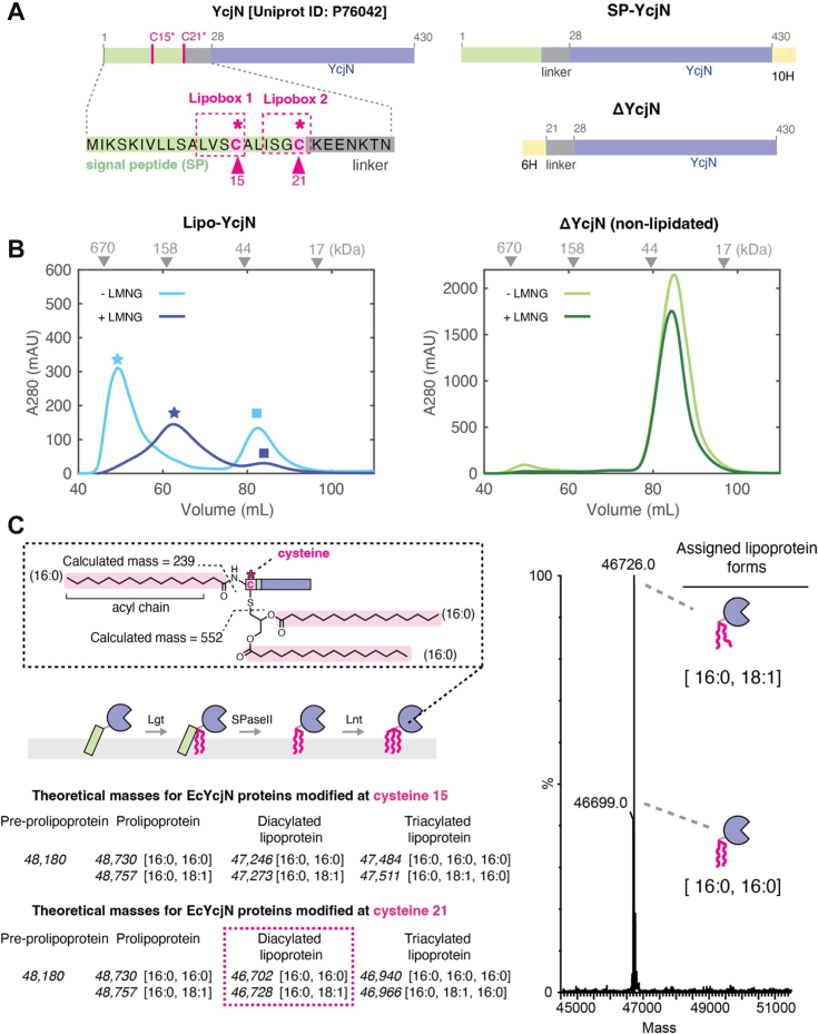
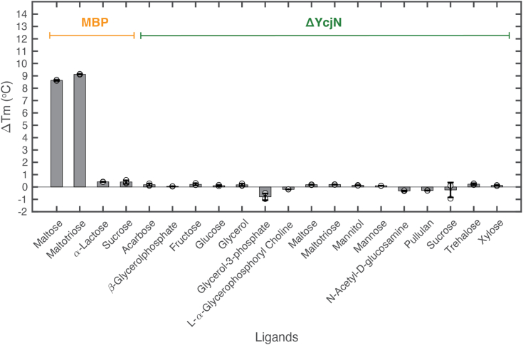
YcjN is a putative substrate binding protein expressed from a cluster of genes involved in carbohydrate import and metabolism in Escherichia coli. Here, we determine the crystal structure of YcjN to a resolution of 1.95 Å, revealing that its three-dimensional structure is similar to substrate binding proteins in subcluster D-I, which includes the well-characterized maltose binding protein. Furthermore, we found that recombinant overexpression of YcjN results in the formation of a lipidated form of YcjN that is posttranslationally diacylated at cysteine 21. Comparisons of size-exclusion chromatography profiles and dynamic light scattering measurements of lipidated and nonlipidated YcjN proteins suggest that lipidated YcjN aggregates in solution via its lipid moiety. Additionally, bioinformatic analysis indicates that YcjN-like proteins may exist in both Bacteria and Archaea, potentially in both lipidated and nonlipidated forms. Together, our results provide a better understanding of the aggregation properties of recombinantly expressed bacterial lipoproteins in solution and establish a foundation for future studies that aim to elucidate the role of these proteins in bacterial physiology.

Keywords: Escherichia coli; X-ray crystallography; bacterial lipoproteins; recombinant protein expression; substrate binding proteins.


Conflict of interest statement

Conflict of interest The authors declare that they have no conflicts of interest with the contents of this article.

Figures

Figure 1
Figure 1
Subcellular localization and posttranslational modification pathways of SBPs. A, diagram of the cell envelope (left panel) and organization of ABC transporter systems in complex with a soluble SBP and a lipoprotein SBP (right panel), respectively. B, nonlipidated protein maturation pathway (left panel) and lipoprotein maturation pathway (right panel). C, representation of the ycj gene cluster in E. coli. The protein encoding genes for the SBP, TMDs, and NBD are colored in light blue, orange, and dark blue, respectively. ABC, ATP-binding cassette; NBD, nucleotide binding domain; SBP, substrate binding protein; TMD, transmembrane domain.
Figure 2
Figure 2
Purification and mass spectrometry analysis of YcjN proteins. A, diagram of the amino acid sequences of YcjN proteins. Full-length YcjN contains two distinct lipoboxes (pink dashed boxes) within its SP (green rectangle). Each lipobox contains a cysteine residue (15 and 21, pink triangles). B, SEC elution profiles of Lipo-YcjN purified in the presence and absence of LMNG are shown in dark and light blue, respectively. SEC elution profiles of ΔYcjN purified in the presence and absence of LMNG are shown in dark and light green, respectively. Peak elution volumes of gel filtration standards are indicated by gray triangles. C, the theoretical masses of YcjN proteins modified at cysteine 15 (top) or cysteine 21 (bottom) are calculated, assuming that triacylation occurs via the canonical triacylation pathway due to the sequential action of three enzymes (Lgt, SPase II, and Lnt). Gray dashed box insert contains illustration of acyl chains attached to a C-terminal cysteine and the average calculated acyl chain masses of a lipoprotein with acyl chain composition [16:0, 16:0, 16:0]. Mass spectrum of the peak fraction (elution volume of 62 ml, dark blue star) of Lipo-YcjN purified in the presence of LMNG. The molecular masses of the major species correspond within 3 Da of the predicted masses of two diacylated YcjN proteins modified at residue 21, one protein with an acyl chain composition [16:0,16:0] and another with [16:0,18:1] (right panel). Assigned lipoprotein forms are shown as illustrations on the right. Lgt, phosphatidylglycerol: prolipoprotein diacylglyceryl transferase; LMNG, 2,2-didecylpropane-1,3-bis-β-D-maltopyranoside; SEC, size-exclusion chromatography; SP, signal peptide; SPase II, type II signal peptidase.
Figure 3
Figure 3
Overall structure of Escherichia coli ΔYcjN. A, ribbon representation of the ΔYcjN structure. The same structure is shown with a 90 x-axis rotation. B, schematic of secondary structure. Subdomains NTD, CTD1, and CTD2 are colored teal, gray, and light blue, respectively. CTD, C-terminal domain; NTD, N-terminal domain.
Figure 4
Figure 4
Comparison of YcjN to other SBPs identified using the Dali Server. A, structures and percent identities to YcjN. The CTD2 domain of each protein is colored blue. B, superposition of SBPs. Each protein color corresponds to that of the protein in panel A. C, SBP subcluster categorization of the aligned proteins. All CTD2 domains (blue) present in subcluster D-I (1ANF, 2ZYK, 4AQ4, and 2UVI) are absent in proteins within subcluster B-I (2FW0, 1GCG, 1URP, and 1GUB) that also bind carbohydrates. D, table of proteins, their size, ligand (∗putative), and species of origin. SBP, substrate binding protein; CTD, C-terminal domain.
Figure 5
Figure 5
NanoDSF thermal shift assay for MBP and ΔYcjN. Bar graph displaying ΔTm for MBP and YcjN with potential ligands. All measurements were performed using final protein concentrations of 1 mg/ml (24 μM MBP and 22 μM ΔYcjN) and ligand concentrations of 5000 μM. Unfilled circles represent individual data points. The height of the bar represents the mean of the data set, and the error bars represent the standard deviation. Measurements were calculated using three technical replicates. MBP, maltose binding protein; nanoDSF, nano-differential scanning fluorimetry.
Figure 6
Figure 6
Bioinformatic analysis of YcjN. A, sunburst diagram of the taxonomic distribution of YcjN homologs. B, the SSN diagram. C, total number of proteins labeled as lipoproteins and nonlipidated proteins in clusters 1 to 4. D, representative diagrams of the putative proteins within clusters 1 to 4 that are frequently encoded near YcjN as determined using the EFI-GNT tool. Putative YcjM, YcjN, YcjO/YcjP proteins are colored in green, purple, and orange, respectively. SSN, sequence similarity network.

Update of

References

    1. Higgins C. ABC transporters: from microorganisms to man. Annu. Rev. Cell Biol. 1992;8:67–113. - PubMed
    1. Hollenstein K., Frei D.C., Locher K.P. Structure of an ABC transporter in complex with its binding protein. Nature. 2007;446:213–216. - PubMed
    1. Auclair S.M., Bhanu M.K., Kendall D.A. Signal peptidase I: cleaving the way to mature proteins. Protein Sci. 2012;21:13–25. - PMC - PubMed
    1. Kovacs-Simon A., Titball R., Michell S.L. Lipoproteins of bacterial pathogens. Infect. Immun. 2011;79:548–561. - PMC - PubMed
    1. Sharaf N.G., Shahgholi M., Kim E., Lai J.Y., VanderVelde D.G., Lee A.T., et al. Characterization of the ABC methionine transporter from Neisseria meningitidis reveals that lipidated MetQ is required for interaction. Elife. 2021;10 - PMC - PubMed

Publication types

MeSH terms

LinkOut - more resources