. 2024 Dec 12;64(6):2400790.
doi: 10.1183/13993003.00790-2024. Print 2024 Dec.

Pathogenic variants in CFAP46, CFAP54, CFAP74 and CFAP221 cause primary ciliary dyskinesia with a defective C1d projection of the central apparatus

Affiliations

Pathogenic variants in CFAP46, CFAP54, CFAP74 and CFAP221 cause primary ciliary dyskinesia with a defective C1d projection of the central apparatus

Kai Wohlgemuth et al. Eur Respir J. .

Abstract

Background: Primary ciliary dyskinesia is a rare genetic disorder caused by insufficient mucociliary clearance leading to chronic airway infections. The diagnostic guideline of the European Respiratory Society primarily recommends an evaluation of the clinical history (e.g. by the PICADAR prediction tool), nasal nitric oxide production rate measurements, high-speed videomicroscopy analysis of ciliary beating and an assessment of ciliary axonemes via transmission electron microscopy. Genetic testing can be implemented as a last step.

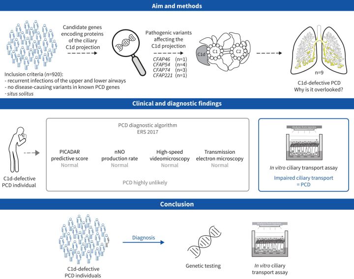
Aims: In this study, we aimed to characterise primary ciliary dyskinesia with a defective C1d projection of the ciliary central apparatus and we evaluated the applicability of the European Respiratory Society diagnostic guideline to this primary ciliary dyskinesia type.

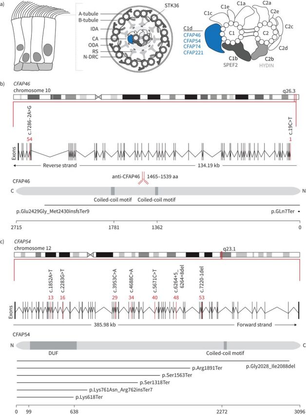
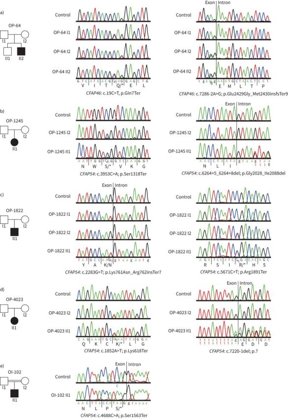
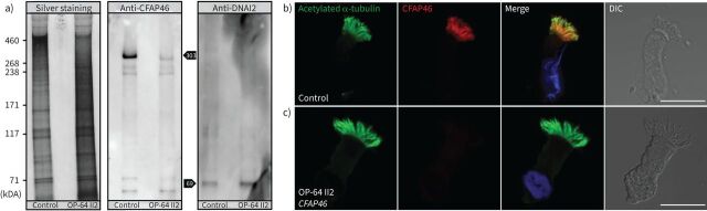
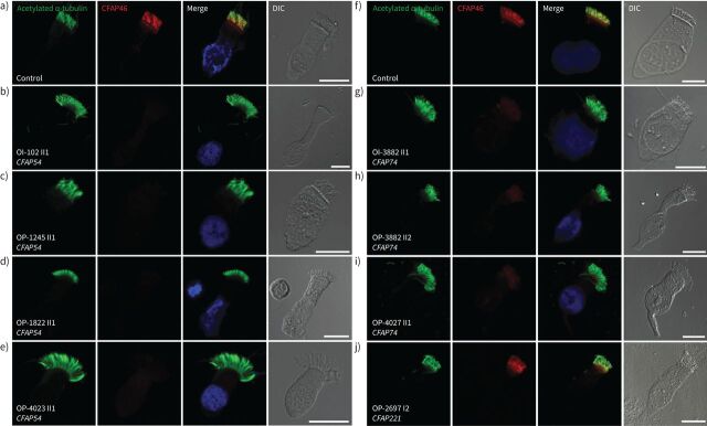
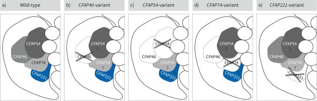
Methods: Using a high-throughput sequencing approach of genes encoding C1d components, we identified pathogenic variants in the novel primary ciliary dyskinesia genes CFAP46 and CFAP54, and the known primary ciliary dyskinesia gene CFAP221. To fully assess this primary ciliary dyskinesia type, we also analysed individuals with pathogenic variants in CFAP74.

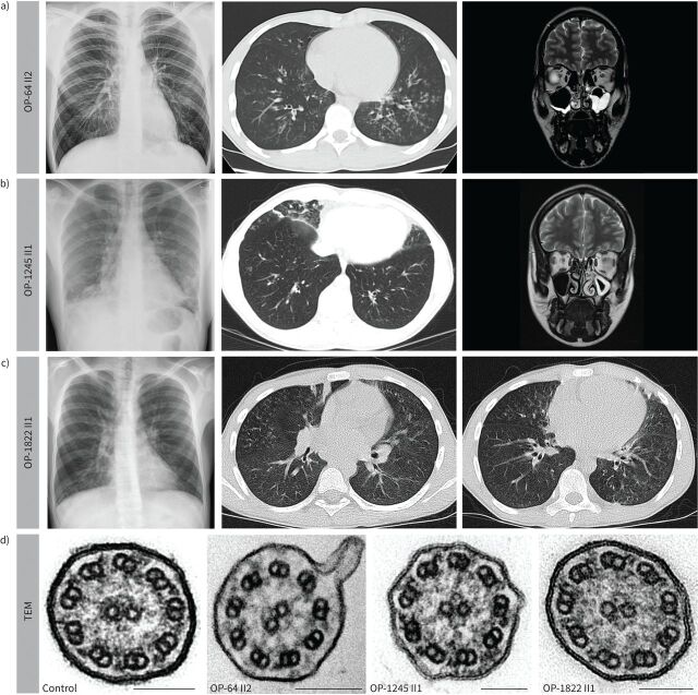
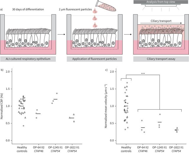
Results: Careful evaluation revealed that C1d-defective primary ciliary dyskinesia is associated with normal situs composition, normal nasal nitric oxide production rates, normal ciliary ultrastructure by transmission electron microscopy and normal ciliary beating by high-speed videomicroscopy analysis. Despite chronic respiratory disease, PICADAR does not reliably detect this primary ciliary dyskinesia type. However, we could show by in vitro ciliary transport assays that affected individuals exhibit insufficient ciliary clearance.

Conclusions: Overall, this study extends the spectrum of primary ciliary dyskinesia genes and highlights that individuals with C1d-defective primary ciliary dyskinesia elude diagnosis when using the current diagnostic algorithm. To enable diagnosis, genetic testing should be prioritised in future diagnostic guidelines.


Conflict of interest statement

Conflict of interest: The authors have no potential conflicts of interest to disclose.

Figures

None
In this study, we examined a cohort of individuals exhibiting recurrent upper and lower airway infections combined with situs solitus (n=920) who had not received a diagnosis previously. Using a high-throughput sequencing approach of genes encoding components of the C1d projection of the central apparatus, we identified pathogenic variants in CFAP46, CFAP54, CFAP74 and CFAP221 as a cause of C1d-defective primary ciliary dyskinesia (PCD) (n=9). Affected individuals easily elude the current European Respiratory Society (ERS) diagnostic algorithm because this PCD type is associated with normal findings on PICADAR, nasal nitric oxide (nNO) production rate, high-speed videomicroscopy and transmission electron microscopy. However, C1d-defective PCD individuals exhibit impaired ciliary transport. In conclusion, only genetic testing and in vitro ciliary transport assays enable diagnosis of C1d-defective PCD.
FIGURE 1
FIGURE 1
Pathogenic variants in CFAP46 and CFAP54 cause primary ciliary dyskinesia with a defective C1d projection. a) Schematic of respiratory epithelium (left), an axonemal 9+2 cross-section (middle) and the ciliary central apparatus (CA) (right). CFAP46, CFAP54, CFAP74 and CFAP221 localise to the C1d projection of the CA. SPEF2 is part of the C1b projection, HYDIN of the C2b projection; STK36 is predicted to localise between the CA and radial spoke (RS). IDA: inner dynein arm; ODA: outer dynein arm; N-DRC: nexin-dynein regulatory complex. b) CFAP46 is located on chromosome 10 and encodes a 2715 amino acid (aa) protein with two coiled-coil motifs. The epitope of the anti-CFAP46 antibody is located between the two coiled-coil motifs and specifically recognises CFAP46. The positions of the identified pathogenic variants in CFAP46 are indicated by exons highlighted in red. Resulting truncated proteins are indicated below. c) CFAP54 is located on chromosome 12 and encodes a 3096 aa protein with a domain of unknown function (DUF) and a coiled-coil motif. The positions of the identified pathogenic variants in CFAP54 are indicated by exons highlighted in red. Resulting truncated proteins are indicated below.
FIGURE 2
FIGURE 2
Sanger verification and segregation analysis of primary ciliary dyskinesia individuals harbouring biallelic pathogenic variants in CFAP46 and CFAP54. a–e) Pedigrees of the five families are indicated. Sanger sequencing confirms compound heterozygosity of the detected variants in OP-64 II2 (a), OP-1245 II1 (b), OP-1822 II1 (c) and OP-4023 II1 (d). OI-102 II1 shows a homozygous variant consistent with homozygosity by descent due to parental consanguinity (e).
FIGURE 3
FIGURE 3
Radiological and ultrastructural findings in primary ciliary dyskinesia individuals with biallelic pathogenic CFAP46 and CFAP54 variants. a) CFAP46-variant individual OP-64 II2 shows situs solitus, mucus plugging and bronchiectasis in the lower lobes on chest X-ray (left panel) and computed tomography (CT) (middle panel). Consistent with chronic rhinosinusitis, magnetic resonance imaging (MRI) (right panel) displays mucosal thickening mainly in the left maxillary sinus and left ethmoidal cells, nasal mucosa swelling and nasal polyps. b) CFAP54-variant individual OP-1245 II1 exhibits situs solitus, basal lung consolidations, mucus plugging and mild bronchiectasis in the middle and lower lobes as well as in the lingula (chest X-ray: left panel; CT: middle panel). MRI scan (right panel) shows swelling of nasal mucosa and mucosal thickening in the left maxillary sinus and ethmoidal cells. Left ethmoidal infundibulum is blocked. c) CFAP54-variant individual OP-1822 II1 displays situs solitus, mucus plugging, dystelectasis and bronchiectasis in the lower lobes (chest X-ray: left panel; CT: right and middle panels). d) All individuals (OP-64 II2, OP-1245 II1 and OP-1822 II1) show normal ciliary ultrastructure in transmission electron microscopy. Scale bars: 100 nm.
FIGURE 4
FIGURE 4
CFAP46 is undetectable in respiratory cilia from the CFAP46-variant individual OP-64 II2. a) Extracts enriched for axonemal proteins from air–liquid interface-cultured respiratory epithelial cells are used for silver staining and immunoblotting. Silver staining demonstrates the integrity of protein recovery after lithium dodecyl sulfate-polyacrylamide gel electrophoresis. Immunoblotting with anti-CFAP46 detects the 303 kDa large isoform of CFAP46 (Q8IYW2-1) in the axonemal extract from a healthy control individual. In the CFAP46-variant individual OP-64 II2, this band is not detectable, consistent with the abnormal immunofluorescence result. The weak band with a size of approximately 300 kDa correlates with a low amount of dysfunctional truncated CFAP46. DNAI2 (69 kDa) is detected as a loading control of protein extracts. b, c) Immunofluorescence microscopy analyses of respiratory epithelial cells are performed using antibodies directed against acetylated α-tubulin (green) and CFAP46 (red). Nuclei are stained with Hoechst 33342 (blue). Scale bars: 10 µm. In healthy control cells, CFAP46 localises along the entire ciliary axoneme (b). In cells of CFAP46-variant individual OP-64 II2, CFAP46 is undetectable in the ciliary axoneme (c). DIC: differential interference contrast.
FIGURE 5
FIGURE 5
Axonemal localisation of CFAP46 in respiratory cilia from CFAP54-, CFAP74- and CFAP221-variant individuals. Immunofluorescence microscopy analyses of respiratory epithelial cells from healthy control individuals (a, f), CFAP54-variant individuals (b–e), CFAP74-variant individuals (g–i) and the CFAP221-variant individual (j). Cells are stained with antibodies directed against acetylated α-tubulin (green) and CFAP46 (red). Nuclei are stained with Hoechst 33342 (blue). Scale bars: 10 µm. a, f) In cells from healthy control individuals, CFAP46 localises along the entire ciliary axoneme. b–e) CFAP46 is not detectable in the ciliary axoneme from the CFAP54-variant individuals OI-102 II1, OP-1245 II1, OP-1822 II1 and OP-4023 II1. g–i) CFAP46 appears to be reduced in the ciliary axoneme from the CFAP74-variant individuals OP-3882 II1, OP-3882 II2 and OP-4027 II1. j) In cells from the CFAP221-variant individual OP-2697 I2, the axonemal localisation of CFAP46 is not affected. DIC: differential interference contrast.
FIGURE 6
FIGURE 6
Model of the composition of the C1d projection in humans. a) Based on our immunofluorescence analyses using anti-CFAP46 antibodies, CFAP46, CFAP54 and CFAP74 form a heterotrimeric subcomplex (grey) within the human wild-type C1d projection. CFAP221 (blue) also belongs to the C1d projection but seems not to be directly involved in this heterotrimeric subcomplex, resembling findings in Chlamydomonas reinhardtii and indicating evolutionary conservation of the C1d projection. Our immunofluorescence analyses reveal the following. b) In a CFAP46-variant individual, CFAP46 is undetectable. c) In CFAP54-variant individuals, CFAP46 is not detectable, indicating that the CFAP46 assembly is dependent on functional CFAP54. d) In CFAP74-variant individuals, CFAP46 appears reduced within the ciliary axoneme, revealing that CFAP74 is involved in the assembly of CFAP46. e) In a CFAP221-variant individual, CFAP46 is not affected, which suggests a distal position of CFAP221 within the C1d projection. (Proteins directly affected by biallelic pathogenic variants are crossed out. Impaired protein assembly is illustrated by a white area with a grey border.)
FIGURE 7
FIGURE 7
In vitro ciliary transport is impaired in CFAP46- and CFAP54-variant individuals. a) To assess the ciliary transport in vitro, respiratory epithelial cells from healthy control individuals and from CFAP46- and CFAP54-variant individuals were cultured at the air–liquid interface (ALI). After 30 days of differentiation, the ALI-cultured respiratory cell layers were used for ciliary transport assays, evaluating the transport of fluorescent particles by ciliary beating from the top view. b) The mean ciliary beat frequencies (CBFs) (indicated by the black bars) in OP-64 II2, OP-1245 II1 and OP-1822 II1 did not differ significantly from healthy controls (p=0.62, t-test). c) In contrast, the mean velocities of fluorescent particles were significantly reduced in the ALI-inserts from OP-64 II2 (37.6%), OP-1245 II1 (53.1%) and OP-1822 II1 (30.6%) (p=0.00022, t-test). Because two different microscope settings were used for the analysis, the data were normalised based on the healthy control pool (Nikon: triangles, Leica: circles). The exact values for CBF (Hz) and ciliary transport velocity (µm·s−1) are shown in supplementary figure S6. Significant differences are indicated. ***: p<0.001.

Comment in

References

    1. Wallmeier J, Nielsen KG, Kuehni CE, et al. . Motile ciliopathies. Nat Rev Dis Primers 2020; 6: 77. doi:10.1038/s41572-020-0209-6 - DOI - PubMed
    1. Horani A, Ferkol TW. Understanding primary ciliary dyskinesia and other ciliopathies. J Pediatr 2021; 230: 15–22. doi:10.1016/j.jpeds.2020.11.040 - DOI - PMC - PubMed
    1. Hannah WB, Seifert BA, Truty R, et al. . The global prevalence and ethnic heterogeneity of primary ciliary dyskinesia gene variants: a genetic database analysis. Lancet Respir Med 2022; 10: 459–468. doi:10.1016/S2213-2600(21)00453-7 - DOI - PMC - PubMed
    1. Ishikawa T. Axoneme structure from motile cilia. Cold Spring Harb Perspect Biol 2017; 9: a028076. doi:10.1101/cshperspect.a028076 - DOI - PMC - PubMed
    1. Samsel Z, Sekretarska J, Osinka A, et al. . Central apparatus, the molecular kickstarter of ciliary and flagellar nanomachines. Int J Mol Sci 2021; 22: 3013. doi:10.3390/ijms22063013 - DOI - PMC - PubMed

LinkOut - more resources