. 2024 Oct 16;38(17-20):915-930.
doi: 10.1101/gad.351913.124.

NEAT1 promotes genome stability via m6A methylation-dependent regulation of CHD4

Affiliations

NEAT1 promotes genome stability via m6A methylation-dependent regulation of CHD4

Victoria Mamontova et al. Genes Dev. .

Abstract

Long noncoding (lnc)RNAs emerge as regulators of genome stability. The nuclear-enriched abundant transcript 1 (NEAT1) is overexpressed in many tumors and is responsive to genotoxic stress. However, the mechanism that links NEAT1 to DNA damage response (DDR) is unclear. Here, we investigate the expression, modification, localization, and structure of NEAT1 in response to DNA double-strand breaks (DSBs). DNA damage increases the levels and N6-methyladenosine (m6A) marks on NEAT1, which promotes alterations in NEAT1 structure, accumulation of hypermethylated NEAT1 at promoter-associated DSBs, and DSB signaling. The depletion of NEAT1 impairs DSB focus formation and elevates DNA damage. The genome-protective role of NEAT1 is mediated by the RNA methyltransferase 3 (METTL3) and involves the release of the chromodomain helicase DNA binding protein 4 (CHD4) from NEAT1 to fine-tune histone acetylation at DSBs. Our data suggest a direct role for NEAT1 in DDR.

Keywords: CHD4; DNA damage response; DNA double-strand breaks; METTL3; NEAT1; long noncoding RNA; paraspeckles.


Figures

Figure 1.
Figure 1.
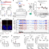
METTL3 stabilizes NEAT1 upon DNA damage in U2OS cells. (A) Scheme (top) and RT-qPCR (bottom) to assess NEAT1 levels in subcellular fractions. (CP) Cytoplasm, (NP) nucleoplasm, (green) primer site (pA) poly-A tail. (B) Imaging (top) and quantitation (bottom) of RNA-FISH signals. (C,D) RT-qPCR assessing NEAT1 upon m6A immunoprecipitation (IP) from total RNA (C) or METTL3 IP from lysates (D). IgG was used as the control. (E) Scheme of pull-down assay (top) and immunoblot (bottom) displaying ectopically expressed FLAG-tagged METTL3 variants upon IP with biotin (BIO)-labeled, immobilized NEAT1 in vitro transcription (IVT) product. (Mock) Nontransfected control. (F) Scheme of SAMfluoro assay (left) and fluorescent counts displaying Resorufin levels (top right) and FLAG-METTL3 variants (immunoblot; bottom right). (SAM) S-adenosyl-L-methionine, (AdoHcy) S-adenosyl-L-homocysteine. (G,H) Browser tracks (G) and quantitation (H) of METTL3 seCLIP-seq reads. (Red box) Region of interest, (arrowhead) transcription start site, (dashed line) background, (red arrowhead) m6A site. (*) P-value < 0.05, (**) P < 0.001; two-tailed t-test. Error bar indicates mean ± SD. (n) Number of replicates or cells.
Figure 2.
Figure 2.
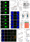
NEAT1 depletion elevates DNA damage and impairs DSB signaling in U2OS cells. (A,B) Imaging (A) and quantitation (B) of neutral comet assay displaying SYBR Gold-stained DNA. (White box) Zoom. (C,D) Imaging (C) and quantitation (D) of 53BP1 signals upon etoposide treatment and chase (Eto. +2 h). (E) Immunoblots for phospho-(p)ATM/ATR substrates and phospho-Thr-68 checkpoint kinase 2 (pCHK2). (ATR) Ataxia telangiectasia and Rad3-related, (ASO) antisense oligonucleotide. Vinculin was used as a control. (F) BLISS DSB count at transcription start/end sites (TSSs/TESs) (top) and region distribution (bottom). (Dashed line) Background (red box) promoter region. (G) Imaging (left) and quantitation (right) of phospho-Ser-139 histone H2A.X (γH2A.X) and Nibrin (NBS1) signals. (4-OHT) 4-hydroxytamoxifen, (white box) zoom. (H,I) ChIP for γH2A.X (H) and histone H2B lys-120 acetylation (H2BK120ac; I) at AsiSI site DS1. (noDSB) Control. (*) P-value < 0.05, (**) P < 0.001; two-tailed t-test or Wilcoxon test (BLISS). Error bar indicates mean ± SD or mean ± ±SEM (BLISS). (n) Number of cells.
Figure 3.
Figure 3.
NEAT1 accumulates at promoter-associated DSBs in U2OS cells. (A) Scheme of CHART assay. (BIO) Biotin, (green) nucleosomes. (B,C) Heat map (B) and browser tracks (C) for NEAT1 CHART-seq read counts. (COs) Capturing oligonucleotides, (red box) quantified region of interest, (arrowhead) transcription start site. (D) Scheme of CRISPR-based NEAT1-labeling (top) and imaging of GFP (bottom left) and PLA (bottom right) signals. (Dashed line) Background. (E) Imaging (top) and quantitation (bottom) of RNA-PLA signals. (Dashed white circle) Nucleus, (dashed line) background. (*) P-value < 0.05, (**) P < 0.001; two-tailed t-test. Error bar indicates mean ± SD. (n) Number of replicates or cells.
Figure 4.
Figure 4.
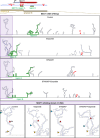
DNA damage alters NEAT1 structure in HEK293 cells. (Top) Scheme of m6A sites and METTL3 binding regions on NEAT1. (Bottom) Visualization of nano-DMS-MaP data from the NEAT1 isoform-overlapping amplicon B. (Green) METTL3 binding region B (triangle position marker).
Figure 5.
Figure 5.
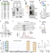
DNA damage impairs CHD4 association with NEAT1 in U2OS cells. (A) NEAT1 interactome analysis. (B) Immunoblots for chromodomain helicase DNA binding protein 4 (CHD4), GFP, SFPQ, and NONO in lysate (input [IN]) of GFP-tagged MS2 coat protein (MCP-GFP)-expressing HEK293:24 × MS2-NEAT1 cells or upon IP with GFP antibody. IgG was used as a control. (C) Scheme of the pull-down assay (left) and an autoradiograph of 32P-γ-ATP end-labeled (32P) IVT product upon IP with immobilized CHD4 (right). IgG was used as a control, (Ab) antibody. (D) Scheme of the pull-down assay (top) and an immunoblot for GFP-tagged CHD4 (CHD4-GFP) in lysate (IN) or upon IP with biotin (BIO)-labeled IVT product (bottom). (IVT T7) IVT SP6 and mouse double-minute 2 homolog (MDM2), which were used as controls. (E) RT-qPCR assessing NEAT1 (top) and immunoblot (bottom) for CHD4 in lysate (IN) and upon IP. Fibrillarin and IgG were used as controls. (F) Immunoblot (IB) for CHD4 (top) and quantitation (bottom) in lysate (IN) or upon sucrose gradient fractionation. (G) RT-qPCR assessing NEAT1 from sucrose gradient fractions. (H) Browser tracks (left) and quantitation (right) of CHD4 seCLIP-seq reads. (Red box) Quantified region of interest (arrowhead) transcription start site.
Figure 6.
Figure 6.
NEAT1 depletion deregulates CHD4 occupancy, histone acetylation, and BRCA1 levels at promoter-associated DSBs in U2OS cells. (A,B) Metagene plots (left) and browser tracks (right) of CHD4 CUT&RUN-seq data at AsiSI sites (dashed line) upon 4-OHT treatment and STM2457 preincubation (A) or NEAT1 depletion (B). (Red box) Quantified region of interest, (arrowhead) transcription start site. The +4-OHT condition is used as benchmark in both A and B. (CE) ChIP for CHD4 (C), histone H3 lys-27 acetylation (H3K27ac) (D), or breast cancer type 1 susceptibility protein BRCA1 (E) at the AsiSI site DS1. (noDSB) Control. (F) Model illustrating our findings. See the text for details. (*) P-value < 0.05, (**) P < 0.001; two-tailed t-test. Error bar indicates mean ± SD. (n) Number of replicates.

References

    1. Adriaens C, Standaert L, Barra J, Latil M, Verfaillie A, Kalev P, Boeckx B, Wijnhoven PW, Radaelli E, Vermi W, et al. 2016. p53 induces formation of NEAT1 lncRNA-containing paraspeckles that modulate replication stress response and chemosensitivity. Nat Med 22: 861–868. 10.1038/nm.4135 - DOI - PubMed
    1. Arnould C, Rocher V, Saur F, Bader AS, Muzzopappa F, Collins S, Lesage E, Le Bozec B, Puget N, Clouaire T, et al. 2023. Chromatin compartmentalization regulates the response to DNA damage. Nature 623: 183–192. 10.1038/s41586-023-06635-y - DOI - PMC - PubMed
    1. Aymard F, Bugler B, Schmidt CK, Guillou E, Caron P, Briois S, Iacovoni JS, Daburon V, Miller KM, Jackson SP, et al. 2014. Transcriptionally active chromatin recruits homologous recombination at DNA double-strand breaks. Nat Struct Mol Biol 21: 366–374. 10.1038/nsmb.2796 - DOI - PMC - PubMed
    1. Blackford AN, Jackson SP. 2017. ATM, ATR, and DNA-PK: the trinity at the heart of the DNA damage response. Mol Cell 66: 801–817. 10.1016/j.molcel.2017.05.015 - DOI - PubMed
    1. Blue SM, Yee BA, Pratt GA, Mueller JR, Park SS, Shishkin AA, Starner AC, Van Nostrand EL, Yeo GW. 2022. Transcriptome-wide identification of RNA-binding protein binding sites using seCLIP-seq. Nat Protoc 17: 1223–1265. 10.1038/s41596-022-00680-z - DOI - PMC - PubMed