

Nanoparticle-mediated delivery of placental gene therapy via uterine artery catheterization in a pregnant rhesus macaque
- PMID: 39362807
- PMCID: PMC11947900
- DOI: 10.1016/j.placenta.2024.09.013
Nanoparticle-mediated delivery of placental gene therapy via uterine artery catheterization in a pregnant rhesus macaque
Abstract
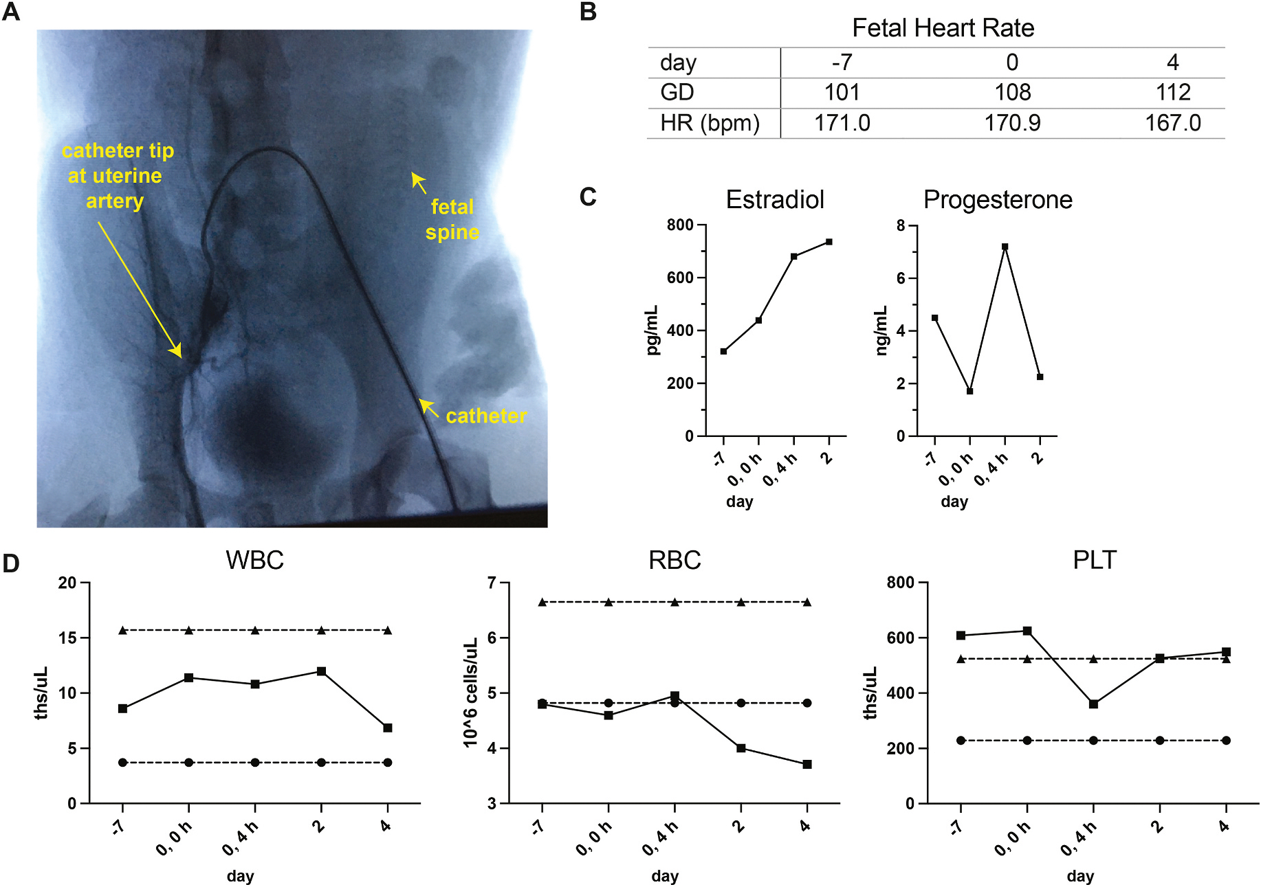
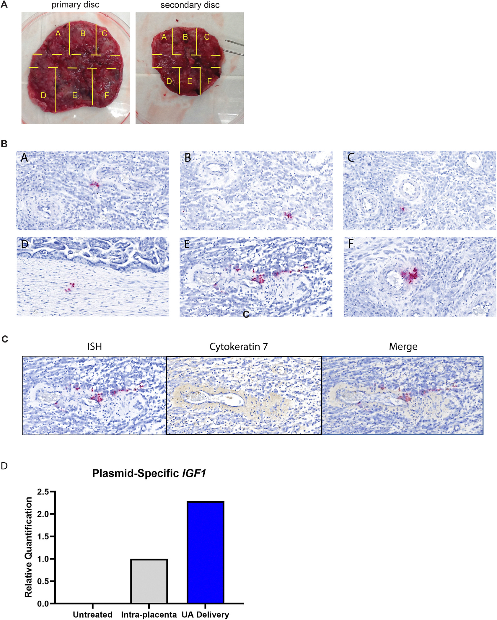
Nanoparticles offer promise as a mechanism to non-invasively deliver targeted placental therapeutics. Our previous studies utilizing intraplacental administration demonstrate efficient nanoparticle uptake into placental trophoblast cells and overexpression of human IGF1 (hIGF1). Nanoparticle-mediated placental overexpression of hIGF1 in small animal models of placental insufficiency and fetal growth restriction improved nutrient transport and restored fetal growth. The objective of this pilot study was to extend these studies to the pregnant nonhuman primate and develop a method for local delivery of nanoparticles to the placenta via maternal blood flow from the uterine artery. Nanoparticles containing hIGF1 plasmid driven by the placenta-specific PLAC1 promoter were delivered to a mid-gestation pregnant rhesus macaque via a catheterization approach that is clinically used for uterine artery embolization. Maternal-fetal interface, fetal and maternal tissues were collected four days post-treatment to evaluate the efficacy of hIGF1 treatment in the placenta. The uterine artery catheterization procedure and nanoparticle treatment was well tolerated by the dam and fetus through the four-day study period following catheterization. Nanoparticles were taken up by the placenta from maternal blood as plasmid-specific hIGF1 expression was detected in multiple regions of the placenta via in situ hybridization and qPCR. The uterine artery catheterization approach enabled successful delivery of nanoparticles to maternal circulation in close proximity to the placenta with no concerns to maternal or fetal health in this short-term feasibility study. In the future, this delivery approach can be used for preclinical evaluation of the long-term safety and efficacy of nanoparticle-mediated placental therapies in a rhesus macaque model.
Keywords: Gene therapy; IGF1; Macaque; Nanoparticles; Placenta.
Copyright © 2024 Elsevier Ltd. All rights reserved.
Conflict of interest statement
Declaration of competing interest None.
Figures
Update of
-
Nanoparticle-mediated delivery of placental gene therapy via uterine artery catheterization in a pregnant rhesus macaque.bioRxiv [Preprint]. 2024 Apr 14:2024.04.10.588902. doi: 10.1101/2024.04.10.588902. bioRxiv. 2024. Update in: Placenta. 2025 Jun 13;166:103-108. doi: 10.1016/j.placenta.2024.09.013. PMID: 38645086 Free PMC article. Updated. Preprint.
References
-
- Hodyl NA, et al. , Child neurodevelopmental outcomes following preterm and term birth: what can the placenta tell us? Placenta 57 (2017) 79–86. - PubMed
-
- De Boo HA, Harding JE, The developmental origins of adult disease (Barker) hypothesis, Aust. N. Z. J. Obstet. Gynaecol. 46 (2006) 4–14. - PubMed
-
- Langley-Evans SC, McMullen S, Developmental origins of adult disease, Med. Princ. Pract. : international journal of the Kuwait University, Health Science Centre 19 (2010) 87–98. - PubMed
-
- Barker D, The developmental origins of adult disease, J. Am. Coll. Nutr. 23 (2004) 588S–595S. - PubMed
MeSH terms
Substances
Grants and funding
LinkOut - more resources
Full Text Sources
Medical
Miscellaneous
