Review
. 2024 Oct 4;9(1):263.
doi: 10.1038/s41392-024-01952-8.

Evolving understanding of autoimmune mechanisms and new therapeutic strategies of autoimmune disorders

Affiliations
Review

Evolving understanding of autoimmune mechanisms and new therapeutic strategies of autoimmune disorders

Yi Song et al. Signal Transduct Target Ther. .

Abstract

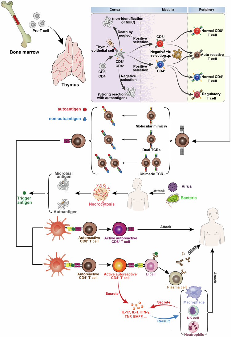
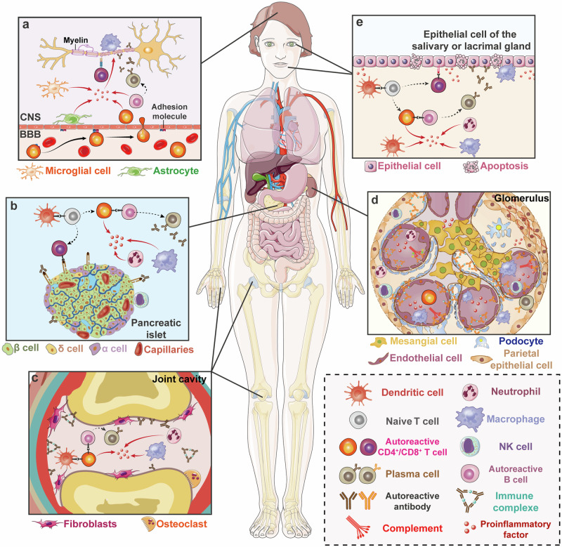
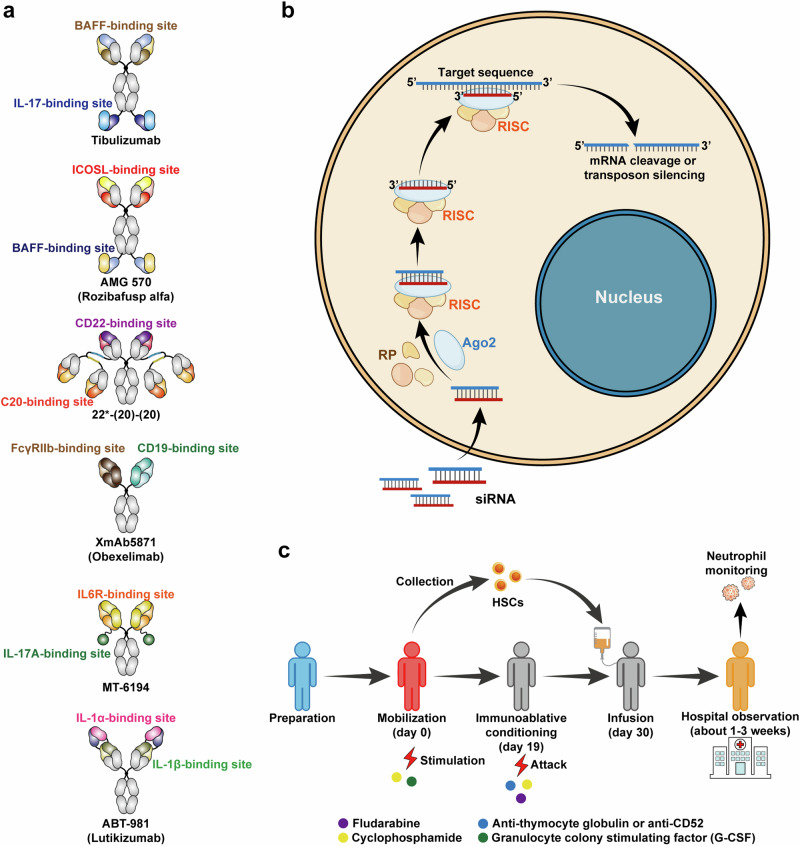
Autoimmune disorders are characterized by aberrant T cell and B cell reactivity to the body's own components, resulting in tissue destruction and organ dysfunction. Autoimmune diseases affect a wide range of people in many parts of the world and have become one of the major concerns in public health. In recent years, there have been substantial progress in our understanding of the epidemiology, risk factors, pathogenesis and mechanisms of autoimmune diseases. Current approved therapeutic interventions for autoimmune diseases are mainly non-specific immunomodulators and may cause broad immunosuppression that leads to serious adverse effects. To overcome the limitations of immunosuppressive drugs in treating autoimmune diseases, precise and target-specific strategies are urgently needed. To date, significant advances have been made in our understanding of the mechanisms of immune tolerance, offering a new avenue for developing antigen-specific immunotherapies for autoimmune diseases. These antigen-specific approaches have shown great potential in various preclinical animal models and recently been evaluated in clinical trials. This review describes the common epidemiology, clinical manifestation and mechanisms of autoimmune diseases, with a focus on typical autoimmune diseases including multiple sclerosis, type 1 diabetes, rheumatoid arthritis, systemic lupus erythematosus, and sjögren's syndrome. We discuss the current therapeutics developed in this field, highlight the recent advances in the use of nanomaterials and mRNA vaccine techniques to induce antigen-specific immune tolerance.


Conflict of interest statement

The authors declare no competing interests.

Figures

Fig. 1
Fig. 1
Pattern diagram of the mechanisms of autoimmune diseases. After differentiation of hematopoietic stem cells, progenitor T cell (pro-T cell) will leave the bone marrow and enter the thymus, and differentiate from double-negative (DN) T cells into double-positive (DP) T cells. Under death by neglect, negative selection, and positive selection via thymic epithelial cells, single positive T cells with low avidity to autoantigens-MHC complexes survive and differentiate into CD4 or CD8 and enter the periphery. However, some autoreactive T cells can avoid these select clearance effects and enter the peripheral. These autoreactive T cells include three types: (1) molecular mimicry, TCR can recognize the autoantigens and foreign antigens similar to autoantigens such as viruses and some bacteria. (2) dual TCRs, one TCR can recognize the non-autoantigens and another can recognize the autoantigens. (3) chimeric TCR, different Vα and Vβ combinations can recognize the autoantigens and non-autoantigens. Viruses, bacteria, and other autoantigens lead to the necrosis of autologous cells and result in the release of autoantigens. Some bacteria similar to autoantigens can induce the activation of these T cells susceptible to autoantigens and promote the autoimmune disease. Besides, the stimulation of external antigens can promote the continuous inflammatory environment and lead to the highly activated immune state of T and B cells. These T cells can secrete various inflammatory cytokines, activate B cells and recruit many immune cells, and induce inflammatory reaction. Eventually this will lead to the occurrence and development of autoimmune diseases. (Part of the figure was modified from Servier Medical Art(http://smart.servier.com/), licensed under a Creative Common Attribution 4.0 Generic License. (https://creativecommons.org/licenses/by/4.0/)
Fig. 2
Fig. 2
Related molecular pathways and membrane surface markers. OX40-OX40L, TRAF2/TRAF5/TRAF6 will induce the form of IKKα/β/γ which further leads to NF-κB entering the nucleus. Besides, OX40-OX40L can promote PI3K/Akt pathway and cause STAT5 to enter the nucleus. CD40-CD40L will recruit various downstream molecules. TRAF1, TRAF2, TRAF3, and TRAF5 bind competitively the one CD40 tail site and TRAF6 can bind to another individually. They can promote the Ras/ERK pathway and the non-classical NF-κB pathway, NIK pathway. Besides, it can promote the TAK1 and MKKs/p38 pathways. CD40-CD40L can start the JAK3/STATs pathway. CD28-B7-1/B7-2 also provides the activation signal. After the tyrosine phosphorylation of the YMNM fragment, the subunit p85 of PI3K binds to YMNM. PI3K will recruit PDK1 and PKB/Akt, and PKB can phosphorylate downstream targets such as mTOR, IκB, GSK3β and Bad after PKB is phosphorylated by PDK1 which leads to an increase of the transcriptional activity of NF-κB and NFAT. Besides, CD28 signal will recruit GRB2/GADs and increase NF-κB, NFAT, and AP1 by Vav catalysis. CTLA-4 also binds B7-1/B7-2, but it transmits the suppression signal to downstream. The specific process is through the inhibition of ZAP70 and PI3K/Akt pathway by recruitment of SHP2 and inhibition of PI3K/Akt pathway by PP2A. The combination with PD-1 and PD-L1 leads to the activation of the tyrosine phosphorylation of the ITIM and ITSM at the tail of PD1. SHP-1 or SHP-2 can bind the ITSM and promote the expression of PTEN which can further inhibit the activation of PI3K/Akt pathways and ZAP70. The SHP2 can also promote the BATF to enter the nucleus. It leads to the inhibition of T cell proliferation and inflammatory progression. This inhibitory process may be somewhat similar to the CTLA-4 pathway
Fig. 3
Fig. 3
Pattern diagram of some typical autoimmune diseases. a Mechanism diagram of MS. Autoreactive T cells enter the CNS through the adhesion molecules on the BBB and trigger local inflammation of the CNS which causes the demyelination reaction and neuronal cell death. b Mechanism diagram of T1D. DCs induce the generation of autoreactive T cells which promote the local inflammation of the pancreas and cause the death of pancreatic β cells which lead to impaired glucose metabolism. c Mechanism diagram of RA. After the activation of induced autoreactive T cells by DCs, various immune cells in the joint cavity begin to execute abnormal programs and fibroblasts will proliferate. The autoreactive antibodies released by B cells can form immune complexes which further expand local inflammation. It ultimately causes the death of osteocytes and osteoarticular injuries. d Mechanism diagram of SLE. It most often involves the kidney, and the pathological change is similar to RA. Immune complexes and complement will deposit in the glomerulus and promote the inflammatory reaction which causes kidney damage finally. e Mechanism diagram of SS. The mechanism of abnormal activation of immune cells is similar to the aforementioned diseases. But it mainly occurs in salivary and lacrimal glands which leads to the epithelial cell death and loss of the function. (Part of the figure was modified from Servier Medical Art(http://smart.servier.com/), licensed under a Creative Common Attribution 4.0 Generic License. (https://creativecommons.org/licenses/by/4.0/)
Fig. 4
Fig. 4
Other new therapeutic strategies to autoimmune diseases. a Some examples of bispecific antibodies in clinical trials. b The schematic diagram of intracellular mechanisms of siRNA. siRNA consists of a guide (antisense) strand and passenger (sense) strand. The former is a functional segment for siRNA and the latter is responsible for transportation and loading. siRNA can combine with RNA-induced silencing complex (RISC) consisting of Argonaute 2 (AGO2), trans-activation response RNA binding protein 2 and DICER1. After the degradation of the passenger strand, the target RNA sequence can be recognized by the guide strand. Eventually, it can induce the silence of the target RNA. c The schematic diagram of hematopoietic stem cell transplantation (HSCT). Before determining transplantation, transplanted patients should be identified. Besides, patients are monitored to prevent flares. Generally, G-CSF and cyclophosphamide (2–4 g/m2) plus uromitexan are applied to the mobilization of HSCs in patients. About 4 or 5 days after mobilization, we collect the peripheral blood stem cells by leukapheresis and these cells are CD34+ in general. The patients can be discharged and wait for the immune conditioning after 1 or 2 weeks. The conditioning process may last for about 10 days. Then HSCs can be infused back into the patients. Patients accepting HSCs are left to observe in the hospital until the neutrophil level returns to normal. After HSCs infusion, the patients’ lymphocytes may decrease extremely but their immune systems can rebuild
Fig. 5
Fig. 5
Timeline of the significant advances in the field of antigen-specific therapy for autoimmune diseases. In 1960, researchers discovered that encephalitogenic protein can suppress EAE progression. Then researchers tried to use modified autoantigens or MHC conjugated autoantigens to treat animal models of autoimmune diseases. In 1998, researchers have tried to use the DNA coding autoantigens to treat EAE. Afterward, the application of nanomaterials gradually emerged in autoantigens transportation, and antigen-specific therapy has experienced a rapid development over the past 20 years. Some researchers also tried to apply the combination of immunosuppressive factors, autoantigens, and nanoparticles for treatment. In 2021, mRNA-LNP technology has been applied for the first time in autoimmune disease models
Fig. 6
Fig. 6
Approaches to deliver autoantigen for the treatment of autoimmune diseases. (1) Whole antigens, peptides, and APL are administered through subcutaneous injection, intravenous injection, intramuscular injection, oral and inhalation. (2) Autoantigens are transported by microbes such as Lactococcus lactis. (3) Microneedles loading antigens target DC cells in the skin. (4) Autoantigens are delivered by hyperbranched polymers. (5) Nanoparticles for delivering autoantigen or pMHC; (6) Combination of autoantigen, Nanoparticles, and immunosuppressive drugs. (7) Gel vaccine with immunosuppressive drugs. (8) Autoantigen transported by extracellular vesicles. (9) Engineered cells modified by autoantigen specificity. (10) Autoantigen-specific tolerogenic cells adoptive transfer. (11) Gene therapies based on DNA-plasmid coding autoantigens. (12) Gene therapies based on mRNA coding autoantigens. Abbreviations: i.m.= intramuscular injection; i.v. intravenous injection, s.c. subcutaneous injection. (Part of the figure was modified from Servier Medical Art(http://smart.servier.com/), licensed under a Creative Common Attribution 4.0 Generic License. (https://creativecommons.org/licenses/by/4.0/)
Fig. 7
Fig. 7
Altered Peptide Ligands (APL) for tolerance induction in TCR-peptides-MHCII. Several amino acid substitutions in key TCR identification positions can cause the signal transmission process obstacles which can affect the immune activation and induce immune tolerance. The yellow circles represent natural amino acids; the red circles represent altered amino acids
Fig. 8
Fig. 8
The framework of the establishment of antigen-specific immune regulatory networks by pMHC II-NPs. pMHC II-NPs can be recognized by pathogenic T cells when enter the lymph node through high endothelial venule (HEV) in the T cell zone. Owing to the absence of costimulatory molecules and the action of IL-10, the pathogenic IFN+CD4+ Th1 will differentiate into memory TR1. The TR1 cells can be amplified and migrate to the specified location before interacting with DCs and cognate B cells. B cells can differentiate into regulatory B cells (Bregs). DCs may dampen the ability of activating pathogenic T cells assisted by relevant anti-inflammatory factors. Meanwhile, the Bregs and TR1 can further regulate the antigen-specific regulatory networks and blunt the autoantigenic and pathogenic cells. The suppression induced by pMHC II-NPs is disease-specific and self-limiting
Fig. 9
Fig. 9
The sketch of antigen peptides delivery by microneedle patch. The microneedle delivery system can deliver antigen peptides to the dermis where there are multiple types of APCs including Langerhans cells, macrophages, and DCs. The abundance of APCs located in the dermis layers makes it an attractive location to deliver antigen peptides for induction of immune tolerance. (Part of the figure was modified from Servier Medical Art(http://smart.servier.com/), licensed under a Creative Common Attribution 4.0 Generic License. (https://creativecommons.org/licenses/by/4.0/)
Fig. 10
Fig. 10
The flowchart of LNP-mRNA and plasmid-DNA vaccines for autoimmune diseases. The encoding of autoantigen is designed by protein and gene databases. DNA sequence fragments encoding the target antigen peptides are inserted into the plasmid vector to synthesize the recombinant plasmids. These plasmids can be used as DNA vaccines after quality control (QC) and purification. Plasmid DNA is transcribed into mRNA by incorporation of the modified bases. Therapeutic mRNA contains 5’cap, 5’UTR, ORF encoding the target protein/peptides, 3’UTR, and Poly(A) tail. Purified mRNA is mixed with LNP (its components are PEG-Lipids, ionizable lipids, helper lipids, and cholesterol) in a Microfluidic mixer to produce the mRNA-LNP vaccines. (Part of the figure was modified from Servier Medical Art(http://smart.servier.com/), licensed under a Creative Common Attribution 4.0 Generic License. (https://creativecommons.org/licenses/by/4.0/)

References

    1. Davidson, A. & Diamond, B. Autoimmune diseases. N. Engl. J. Med.345, 340–350 (2001). - PubMed
    1. Pisetsky, D. S. Pathogenesis of autoimmune disease. Nat. Rev. Nephrol.19, 509–524 (2023). - PMC - PubMed
    1. Autoimmune Disease List, <https://autoimmune.org/disease-information/> (2024).
    1. Fugger, L., Jensen, L. T. & Rossjohn, J. Challenges, progress, and prospects of developing therapies to treat autoimmune diseases. Cell181, 63–80 (2020). - PubMed
    1. Wang, L., Wang, F. S. & Gershwin, M. E. Human autoimmune diseases: a comprehensive update. J. Intern. Med.278, 369–395 (2015). - PubMed

Publication types

LinkOut - more resources