

PRAME expression in fibrosarcomatous dermatofibrosarcoma protuberans
- PMID: 39362949
- PMCID: PMC11450026
- DOI: 10.1038/s41598-024-74556-5
PRAME expression in fibrosarcomatous dermatofibrosarcoma protuberans
Abstract
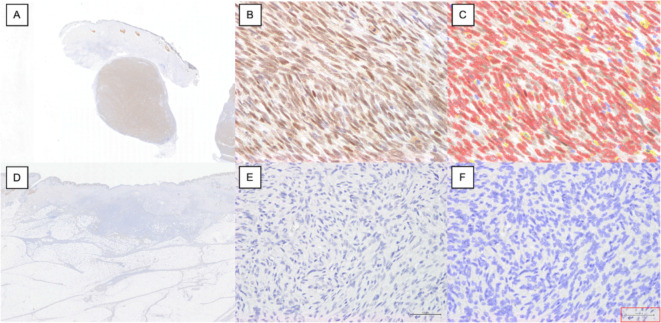
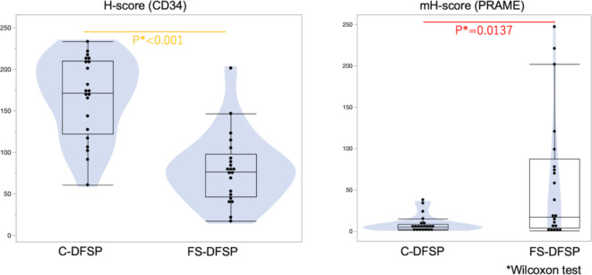
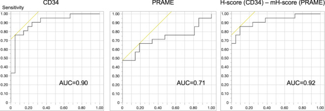
PRAME (PReferentially expressed Antigen in MElanoma) was first identified as a malignant melanoma-specific antigen. Recently, a few cases of fibrosarcomatous dermatofibrosarcoma protuberans (FS-DFSP) were shown to have positivity for PRAME, while conventional dermatofibrosarcoma protuberans (C-DFSP) was negative. Because PRAME may be of diagnostic utility in FS-DFSP and is raising expectations as a new immunotherapy target, we examined the positivity of PRAME in FS-DFSP. Twenty-one cases of FS-DFSP and age/sex/location-matched cases of C-DFSP as a control group were examined by immunohistochemistry for CD34 and PRAME. The results were then evaluated by H-score, which was objectively and semi-quantitatively calculated using the open-source bioimaging analysis software QuPath. The results revealed that the PRAME H-score in FS-DFSP was significantly higher than that in C-DFSP (p = 0.0137). As for CD34, the H-score in FS-DFSP was significantly lower than that in C-DFSP (p < 0.001). Using these two immunohistochemical analyses in combination, the sensitivity and specificity for the diagnosis of FS-DFSP were 86% and 90%, respectively. Double staining of CD34 and PRAME revealed that PRAME-positive and CD34-positive areas did not overlap. This is the largest study to examine PRAME expression in FS-DFSP, and it confirmed the usefulness of PRAME in diagnosing this condition.
Keywords: Bioimaging analysis; CD34; Fibrosarcomatous dermatofibrosarcoma protuberans; PRAME; QuPath.
© 2024. The Author(s).
Conflict of interest statement
The authors declare that they have no significant relationships with, or financial interest in, any commercial entities pertaining to this article.
Figures


References
-
- Doolan, P. et al. Prevalence and prognostic and predictive relevance of PRAME in breast cancer. Breast Cancer Res. Treat.109, 359–365 (2008). - PubMed
-
- Šafanda, A. et al. Immunohistochemical expression of PRAME in 485 cases of epithelial tubo-ovarian tumors. Virchows Arch.483, 509–516 (2023). - PubMed
-
- Cammareri, C. et al. PRAME immunohistochemistry in soft tissue tumors and mimics: a study of 350 cases highlighting its imperfect specificity but potentially useful diagnostic applications. Virchows Arch.483, 145–156 (2023). - PubMed
MeSH terms
Substances
LinkOut - more resources
Full Text Sources
Molecular Biology Databases
Research Materials
