. 2024 Oct 4;14(1):23044.
doi: 10.1038/s41598-024-73082-8.

Optogenetic targeting of cortical astrocytes selectively improves NREM sleep in an Alzheimer's disease mouse model

Affiliations

Optogenetic targeting of cortical astrocytes selectively improves NREM sleep in an Alzheimer's disease mouse model

Qiuchen Zhao et al. Sci Rep. .

Abstract

Alzheimer's disease (AD) is a progressive neurodegenerative condition marked by memory impairments and distinct histopathological features such as amyloid-beta (Aβ) accumulations. Alzheimer's patients experience sleep disturbances at early stages of the disease. APPswe/PS1dE9 (APP) mice exhibit sleep disruptions, including reductions in non-rapid eye movement (NREM) sleep, that contribute to their disease progression. In addition, astrocytic calcium transients associated with a sleep-dependent brain rhythm, slow oscillations prevalent during NREM sleep, are disrupted in APP mice. However, at present it is unclear whether restoration of circuit function by targeting astrocytic activity could improve sleep in APP mice. To that end, APP mice expressing channelrhodopsin-2 (ChR2) targeted to astrocytes underwent optogenetic stimulation at the slow oscillation frequency. Optogenetic stimulation of astrocytes significantly increased NREM sleep duration but not duration of rapid eye movement (REM) sleep. Optogenetic treatment increased delta power and reduced sleep fragmentation in APP mice. Thus, optogenetic activation of astrocytes increased sleep quantity and improved sleep quality in an AD mouse model. Astrocytic activity provides a novel therapeutic avenue to pursue for enhancing sleep and slowing AD progression.

Keywords: Alzheimer’s disease; Astrocyte; Delta power; EEG; NREM; Optogenetics; REM; Sleep; Sleep fragmentation; Slow oscillations; Slow waves; Wake.


Conflict of interest statement

The authors declare no competing interests.

Figures

Fig. 1
Fig. 1
Schematic description of the experimental sleep recording and optogenetic stimulation setup. (A) Viral injection strategy to target astrocytes with AAV8-GFAP104-ChR2-mCherry (ChR2-mCherry) or AAV8-GFAP104-mCherry (mCherry). (B) Schematic view showing placement of EEG/EMG implant. Placement of EEG, EMG electrodes, and fiber-optic cannula on the skull. EMG electrodes were placed within the nuchal musculature. (C) Experimental design. Following AAV infusion and cannula installation, sleep recordings were acquired prior and during continuous optogenetic stimulation (400 ms, 0.6 Hz, 24 h/day). (D) Diagram showing placement of EEG/EMG implant. The processed signal was transferred wirelessly to acquisition software. (E) Representative EEG and EMG traces during NREM, REM, and Wake states.
Fig. 2
Fig. 2
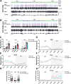
Optogenetic stimulation of astrocytes increased NREM sleep in APP mice. (A, B) Overall 24-hour sleep pattern and sleep architecture of an APP-ChR2 mouse before (A) and during (B) optogenetic stimulation (APP-ChR2-opto). (C) Average time spent in each sleep-wake cycle stage (NREM, REM and Wake) during 24-hour, 12-hour dark phase and 12-hour light phase in APP-ChR2 mice before and during optogenetic stimulation. (D-F) Time course of the changes in REM (D), NREM (E) and Wake states (F) in APP-ChR2 mice before and during optogenetic stimulation. (G) Counts of locomotor activity in APP-ChR2 mice before and during optogenetic stimulation. (H) Time course of locomotor activity in APP-ChR2 mice before and during optogenetic stimulation. All data are expressed as mean ± standard error. Data points represented by filled circles indicate measurements obtained from male mice, while filled triangles denote data from female mice. Paired t-tests were used. The number of mice examined: APP-ChR2 = 6 mice. *P < 0.05, **P < 0.01, and ***P < 0.001. n.s., not significant.
Fig. 3
Fig. 3
Optogenetic stimulation of astrocytes increased delta power during sleep and promoted sleep integrity in APP mice. (A, B) The average EEG power density in the delta (0.5–4 Hz), theta (4–8 Hz), alpha (8–12 Hz), sigma (12–16 Hz), and beta (16–24 Hz) bands during NREM and REM sleep in the 24-hour, 12-hour light phase and 12-hour dark phase in APP-ChR2 mice before and during optogenetic stimulation (APP-ChR2-opto). (C-H) Relative power spectral density of NREM and REM sleep during 24-hour, 12-hour light phase, and 12-hour dark phase in APP-ChR2 mice before and during optogenetic stimulation. (I, J) Average bout count (I) and length (J) in each sleep-wake cycle stage (NREM, REM, and Wake) during the 24-hour, 12-hour dark phase, and 12-hour light phase in APP-ChR2 mice before and during optogenetic stimulation. All data are expressed as means ± standard error. Data points represented by filled circles indicate measurements obtained from male mice, while filled triangles denote data from female mice. Paired t-tests were used. The number of mice examined: APP-ChR2 = 6 mice. *P < 0.05 and **P < 0.01. n.s., not significant.
Fig. 4
Fig. 4
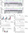
Light stimulation of mCherry in the absence of ChR2 did not significantly affect NREM sleep in APP mice. (A, B) Overall 24-hour sleep pattern and sleep architecture of APP-mCherry mice before (A) and during (B) light stimulation (APP-mCherry-opto). (C) Average time spent in each sleep-wake cycle stage (NREM, REM, and Wake) during the 24-hour, 12-hour dark phase and 12-hour light phase in APP-mCherry mice before and during light stimulation. (D-F) Time course of the changes in REM (D), NREM (E), and Wake states (F) in APP-mCherry mice before and during light stimulation. (G) Counts of the locomotor activity in APP-mCherry mice before and during light stimulation. (H) Time course of the locomotor activity in APP-mCherry mice before and during light stimulation. All data are expressed as mean ± standard error. Data points represented by filled circles indicate measurements obtained from male mice, while filled triangles denote data from female mice. Paired t-tests were used. The number of mice examined: APP-mCherry = 6 mice. n.s., not significant.
Fig. 5
Fig. 5
Light stimulation of mCherry in the absence of ChR2 did not significantly affect power during sleep and sleep integrity in APP mice. (A, B) The average EEG power density in the delta (0.5–4 Hz), theta (4–8 Hz), alpha (8–12 Hz), sigma (12–16 Hz), and beta (16–24 Hz) bands during NREM and REM sleep in 24-hour, 12-hour light phase, and 12-hour dark phase in APP-mCherry mice before and during light stimulation (APP-mCherry-opto). (C-H) Relative power spectral density of NREM and REM sleep during 24-hour, 12-hour light phase, and 12-hour dark phase in APP-mCherry mice before and during light stimulation. (I, J) Average bout count (I) and length (J) in each sleep-wake cycle stage (NREM, REM, and Wake) during 24-hour, 12-hour light phase, and 12-hour dark phase in APP-mCherry mice before and during light stimulation. All data are expressed as means ± standard error. Data points represented by filled circles indicate measurements from male mice, while filled triangles denote data from female mice. Paired t-tests were used. The number of mice examined: APP-mCherry = 6 mice. n.s., not significant.

References

    1. Jack, C. R. et al. NIA-AA research framework: Toward a biological definition of Alzheimer’s disease. Alzheimers Dement.14, 535–562 (2018). - PMC - PubMed
    1. Knopman, D. S. et al. Alzheimer disease. Nat. Rev. Dis. Primers7, 33 (2021). - PMC - PubMed
    1. Sarlus, H. & Heneka, M. T. Microglia in Alzheimer’s disease. J. Clin. Invest.127, 3240–3249 (2017). - PMC - PubMed
    1. Hardy, J. & Selkoe, D. J. The amyloid hypothesis of Alzheimer’s disease: Progress and problems on the road to therapeutics. Science297, 353–356 (2002). - PubMed
    1. Liew, S. C. & Aung, T. Sleep deprivation and its association with diseases—a review. Sleep. Med.77, 192–204 (2021). - PubMed

Substances

LinkOut - more resources