Comparative Study
. 2024 Oct 3;14(1):23032.
doi: 10.1038/s41598-024-74228-4.

Proteomic analysis of plasma-derived extracellular vesicles: pre- and postprandial comparisons

Affiliations
Comparative Study

Proteomic analysis of plasma-derived extracellular vesicles: pre- and postprandial comparisons

Alejandra P Garza et al. Sci Rep. .

Abstract

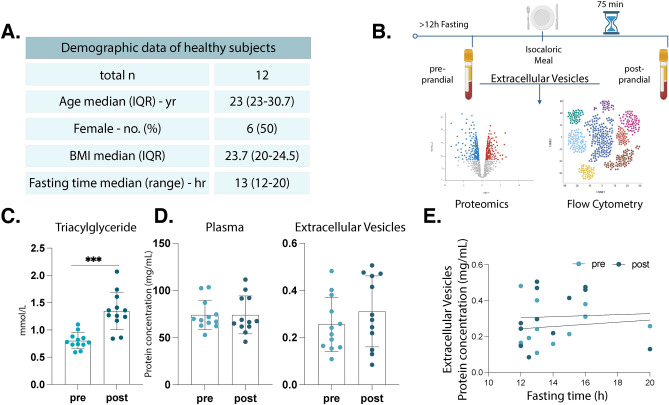
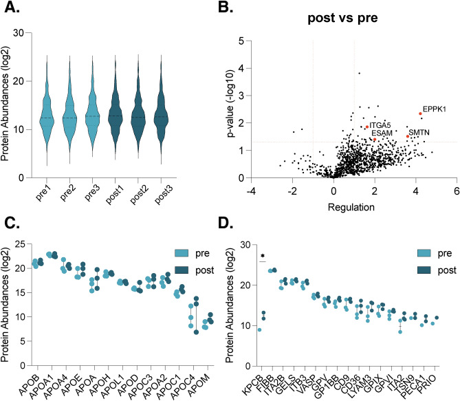
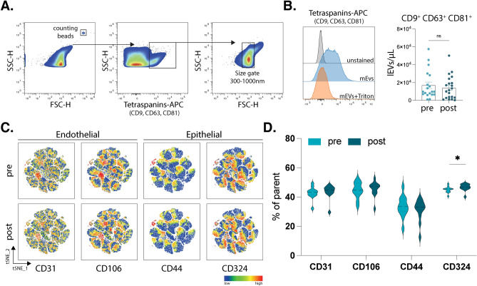
Extracellular vesicles (EVs) are key in intercellular communication, carrying biomolecules like nucleic acids, lipids, and proteins. This study investigated postprandial characteristics and proteomic profiles of blood-derived EVs in healthy individuals. Twelve participants fasted overnight before baseline assessments. After consuming a controlled isocaloric meal, EVs were isolated for proteomic and flow cytometric analysis. Plasma triacylglyceride levels confirmed fasting completion, while protein concentrations in plasma and EVs were monitored for postprandial stability. Proteomic analysis identified upregulated proteins related to transport mechanisms and epithelial/endothelial functions postprandially, indicating potential roles in physiological responses to nutritional intake. Enrichment analyses revealed vesicle-related pathways and immune system processes. Flow cytometry showed increased expression of CD324 on CD9+CD63+CD81+ large extracellular vesicles postprandially, suggesting an epithelial origin. These findings offer valuable insights into postprandial EV dynamics and their potential physiological significance, highlighting the need for stringent fasting guidelines in EV studies to account for postprandial effects on EV composition and function.

Keywords: Extracellular vesicles; Fasting; Liquid biopsy; Plasma; Postprandial; Proteomics.


Conflict of interest statement

The authors declare no competing interests.

The authors declare that they have no competing interests.

Figures

Fig. 1
Fig. 1
Experimental design and pre-requisites for assessing postprandial changes in healthy individuals. Detailed demographic information of the healthy participants (A). Schematic representation of the experimental setup, wherein blood samples were collected from healthy participants before and 75 min after consumption of an isocaloric meal. Samples were then processed to obtain plasma and plasma EVs, of which the later were used for detailed characterization by proteomics and flow cytometry (B). Bar chart illustrating the triacylglycerides levels in plasma before and after food intake, indicating postprandial changes (C). Bar plots displaying total protein concentrations in plasma and plasma-EVs, both pre- and postprandially (D). Correlation plot demonstrating no correlation between fasting time and total protein concentration in EV samples both in pre- and postprandial time points (E). n = 12, all samples were measured in technical triplicates. Statistical analyses were performed using paired t-test (CD) and Pearson correlation (E), respectively. P values: * for p ≤ 0.05; ** for p ≤ 0.001; *** for p ≤ 0.0001.
Fig. 2
Fig. 2
Proteomic analyses of fasted and postprandial isolated extracellular vesicles. Distribution of relative protein abundances (log2) in isolated EV samples (A). Volcano plots showing protein regulations (− log10) in EVs isolated post- vs preprandially (B). Protein abundances of apolipoproteins (log2) in EV samples comparing pre- and postprandial conditions (C). Platelet protein abundances (log2) in EV samples comparing pre- and postprandial conditions (D). n = 3, all samples were measured in technical triplicates; P values are given in − log10. Student’s t-test using an FDR of 0.05 (= 1.3 − log10) and a up- or down regulation of < − 2 or > 2 (≤ − 1 or > 1 log2).
Fig. 3
Fig. 3
Enrichment analyses of significantly regulated proteins in the posprandial state. Network analysis showing proteins annotated as “extracellular exosome” in red, names are given in bold (A). Enrichment analyses showing enriched reactome pathways and biological processes (GO) analyses revealed multiple enriched biological processes (p-values given in − log2) in postprandial EVs. Size of the dots indicate number of proteins; color of the dots indicates strength of the enriched processes and pathways (B,C). Analyses were performed using the STRING database.
Fig. 4
Fig. 4
Flow cytometry of plasma extracellular vesicles. Representative gating strategy of plasma derived lEVs showing first counting beads as reference (left graph). Selection of events positive for tetraspanin markers (CD9, CD63 and CD81) was followed by selection of events based on a size gate established with silica beads, additionally showing sensitivity to Triton X and an unstained control (A). Bar charts showing absolute number of lEVs per microliter comparing pre- and postprandial timepoints based on measurement with counting beads (B). t-distributed stochastic neighbor embedding (t-SNE) plots showing the mean fluorescence intensity of CD31, CD106, CD44, and CD324 in a total of 80,400 positive events (based on CD9+ , CD63+ CD81+ events and size) in each pre- and postprandial states (C). Violin plots showing the frequency of positive events for CD31 and CD106 (endothelial markers) as well as CD44 and CD324 (epithelial markers) in pre- vs postprandial states (D). n = 12, all samples were measured in technical duplicates. Statistical analyses were performed using paired t-test. P values: * for p ≤ 0.05; ** for p ≤ 0.001; *** for p ≤ 0.0001.).

Similar articles

Cited by

References

    1. Kumar, M. A. et al. Extracellular vesicles as tools and targets in therapy for diseases. Signal Transduct. Targ. Ther.9, 1–41 (2024). - PMC - PubMed
    1. Shah, R., Patel, T. & Freedman, J. E. Circulating extracellular vesicles in human disease. N. Engl. J. Med.379, 958–966 (2018). - PubMed
    1. Lyu, C., Sun, H., Sun, Z., Liu, Y. & Wang, Q. Roles of exosomes in immunotherapy for solid cancers. Cell Death Dis.2024(15), 1–21 (2024). - PMC - PubMed
    1. Buzas, E. I. The roles of extracellular vesicles in the immune system. Nat. Rev. Immunol.23, 236–250 (2022). - PMC - PubMed
    1. Van Niel, G., D’Angelo, G. & Raposo, G. Shedding light on the cell biology of extracellular vesicles. Nat. Rev. Mol. Cell Biol.19, 213–228 (2018). - PubMed

Publication types

LinkOut - more resources