. 2024 Oct 3;7(1):1254.
doi: 10.1038/s42003-024-06938-4.

Placental extracellular vesicles promote cardiomyocyte maturation and fetal heart development

Affiliations

Placental extracellular vesicles promote cardiomyocyte maturation and fetal heart development

Mariyan J Jeyarajah et al. Commun Biol. .

Abstract

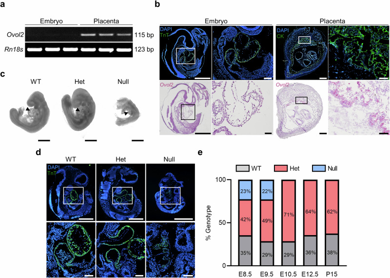
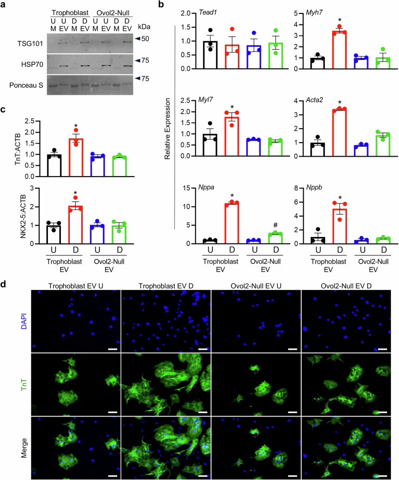
Congenital heart defects are leading causes of neonatal mortality and are often associated with placental abnormalities, but mechanisms linking placenta and heart development are poorly understood. Herein, we investigated a potential signaling network connecting the placenta and nascent heart in mice. We found that fetal hearts exposed to media conditioned by placental tissue or differentiated wild-type trophoblast stem (TS) cells, but not undifferentiated TS cells, showed increased heart rate and epicardial cell outgrowth. This effect was not observed when hearts were exposed to media from TS cells lacking OVO-Like 2, a transcription factor required for trophoblast differentiation and placental development. Trophoblasts released abundant extracellular vesicles into media, and these vesicles were sufficient to mediate cardio-promoting effects. Our findings provide a potential mechanism whereby the placenta communicates with the fetal heart to promote cardiac morphogenesis, and offers insight into the link between poor placentation and a higher incidence of heart defects.


Conflict of interest statement

The authors declare no competing interests.

Figures

Fig. 1
Fig. 1. Ovol2 deficiency results in poor heart formation and embryonic lethality.
a RT-PCR showing Ovol2 and Rn18s in the mouse embryo and placenta on E9.5. b Immunohistochemistry showing Troponin T (TnT; green, upper left images) and Cytokeratin (CK; green, upper right images) in the E9.5 embryo and placenta, respectively. DAPI was used to stain nuclei. In the bottom panels, in situ hybridization was used to demarcate Ovol2 (pink) expression in the embryo and placenta. Boxes in the lower magnification images (left panels) show the location of higher magnification images (right panels) for both the embryo and placenta. Please note that Ovol2-positive cells are only detectable within the placenta. c Phase-contrast images of Ovol2+/+ (WT), Ovol2+/- (Het) and Ovol2-/- (Null) embryos. Images were captured using a Leica DMi1 inverted microscope. Arrowheads indicate the fetal heart. d Immunohistochemistry showing TnT (green) in WT, Het, and Null embryos. Boxes in the lower magnification images (top panels) show the location of higher magnification images (bottom panels). e Quantification of the percent of WT, Het, and Null embryos present at various gestational timepoints (n = 4 pregnant mice; actual number of embryos/pups of each genotype are presented in Supplementary Table 1). Scale bars represent 500 µm for low magnification images in (b) and (d), and the images in (c). Scale bars for high magnification images in (b) and (d) represent 50 µm. Uncropped images of DNA gels are provided in Supplementary Fig. 7.
Fig. 2
Fig. 2. Medium conditioned by placental tissue and differentiated trophoblasts promotes fetal heart development and cardiomyocyte growth.
a Schematic indicating four culture conditions: fetal hearts cultured in unconditioned media (CTRL), fetal hearts cultured in media conditioned by E15.5 placental tissue (Placental CM), fetal hearts cultured in media conditioned by 5-day differentiated Ovol2+/+ TS cells (Trophoblast CM), and fetal hearts cultured in media conditioned by 5-day differentiated Ovol2-/- TS cells (Ovol2-Null CM). b Average number of beats per minute when fetal hearts were exposed to CTRL, Placental CM, Trophoblast CM, or Ovol2-Null CM for 24 and 48 h. c Quantification of epicardial cell outgrowth from fetal hearts exposed to CTRL, Placental CM, Trophoblast CM, or Ovol2-Null CM for 24 and 48 h. c Representative images of epicardial cell outgrowths at 24 h. A red dashed line delineates fetal heart borders and arrows demarcate examples of outgrowths. d Expression of six genes (Tead1, Myh7, Myl7, Acta2, Nppa, and Nppb) associated with cardiac development in cardiomyocytes exposed to CTRL, Placental CM, Trophoblast CM, or Ovol2-Null CM. e Western blot showing levels of Troponin T (TnT) and NKX2-5 in cardiomyocytes treated with CTRL, Placental CM, Trophoblast CM, or Ovol2-Null CM. ACTB was used as a loading control. Quantification of relative band intensity is shown to the right of the representative blots. f Immunofluorescence for TnT (green) in cardiomyocytes treated with CTRL, Placental CM, Trophoblast CM, or Ovol2-Null CM. DAPI was used to demarcate nuclei. Values significantly different from CTRL (N = 6 biologically independent samples, P < 0.05) are denoted with an asterisk (*); values significantly increased from CTRL but decreased from Placental CM are denoted with a number sign (#). One-way ANOVA followed by Tukey’s post-hoc test was used to determine statistical significance. Scale bar = 100 µm. Graphs represent means ± SEM. Uncropped images of western blots are provided in Supplementary Fig. 8.
Fig. 3
Fig. 3. Placental EVs promote fetal heart development and cardiomyocyte growth.
a Schematic showing procedure used to isolate EVs from unconditioned media (CTRL) and media conditioned by E15.5 placental tissue (Placental CM). b Western blot for EV markers TSG101 and HSP70 in media supernatants and EVs isolated from CTRL and Placental CM. Ponceau S was used to verify total protein. c Quantification of fetal heart rate and epicardial cell outgrowth from embryonic hearts exposed to supernatant from either unconditioned media (M-C) or Placental CM (M-P); or varying concentrations of CTRL (from FBS) or Placental EVs (1, 5, or 10 µg/mL) for 48 h. d Expression of six genes (Tead1, Myh7, Myl7, Acta2, Nppa, and Nppb) associated with cardiac development in cardiomyocytes treated with EVs (10 µg/mL) isolated from CTRL or Placental CM. e Western blot showing levels of Troponin T (TnT) and NKX2-5 in cardiomyocytes treated with CTRL or Placental EVs (10 µg/mL). ACTB was used as a loading control. Quantification of relative band intensity is found to the right of the representative blots. f Immunofluorescence for TnT (green) in cardiomyocytes treated with CTRL or Placental EVs (10 µg/mL). DAPI was used to demarcate nuclei. Values significantly different from CTRL media supernatants (N = 5 independent experiments, P < 0.05), or from CTRL EVs (N = 3 independent experiments, P < 0.05) are denoted with an asterisk (*), using one-way ANOVA followed by Tukey’s post-hoc test (c) or two-tailed Student’s t-test (d and e). Scale bar = 100 µm. Graphs represent means ± SEM. Uncropped images of western blots are provided in Supplementary Fig. 8.
Fig. 4
Fig. 4. Placental EVs are present in fetal and maternal plasma.
a Schematic showing experimental design. Plasma was isolated from non-pregnant virgin mice, pregnant dams, or fetuses on E18.5. Plasma was then incubated with syncytin-A-FITC and CD9-PE antibodies, and subjected to nanoflow cytometry analysis. b Quantification of plasma EVs positive for both syncytin-A-FITC and CD9-PE antibodies. c Representative nanoflow cytometry plots showing an unstained control (pooled plasma), and syncytin-A-FITC and CD9-PE positive EVs isolated from plasma from non-pregnant virgin mice, pregnant dams, or fetuses on E18.5. Values significantly different from non-pregnant mice (N = 3 different mice, P < 0.05) are denoted with an asterisk (*), using one-way ANOVA followed by Tukey’s post-hoc test. Graphs represent means ± SEM.
Fig. 5
Fig. 5. EVs from media conditioned by differentiated trophoblasts promote cardiomyocyte growth.
a Western blot for EV markers TSG101 and HSP70 in media supernatants (M) and EVs isolated from media conditioned by Ovol2+/+ TS cells (Trophoblast) and Ovol2-/- (Ovol2-Null) TS cells cultured in stem or differentiation media. Ponceau S was used to verify total protein. U=cells exposed to stem media (undifferentiated), D=cells exposed to differentiation media for 5 days. b Expression of six genes (Tead1, Myh7, Myl7, Acta2, Nppa, and Nppb) associated with cardiac development in cardiomyocytes treated with EVs (10 µg/mL) isolated from CM of Ovol2+/+ and Ovol2-Null TS cells cultured in undifferentiated and differentiated states. c Relative band intensity from western blots for Troponin T (TnT) and NKX2-5 in comparison to ACTB in cardiomyocytes exposed to EVs (10 µg/mL) isolated from CM of Ovol2+/+ and Ovol2-Null TS cells cultured in undifferentiated and differentiated states. d Immunofluorescence for TnT (green) in cardiomyocytes treated with EVs (10 µg/mL) isolated from CM of Ovol2+/+ and Ovol2-Null TS cells cultured in undifferentiated and differentiated states. DAPI was used to stain nuclei. Values significantly different from undifferentiated Trophoblast EVs (N = 3 independent experiments, P < 0.05) are denoted with an asterisk (*); values different from both undifferentiated trophoblast EVs and differentiated trophoblast EVs are denoted with a number sign (#). A one-way ANOVA followed by Tukey’s post-hoc test was used to determine statistical significance, using values from the undifferentiated Trophoblast EVs normalized to 1 as the control condition. Scale bar = 100 µm. Graphs represent means ± SEM. Uncropped images of western blots are provided in Supplementary Fig. 8.

References

    1. Linask, K. K. The heart-placenta axis in the first month of pregnancy: induction and prevention of cardiovascular birth defects. J. Pregnancy2013, (2013). - PMC - PubMed
    1. Maslen, C. L. Recent Advances in Placenta-Heart Interactions. Front. Physiol.9, 735 (2018). - PMC - PubMed
    1. Seo, D. M. et al. The outcome of open heart surgery for congenital heart disease in infants with low body weight less than 2500 g. Pediatr. Cardiol.32, 578–584 (2011). - PubMed
    1. Best, K. E., Tennant, P. W. G. & Rankin, J. Survival, by Birth Weight and Gestational Age, in Individuals With Congenital Heart Disease: A Population-Based Study. J. Am. Heart Assoc.6, e005213 (2017). - PMC - PubMed
    1. Suluba, E., Shuwei, L., Xia, Q. & Mwanga, A. Congenital heart diseases: genetics, non-inherited risk factors, and signaling pathways. Egypt. J. Med. Hum. Genet. 2020 21121, 1–12 (2020).

LinkOut - more resources