. 2024 Oct 3;12(1):156.
doi: 10.1186/s40478-024-01867-z.

Targeting the TDP-43 low complexity domain blocks spreading of pathology in a mouse model of ALS/FTD

Affiliations

Targeting the TDP-43 low complexity domain blocks spreading of pathology in a mouse model of ALS/FTD

Elodie Chevalier et al. Acta Neuropathol Commun. .

Abstract

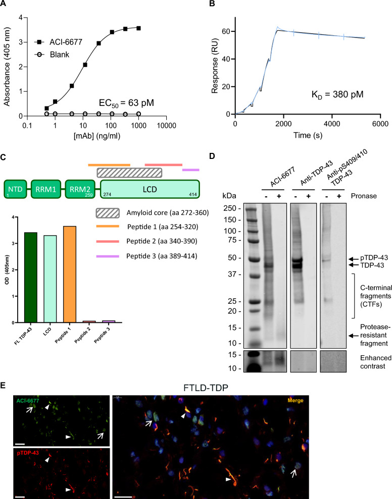
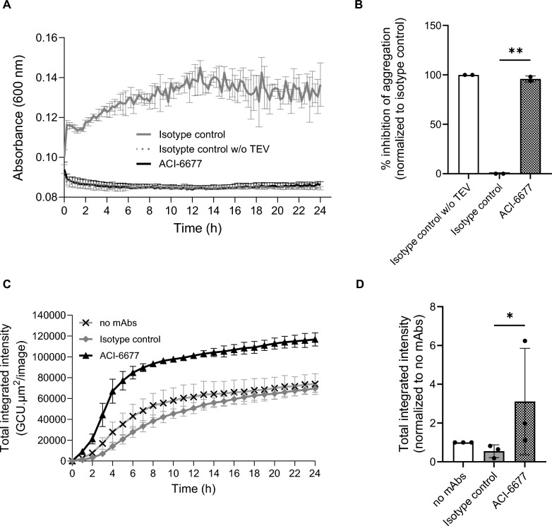
Abnormal cytoplasmic localization and accumulation of pathological transactive response DNA binding protein of 43 kDa (TDP-43) underlies several devastating diseases such as amyotrophic lateral sclerosis (ALS) and frontotemporal lobar degeneration with TDP-43 pathology (FTLD-TDP). A key element is the correlation between disease progression and spatio-temporal propagation of TDP-43-mediated pathology in the central nervous system. Several lines of evidence support the concept of templated aggregation and cell to cell spreading of pathological TDP-43. To further investigate this mechanism in vivo, we explored the efficacy of capturing and masking the seeding-competent region of extracellular TDP-43 species. For this, we generated a novel monoclonal antibody (mAb), ACI-6677, that targets the pathogenic protease-resistant amyloid core of TDP-43. ACI-6677 has a picomolar binding affinity for TDP-43 and is capable of binding to all C-terminal TDP-43 fragments. In vitro, ACI-6677 inhibited TDP-43 aggregation and boosted removal of pathological TDP-43 aggregates by phagocytosis. When injecting FTLD-TDP brain extracts unilaterally in the CamKIIa-hTDP-43NLSm mouse model, ACI-6677 significantly limited the induction of phosphorylated TDP-43 (pTDP-43) inclusions. Strikingly, on the contralateral side, the mAb significantly prevented pTDP-43 inclusion appearance exemplifying blocking of the spreading process. Taken together, these data demonstrate for the first time that an immunotherapy targeting the protease-resistant amyloid core of TDP-43 has the potential to restrict spreading, substantially slowing or stopping progression of disease.

Keywords: ALS; FTD; Immunotherapy; Neuropathology; Pathomechanism; Spreading; TDP-43.


Conflict of interest statement

T.A. and T.S. are coinventors on a patent application, publication number WO2020/234473. R.O., T.A. and T.S. are coinventors on a patent application, publication number WO2022/034228. E.C., M.R., R.O., A.F., K.P., A.P., M.V., T.S. are employees of AC Immune and entitled to options and/or shares. M.A. and T.A. were employee of AC Immune at the time of this study. The other authors declare no competing interests.

Figures

Fig. 1
Fig. 1
ACI-6677 binds with high affinity to the low complexity domain of TDP-43. A The binding of ACI-6677 to human TDP-43 was measured by ELISA and B measured by SPR; blue line depicts the experimental data and black line depicts the data fit to the model; C Epitope mapping of ACI-6677 using various peptides derived from the human TDP-43 sequence (coverage indicated by a bar above the cartoon of the TDP-43 domains) indicates binding to the LCD and to peptide 1 (aa 254–320); NTD, N-terminal domain; RRM, RNA Recognition Motif; LCD, Low Complexity Domain; D Immunoblotting with ACI-6677 or two control antibodies, an anti-total TDP-43 mAb and an anti-pTDP-43 (specific for pS409/pS410) mAb, on sarkosyl-insoluble brain extracts prepared from frontal cortex of cases with FTLD-TDP type A before (−) or after (+) limited proteolysis (revealing the protease resistant fragments) to determine the binding of ACI-6677 to pTDP-43, TDP-43, C-terminal fragments (CTFs) and the protease resistant fragments. Bottom panels with enhanced contrast included to show the binding to protease resistant fragments; molecular weight (kDa) markers shown on the left; E Immunolabeling with ACI-6677 (green), an anti-p-TDP-43 mAb (red) and DAPI (cell nuclei, blue) demonstrates binding of ACI-6677 to pTDP-43 (arrowheads) in sections from a FTLD-TDP type A patient (temporal cortex). Arrows indicate areas of physiological nuclear TDP-43. Scale bar = 20 μm
Fig. 2
Fig. 2
ACI-6677 inhibits human TDP-43 aggregation and promotes phagocytic clearance by microglia. A de novo aggregation was assessed using 2.5 μM of ACI-6677 in an assay where the TEV protease is added to cleave MBP-tagged TDP-43, initiating the aggregating process. An isotype control mAb in the presence or absence of the TEV protease provides the kinetic profiles for maximal or no aggregation, respectively; Data represent mean ± SD of 3 independent replicates. B Quantification of percent aggregation inhibition at 24 h from 2 independent experiments normalized to the isotype control without TEV protease. Data shown as mean ± SD reported using a ratio-paired t-test **p < 0.01; C Phagocytosis assay with mouse microglia of ACI-6677-TDP-43 immune complexes over 24 h; the total integrated intensity (expressed as green calibrated unit (GCU) x mm2 per image) of fluorescent pHrodo™-labeled TDP-43 (30 nM) internalized at each timepoint is reported after incubation with 30 nM of ACI-6677 or isotype control mAb. Data represents mean ± SD from three technical replicates; D Quantification of total integrated intensity at 6 h from three independent experiments normalized to the no mAbs control. Data shown as mean ± SD reported using a ratio-paired t-test *p < 0.05
Fig. 3
Fig. 3
ACI-6677 reduces spreading of TDP-43 pathology in CamKIIa-hTDP-43NLSm transgenic mice inoculated in one hemisphere with FTLD-TDP-derived brain extracts. A Immunohistochemistry post 13 weeks of treating the CamKIIa-hTDP-43NLSm mice with ACI-6677 (bottom), the isotype control mAb (middle) or untreated mice (top, no extracts) was performed using an anti-pTDP-43 mAb (red) and anti-NeuN mAb (yellow); the ipsilateral and contralateral hippocampal regions are indicated and representative low and high (scale bar = 40 μm) magnification provided; B, C Quantification of the level of pTDP-43 (mean staining density) in the ipsilateral (B) and contralateral (C) hippocampi of ACI-6677 (n = 13) versus isotype control (n = 14) treated mice; background level of pathology in transgenic mice ("no extracts", n = 8) injected in the hippocampus with PBS and not treated with antibodies is provided. Data represent mean ± SEM. A one-way ANOVA followed by a Tukey post-hoc test for multiple comparisons was used. **p < 0.01; ***p < 0.001; ****p < 0.0001; D Correlation of the pTDP-43 levels with the concentration of ACI-6677 (measured in plasma 24 h post the last dose) for the ipsilateral (r2 = 0.37 and p = 0.06) and contralateral (r2 = 0.23 and p = 0.17) regions

References

    1. Afroz T, Chevalier E, Audrain M, Dumayne C, Ziehm T, Moser R et al (2023) Immunotherapy targeting the C-terminal domain of TDP-43 decreases neuropathology and confers neuroprotection in mouse models of ALS/FTD. Neurobiol Dis 179:106050. 10.1016/j.nbd.2023.106050 - PubMed
    1. Afroz T, Perez-Berlanga M, Polymenidou M (2019) Structural transition, function and dysfunction of TDP-43 in neurodegenerative diseases. Chimia (Aarau) 73:380–390. 10.2533/chimia.2019.380 - PubMed
    1. Arai T, Hasegawa M, Akiyama H, Ikeda K, Nonaka T, Mori H et al (2006) TDP-43 is a component of ubiquitin-positive tau-negative inclusions in frontotemporal lobar degeneration and amyotrophic lateral sclerosis. Biochem Biophys Res Commun 351:602–611. 10.1016/j.bbrc.2006.10.093 - PubMed
    1. Arseni D, Chen R, Murzin AG, Peak-Chew SY, Garringer HJ, Newell KL et al (2023) TDP-43 forms amyloid filaments with a distinct fold in type A FTLD-TDP. Nature 620:898–903. 10.1038/s41586-023-06405-w - PMC - PubMed
    1. Arseni D, Hasegawa M, Murzin AG, Kametani F, Arai M, Yoshida M et al (2021) Structure of pathological TDP-43 filaments from ALS with FTLD. Nature. 10.1038/s41586-021-04199-3 - PMC - PubMed

Publication types

MeSH terms

LinkOut - more resources