. 2024 Nov;13(29):e2400804.
doi: 10.1002/adhm.202400804. Epub 2024 Oct 4.

Targeted Therapy of Osteoarthritis via Intra-Articular Delivery of Lipid-Nanoparticle-Encapsulated Recombinant Human FGF18 mRNA

Affiliations

Targeted Therapy of Osteoarthritis via Intra-Articular Delivery of Lipid-Nanoparticle-Encapsulated Recombinant Human FGF18 mRNA

Mengze Sun et al. Adv Healthc Mater. 2024 Nov.

Abstract

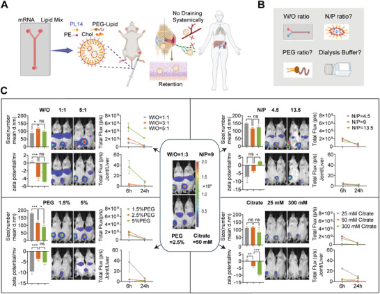
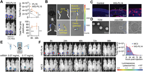
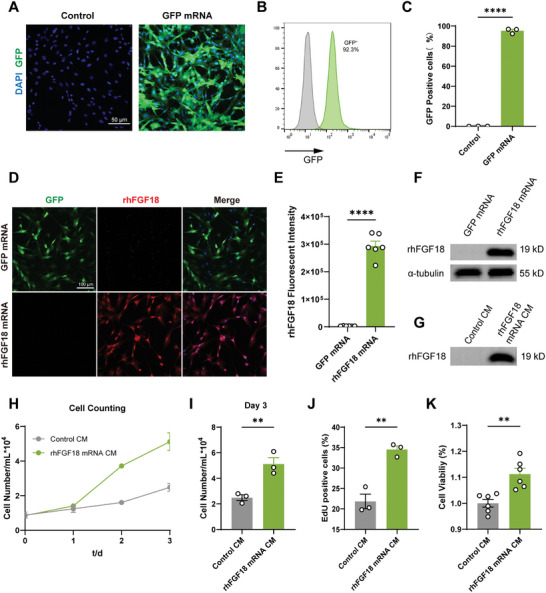
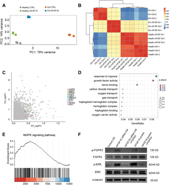
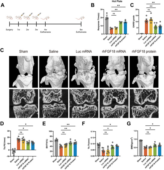
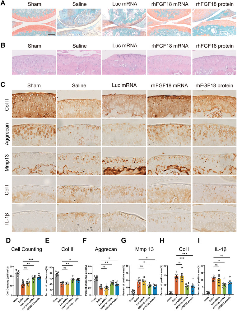
Fibroblast growth factor 18 (FGF18) emerges as a promising therapeutic target for osteoarthritis (OA). In this study, a novel articular cavity-localized lipid nanoparticle (LNP) named WG-PL14 is developed. This optimized formulation has a nearly 30-fold increase in mRNA expression as well as better articular cavity enrichment compared to commercial lipids MC3 when performing intra-articular injection. Then, a mRNA sequence encoding recombinant human FGF18 (rhFGF18) for potential mRNA therapy in OA is optimized. In vitro assays confirm the translation of rhFGF18 mRNA into functional proteins within rat and human chondrocytes, promoting cell proliferation and extracellular matrix (ECM) synthesis. Subsequently, the therapeutic efficacy of the LNP-rhFGF18 mRNA complex is systematically assessed in a mouse OA model. The administration exhibits several positive outcomes, including an improved pain response, upregulation of ECM-related genes (e.g., AGRN and HAS2), and remodeling of subchondral bone homeostasis compared to a control group. Taken together, these findings underscore the potential of localized LNP-rhFGF18 mRNA therapy in promoting the regeneration of cartilage tissue and mitigating the progression of OA.

Keywords: lipid nanoparticles; mRNA; osteoarthritis; recombinant human FGF18.


Conflict of interest statement

The authors declare no conflict of interest.

Figures

Figure 1
Figure 1
Formulation optimizations of PL14 LNPs to improve the specific accumulation of LNPs and mRNA in the articular cavity while minimizing expression in the liver. A) Schematic illustration of the preparation method of nanoparticles. B) Illustration of enhanced accumulation of Luc‐mRNA LNPs in cartilage cavity by adjusting the water‐to‐ethanol ratio (W/O ratio), the N/P ratio, the percentage of PEG, and the concentration of dialysis buffer. C) The size, zeta potential, and bioluminescence images of ICR mice (n = 3) after intra‐articular injection of LNP‐Luc mRNA (2 µg mRNA) in different formulations. The unit of the fluorescent scale bar is photons/sec/cm2/sr, the radiance scale of 1.0 × 107–2 × 108. Data were represented as mean ± SEM. Statistical significance was calculated via one‐way ANOVA, followed by Dunnett's multiple comparisons test (* p < 0.05, ** p < 0.01, and *** p < 0.001).
Figure 2
Figure 2
Comparison of the top performing WG‐PL14 LNPs with MC3 LNPs for intra‐articular injections. A) Representative biodistribution image of luciferase expression for original and optimized formulation LNP. B) The biodistribution image of luciferase expression in articular cartilage as measured with an IVIS imaging system 24 h after intra‐articular injection of 2 µg of total mRNA. C) Immunofluorescence staining for Luc in mouse chondrocytes after intra‐articular injection of LNP‐Luc mRNA (2 µg mRNA). The yellow arrows indicate the luminescent zone. D) Representative TEM and STEM of LNPs. E) The biodistribution image of luciferase expression in articular cartilage after intra‐articular injection of concentrated and unconcentrated LNP‐Luc mRNA (2 µg mRNA). F) The comparison between WG‐PL14 and MC3 in vivo expression, after injection of LNP‐Luc mRNA (2 µg mRNA), then imaged in 6, 24, 48, 72, and 96 h. Data were represented as mean ± SEM. Statistical significance was calculated by t‐test. The unit of the fluorescent scale bar is photons/sec/cm2/sr, the F radiance scale of 1.0 × 107–2 × 108 (* p < 0.05, ** p < 0.01, and *** p < 0.001).
Figure 3
Figure 3
In vitro transfection and characterization of GFP and rhFGF18 mRNA. A) Representative images of rat primary chondrocytes 24 h post‐GFP mRNA transfection. B) Flow cytometry analysis of rat primary chondrocytes 24 h post‐GFP mRNA transfection. C) Statistical analysis of GFP positive cells between control and GFP mRNA group. D) Representative images of immunofluorescence of rat primary chondrocytes 24 h post‐rhFGF18 mRNA transfection or GFP mRNA transfection. E) Quantification of the immunofluorescence images. Western blot analysis of F) total protein and G) supernatant of rat primary chondrocytes 24 h post‐rhFGF18 mRNA transfection or GFP mRNA transfection. Rat primary chondrocytes cultured with control CM and rhFGF18 mRNA CM and perform H) cell counting on day 1,2,3, I) statistical analysis of cell numbers on day 3, J) Quantification of flow cytometry analysis on EdU positive cells, K) CCK8 assays to assess cell viability. Data are shown as the mean ± SEM. Student's t‐test was used for C), E), I), J), and K). * p < 0.05, ** p < 0.01, and *** p < 0.001.
Figure 4
Figure 4
Transcriptome analysis of human primary chondrocytes after rhFGF18 mRNA CM stimulation. A) Principal component analysis (PCA). B) Pairwise correlation based on Pearson coefficients between all samples. C) Genes co‐upregulated in chondrocytes from healthy or osteoarthritis areas after stimulation of rhFGF18 mRNA CM. D) Gene ontology analysis between chondrocytes cultured with rhFGF18 mRNA CM and control CM. E) Gene set enrichment analysis (GSEA) reveals MAPK signaling pathway‐related genes enriched in chondrocytes cultured with rhFGF18 mRNA CM. F) Western blot analysis on phosphorylation of key components of MAPK signaling pathway.
Figure 5
Figure 5
WG‐PL14‐rhFGF18 mRNA prevents against the progression of post‐traumatic OA in vivo. A) Schematic diagram of the timeline of intra‐articular injection of LNP‐rhFGF18 mRNA and subsequent analysis. B) Pain response time after placing the mice onto a 55 °C hot plate meter at 8 weeks post‐surgery. C) Representative micro‐CT (µCT) images of the joints from mice treated with saline, LNP‐luc mRNA, LNP‐rhFGF18 mRNA, or rhFGF18 protein at 8 weeks after sham or ACLT surgery. n = 6 for each group. Quantitative µCT analysis of tibia subchondral bone parameters including D) trabecular bone pattern factor (Tb.Pf), E) trabecular bone volume per tissue volume (BV/TV), and F) trabecular thickness (Tb.Th) and G) bone mineral density (BMD) in each group at 8 weeks postoperatively. n = 6 for each group. Data are shown as the mean ± SEM. One‐way ANOVA was used for D), E), F), and G). * p < 0.05, ** p < 0.01, and *** p < 0.001.
Figure 6
Figure 6
LNP‐rhFGF18 mRNA intra‐articular injection enhanced chondrocyte proliferation and matrix anabolism. A) Safranin O‐fast green staining, B) H&E staining, and C) IHC staining for Collagen II, Aggrecan, Mmp13, Collagen I, and IL‐1β of mouse knees at 8 weeks post intra‐articular injection of LNP‐rhFGF18 mRNA. Quantification of D) cell numbers in H&E staining and positive area in IHC staining of E) Collagen II, F) Aggrecan, G) Mmp13, H) Collagen I and I) IL‐1β. n = 6 in each group. “n” represents the number of mice. Data are shown as the mean ± SEM. Scale bars, 200 µm for A) and 50 µm for B) and C). One‐way ANOVA was used for D), E), F), G), H), and I). * p < 0.05, ** p < 0.01, and *** p < 0.001.

Similar articles

Cited by

References

    1. Hunter D. J., Bierma‐Zeinstra S., Lancet 2019, 393, 1745. - PubMed
    1. Wallace I. J., Worthington S., Felson D. T., Jurmain R. D., Wren K. T., Maijanen H., Woods R. J., Lieberman D. E., Proc. Natl. Acad. Sci. 2017, 114, 9332. - PMC - PubMed
    1. Safiri S., Kolahi A.‐A., Smith E., Hill C., Bettampadi D., Mansournia M. A., Hoy D., Ashrafi‐Asgarabad A., Sepidarkish M., Almasi‐Hashiani A., Collins G., Kaufman J., Qorbani M., Moradi‐Lakeh M., Woolf A. D., Guillemin F., March L., Cross M., Ann. Rheum. Dis. 2020, 79, 819. - PubMed
    1. Latourte A., Kloppenburg M., Richette P., Nat Rev Rheumatol 2020, 16, 673. - PubMed
    1. Dai L., Zhang X., Hu X., Liu Q., Man Z., Huang H., Meng Q., Zhou C., Ao Y., Mol. Ther. 2015, 23, 1331. - PMC - PubMed

MeSH terms