. 2024 Sep 30;15(41):17232-17244.
doi: 10.1039/d4sc03295k. Online ahead of print.

Dynamical responses predict a distal site that modulates activity in an antibiotic resistance enzyme

Affiliations

Dynamical responses predict a distal site that modulates activity in an antibiotic resistance enzyme

Michael Beer et al. Chem Sci. .

Abstract

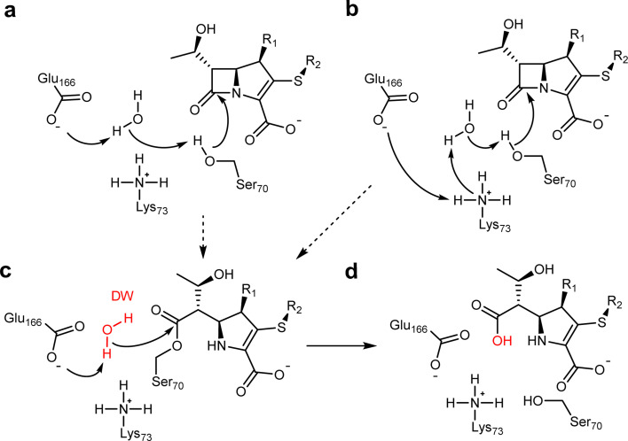
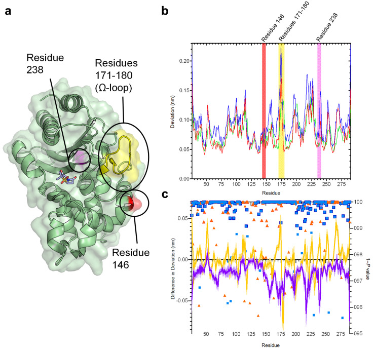
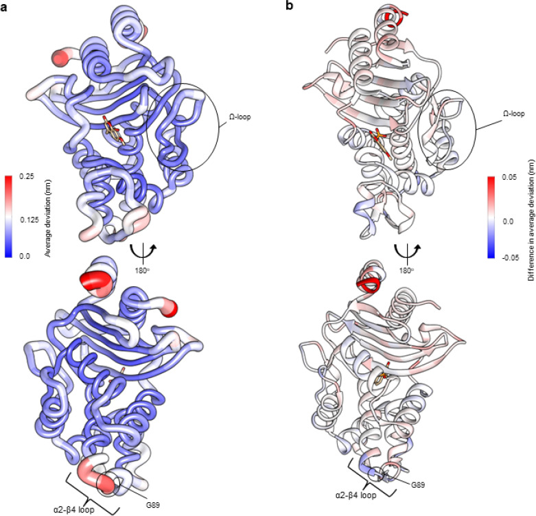
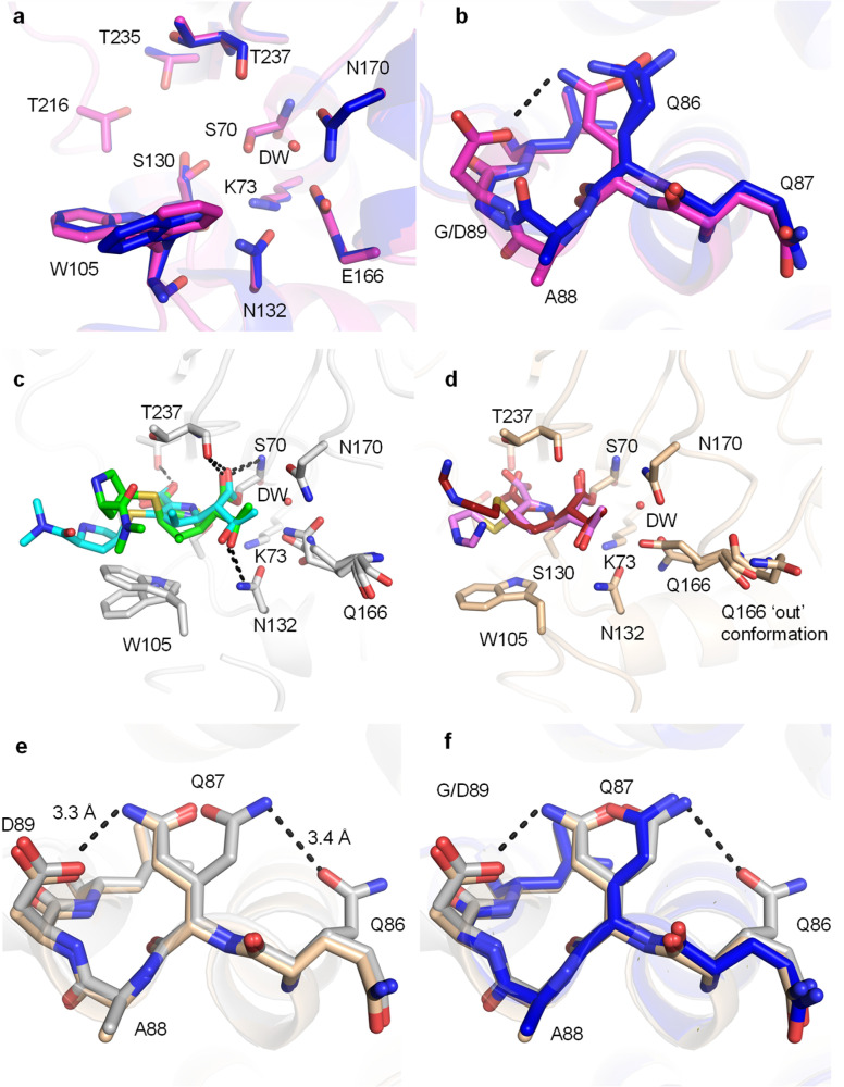
β-Lactamases, which hydrolyse β-lactam antibiotics, are key determinants of antibiotic resistance. Predicting the sites and effects of distal mutations in enzymes is challenging. For β-lactamases, the ability to make such predictions would contribute to understanding activity against, and development of, antibiotics and inhibitors to combat resistance. Here, using dynamical non-equilibrium molecular dynamics (D-NEMD) simulations combined with experiments, we demonstrate that intramolecular communication networks differ in three class A SulpHydryl Variant (SHV)-type β-lactamases. Differences in network architecture and correlated motions link to catalytic efficiency and β-lactam substrate spectrum. Further, the simulations identify a distal residue at position 89 in the clinically important Klebsiella pneumoniae carbapenemase 2 (KPC-2), as a participant in similar networks, suggesting that mutation at this position would modulate enzyme activity. Experimental kinetic, biophysical and structural characterisation of the naturally occurring, but previously biochemically uncharacterised, KPC-2G89D mutant with several antibiotics and inhibitors reveals significant changes in hydrolytic spectrum, specifically reducing activity towards carbapenems without effecting major structural or stability changes. These results show that D-NEMD simulations can predict distal sites where mutation affects enzyme activity. This approach could have broad application in understanding enzyme evolution, and in engineering of natural and de novo enzymes.


Conflict of interest statement

There are no conflicts to declare.

Figures

Fig. 1
Fig. 1. β-Lactam hydrolysis catalysed by class A β-lactamases. Substrate shown is a generalised carbapenem. Acylation (dashed arrows) catalysed by Glu166 (a) or Glu166/Lys73 (b) general bases forms the covalent acyl–enzyme (c). Deacylation requires nucleophilic attack of the deacylating water molecule (DW, red), activated by proton transfer to Glu166, on the acyl–enzyme carbonyl, to regenerate the enzyme and liberate the hydrolysed β-lactam (d).
Fig. 2
Fig. 2. Residue deviations in SHV-1, -2 and -38 from D-NEMD simulations. (a) Cartoon of SHV-1 β-lactamase, showing positions of mutations in SHV-2 (SHV-1 G238S, pink) and SHV-38 (SHV-1 A146V, red), and part of the Ω-loop (residues 171–180, yellow) close to the active site. Sulbactam ligand is shown as sticks. (b) Per-residue deviations calculated using the Kubo–Onsager relation for SHV-1 (red), SHV-2 (blue) and SHV-38 (green). Residues 146 (red bar), 171–180 (yellow bar) and 238 (pink bar) are highlighted. Deviations are calculated by averaging Cα RMSD values between perturbed (i.e. after removal of bound sulbactam ligand) and unperturbed systems 5 ns after perturbation for each residue (c) differences in deviations 5 ns after perturbation, highlighting the effect of mutations on the communication network. Plots show SHV-1 vs. SHV-2 (purple) and SHV-1 vs. SHV-38 (yellow). Negative values indicate that SHV-2 or SHV-38 have greater Cα deviations than SHV-1 at specified residues. Error bars (one standard error of difference) are displayed above and below difference plots (lighter shading). Symbols show 1 − P values ≥0.95 (right axis, indicating statistical significance) for differences in deviation for SHV-1 vs. SHV-2 (blue squares) and SHV-1 vs. SHV-38 (orange triangles). Values highlighted with a navy (SHV-1 vs. SHV-2) or orange (SHV-2 vs. SHV-38) outline remain significant after false discovery rate corrections.
Fig. 3
Fig. 3. Residue deviations for KPC-2 and KPC-2G89D from D-NEMD simulations. Deviations are calculated from the differences in Cα positions between perturbed and unperturbed systems (nonequilibrium vs. equilibrium simulations at equivalent time points) for each residue using the Kubo–Onsager relation. Cα deviations were then averaged over all 200 simulations (non-equilibrium simulations started from snapshots of 5 equilibrium simulations taken every 5 ns from 50 ns to 250 ns, Fig. S2 and S7†). (a) Residue deviations for KPC-2 5 ns after deletion of the active site ligand rendered onto KPC-2 crystal structure (PDB ID 6D16 (ref. 28)). (b) Difference in average residue deviations between KPC-2 and KPC-2G89D 5 ns after deletion of the active site ligand rendered onto KPC-2 crystal structure (PDB ID 6D16). Positive values (red) indicate where Cα deviations were greater in KPC-2, negative values (blue) indicate where Cα deviations were greater in KPC-2G89D. Many difference values are statistically significant, due to the large number of replicates obtained using the D-NEMD approach (Fig. S2 and S8†). The KPCG89D structure was created using the ColabFold tool (Fig. S9†). The active site ligand (compound) is shown in stick form to highlight the active site region.
Fig. 4
Fig. 4. Crystal structures of the KPC-2G89D mutant compared with KPC-2. (a) Active sites of uncomplexed (apo) KPC-2G89D (magenta, this work) and KPC-2 (PDB ID 5UL8, blue). Residues implicated in catalysis are shown as sticks, and the deacylating water molecule (DW) as a red sphere. (b) α2–β4 loops of KPC-2G89D (magenta) and KPC-2. The additional hydrogen bond between D89 and Q86 in the KPC-2G89D structure is highlighted (black, dashed line). (c) Active site of the KPC-2G89D/E166Q : meropenem acyl–enzyme (side chain carbon atoms in grey, Δ2 tautomer in cyan and Δ1-(2R) in green, Fig. S15–S17†). Hydrogen bonds between the enzyme and meropenem are shown (black, dashed lines). (d) Active site of the KPC-2G89D/E166Q : imipenem acyl–enzyme (side chain carbon atoms tan, Δ1-(2R) tautomer in red and Δ1-(2S) tautomer in pink, Fig. S14–S17†). Hydrogen bonds are those observed in the KPC-2G89D/E166Q : meropenem complex (distances between atoms differ, Fig. S16†). Note multiple conformations of Gln166 in both KPC-2G89D/E166Q : carbapenem complexes, in particular the ‘out’ conformation of Gln166 in the KPC-2G89D/E166Q : imipenem acyl–enzyme. (e) Architecture of the α2–β4 loop in KPC-2G89D/E166Q : meropenem acyl–enzyme complex (grey) and the KPC-2G89D/E166Q : imipenem acyl–enzyme complex (tan). (f) α2–β4 loop in the KPC-2G89D/E166Q : meropenem derived acyl–enzyme complex (grey) and the KPC-2G89D/E166Q : imipenem derived acyl–enzyme complex (tan) and KPC-2 : meropenem (PDB 8AKL, blue).

Similar articles

Cited by

References

    1. Murray C. J. L. Ikuta K. S. Sharara F. et al., Global burden of bacterial antimicrobial resistance in 2019: a systematic analysis. Lancet. 2022;399:629–655. doi: 10.1016/S0140-6736(21)02724-0. doi: 10.1016/S0140-6736(21)02724-0. - DOI - DOI - PMC - PubMed
    1. Livermore D. M. Woodford N. The beta-lactamase threat in Enterobacteriaceae, Pseudomonas and Acinetobacter. Trends Microbiol. 2006;14:413–420. doi: 10.1016/j.tim.2006.07.008. doi: 10.1016/j.tim.2006.07.008. - DOI - DOI - PubMed
    1. Tooke C. L. Hinchliffe P. Bragginton E. C. et al., β-Lactamases and β-Lactamase Inhibitors in the 21st Century. J. Mol. Biol. 2019;431:3472–3500. doi: 10.1016/j.jmb.2019.04.002. doi: 10.1016/j.jmb.2019.04.002. - DOI - DOI - PMC - PubMed
    1. Eiamphungporn W. Schaduangrat N. Malik A. A. et al., Tackling the Antibiotic Resistance Caused by Class A β-Lactamases through the Use of β-Lactamase Inhibitory Protein. Int. J. Mol. Sci. 2018;19:2222. doi: 10.3390/ijms19082222. doi: 10.3390/ijms19082222. - DOI - DOI - PMC - PubMed
    1. Palzkill T. Structural and Mechanistic Basis for Extended-Spectrum Drug-Resistance Mutations in Altering the Specificity of TEM, CTX-M, and KPC β-lactamases. Front. Mol. Biosci. 2018;5:1–19. doi: 10.3389/fmolb.2018.00016. doi: 10.3389/fmolb.2018.00001. - DOI - DOI - PMC - PubMed

LinkOut - more resources