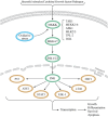
The JNK signaling pathway in intervertebral disc degeneration
- PMID: 39364138
- PMCID: PMC11447294
- DOI: 10.3389/fcell.2024.1423665
The JNK signaling pathway in intervertebral disc degeneration
Abstract
Intervertebral disc degeneration (IDD) serves as the underlying pathology for various spinal degenerative conditions and is a primary contributor to low back pain (LBP). Recent studies have revealed a strong correlation between IDD and biological processes such as Programmed Cell Death (PCD), cellular senescence, inflammation, cell proliferation, extracellular matrix (ECM) degradation, and oxidative stress (OS). Of particular interest is the emerging evidence highlighting the significant involvement of the JNK signaling pathway in these fundamental biological processes of IDD. This paper explores the potential mechanisms through the JNK signaling pathway influences IDD in diverse ways. The objective of this article is to offer a fresh perspective and methodology for in-depth investigation into the pathogenesis of IDD by thoroughly examining the interplay between the JNK signaling pathway and IDD. Moreover, this paper summarizes the drugs and natural compounds that alleviate the progression of IDD by regulating the JNK signaling pathway. This paper aims to identify potential therapeutic targets and strategies for IDD treatment, providing valuable insights for clinical application.
Keywords: JNK path; cell proliferation, inflammation; cellular senescence (CS); extracellar matrix (ECM) degradation; intervertebral disc degeneration (IDD); oxidative stress (OS); programmed cell death (PCD).
Copyright © 2024 Liu, Gao, Wang, Xie, Gao and Wu.
Conflict of interest statement
The authors declare that the research was conducted in the absence of any commercial or financial relationships that could be construed as a potential conflict of interest.
Figures


Similar articles
-
Unraveling the mechanisms of intervertebral disc degeneration: an exploration of the p38 MAPK signaling pathway.Front Cell Dev Biol. 2024 Jan 19;11:1324561. doi: 10.3389/fcell.2023.1324561. eCollection 2023. Front Cell Dev Biol. 2024. PMID: 38313000 Free PMC article. Review.
-
Pharmacological network analysis of the functions and mechanism of kaempferol from Du Zhong in intervertebral disc degeneration (IDD).J Orthop Translat. 2023 Mar 3;39:135-146. doi: 10.1016/j.jot.2023.01.002. eCollection 2023 Mar. J Orthop Translat. 2023. PMID: 36909862 Free PMC article.
-
Targeting nucleus pulposus cell death in the treatment of intervertebral disc degeneration.JOR Spine. 2024 Dec 18;7(4):e70011. doi: 10.1002/jsp2.70011. eCollection 2024 Dec. JOR Spine. 2024. PMID: 39703198 Free PMC article. Review.
-
MAPK /ERK signaling pathway: A potential target for the treatment of intervertebral disc degeneration.Biomed Pharmacother. 2021 Nov;143:112170. doi: 10.1016/j.biopha.2021.112170. Epub 2021 Sep 15. Biomed Pharmacother. 2021. PMID: 34536759 Review.
-
Targeted therapy for intervertebral disc degeneration: inhibiting apoptosis is a promising treatment strategy.Int J Med Sci. 2021 May 27;18(13):2799-2813. doi: 10.7150/ijms.59171. eCollection 2021. Int J Med Sci. 2021. PMID: 34220308 Free PMC article. Review.
Cited by
-
Targeting skeletal interoception: a novel mechanistic insight into intervertebral disc degeneration and pain management.J Orthop Surg Res. 2025 Feb 12;20(1):159. doi: 10.1186/s13018-025-05577-7. J Orthop Surg Res. 2025. PMID: 39940003 Free PMC article. Review.
References
-
- Adams M. A., Stefanakis M., Dolan P. (2010). Healing of a painful intervertebral disc should not be confused with reversing disc degeneration: implications for physical therapies for discogenic back pain. Clin. Biomech. (Bristol, Avon) 25 (10), 961–971. 10.1016/j.clinbiomech.2010.07.016 - DOI - PubMed
-
- Bai X., Yao M., Zhu X., Lian Y., Zhang M. (2023). Baicalin suppresses interleukin-1β-induced apoptosis, inflammatory response, oxidative stress, and extracellular matrix degradation in human nucleus pulposus cells. Immunopharmacol. Immunotoxicol. 45 (4), 433–442. 10.1080/08923973.2023.2165942 - DOI - PubMed
Publication types
LinkOut - more resources
Full Text Sources
Research Materials
Miscellaneous

