

Intralesional gene expression profile of JAK-STAT signaling pathway and associated cytokines in Leishmania tropica- infected patients
- PMID: 39364404
- PMCID: PMC11446769
- DOI: 10.3389/fimmu.2024.1436029
Intralesional gene expression profile of JAK-STAT signaling pathway and associated cytokines in Leishmania tropica- infected patients
Abstract
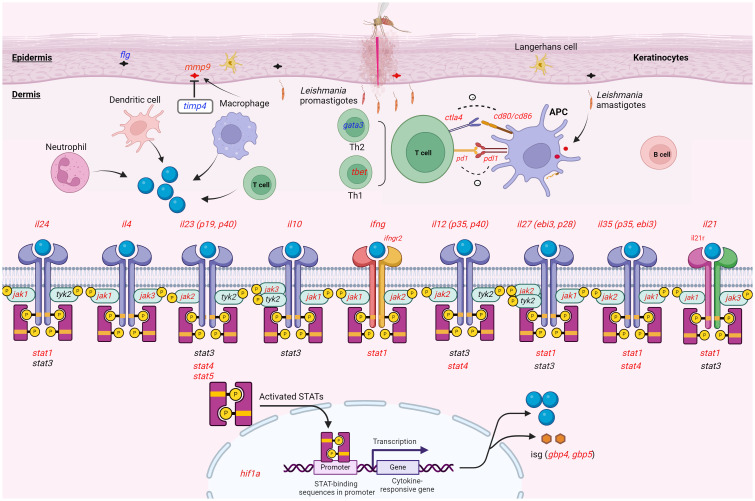
Background: The JAK-STAT signaling pathway is a central cascade of signal transduction for the myriad of cytokines in which dysregulation has been implicated in progression of inflammatory and infectious diseases. However, the involvement of this pathway in human cutaneous leishmaniasis (CL) due to Leishmania (L.) tropica warrants further investigation.
Methods: This study sought to investigate differential gene expression of several cytokines and their associated jak-stat genes in the lesions of L. tropica-infected patients byquantitative Real-Time PCR. Further, the expression of five inhibitory immune checkpoint genes was evaluated.
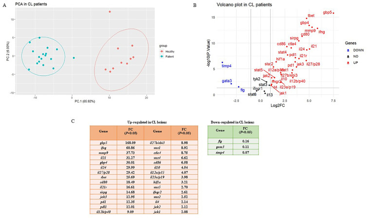
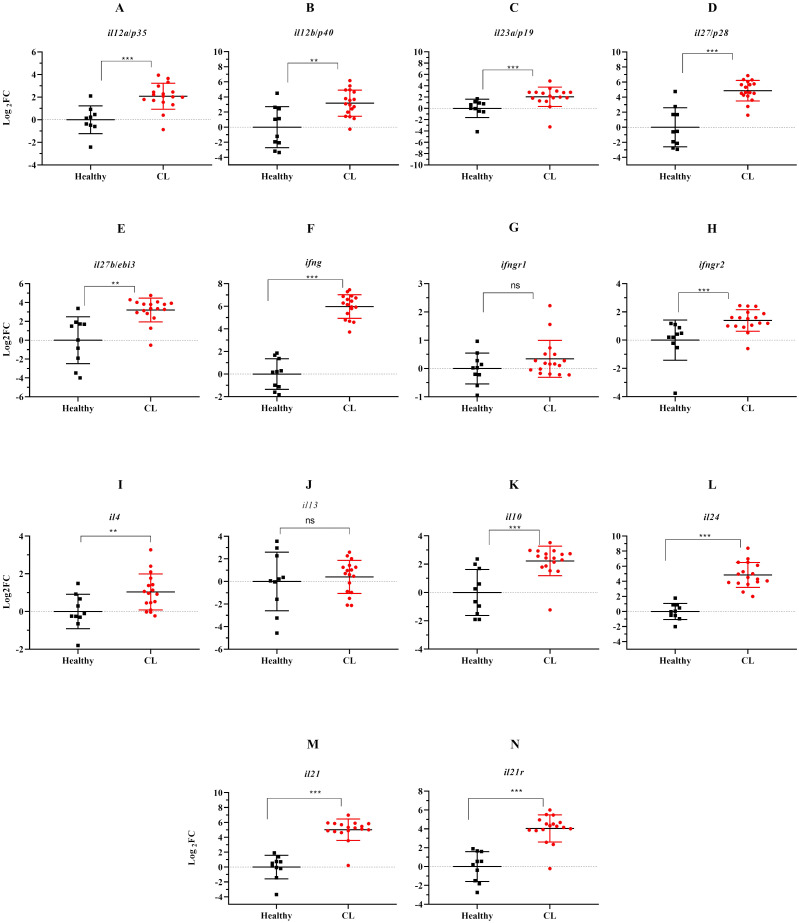
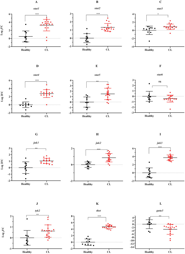
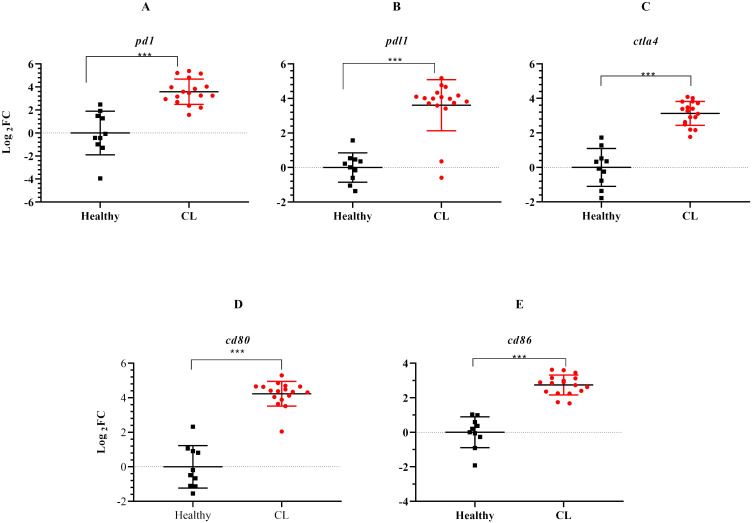
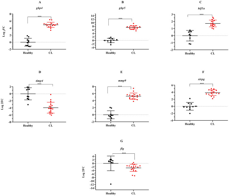
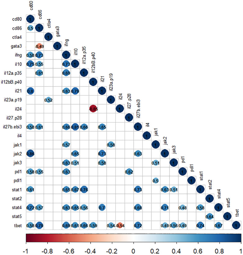
Results: Results showed that the gene expression levelsof both Th1 (ifng, il12, il23) and Th2 (il4, il10) types cytokines were increased in the lesion of studied patients. Further, elevated expression levels of il35, il21, il27 and il24 genes were detected in the lesions of CL patients. Notably, the expression of the majority of genes involved in JAK/STAT signaling pathway as well as checkpoint genes including pdl1, ctla4 and their corresponding receptors was increased.
Conclusion: Our finding revealed dysregulation of cytokines and related jak-stat genes in the lesion of CL patients. These results highlight the need for further exploration of the functional importance of these genes in the pathogenesis of, and immunity to, CL.
Keywords: JAK-STAT signaling pathway; Leishmania tropica; cutaneous leishmaniasis; cytokines; immune checkpoints.
Copyright © 2024 Hadifar, Masoudzadeh, Heydari, Mashayekhi Goyonlo, Kerachian, Daneshpazhooh, Sadeghnia, Tootoonchi, Erfanian Salim, Rafati and Harandi.
Conflict of interest statement
The authors declare that the research was conducted in the absence of any commercial or financial relationships that could be construed as a potential conflict of interest. The author(s) declared that they were an editorial board member of Frontiers, at the time of submission. This had no impact on the peer review process and the final decision.
Figures
Similar articles
-
Comparative gene expression pattern of immune-related genes using dual-color RT-MLPA in the lesions of cutaneous leishmaniasis caused by L. major and L. tropica.PLoS Negl Trop Dis. 2025 Mar 18;19(3):e0012812. doi: 10.1371/journal.pntd.0012812. eCollection 2025 Mar. PLoS Negl Trop Dis. 2025. PMID: 40100809 Free PMC article.
-
Integrated analysis of lncRNA and mRNA expression profiles in cutaneous leishmaniasis lesions caused by Leishmania tropica.Front Cell Infect Microbiol. 2024 Nov 21;14:1416925. doi: 10.3389/fcimb.2024.1416925. eCollection 2024. Front Cell Infect Microbiol. 2024. PMID: 39639867 Free PMC article.
-
The effects of CCR7 and related signaling pathways on Leishmania major -infected human dendritic cells.J Cell Physiol. 2019 Aug;234(8):13145-13156. doi: 10.1002/jcp.27985. Epub 2018 Dec 24. J Cell Physiol. 2019. PMID: 30584667
-
Potential Natural Biomolecules Targeting JAK/STAT/SOCS Signaling in the Management of Atopic Dermatitis.Molecules. 2022 Jul 21;27(14):4660. doi: 10.3390/molecules27144660. Molecules. 2022. PMID: 35889539 Free PMC article. Review.
-
The JAK/STAT signaling pathway: from bench to clinic.Signal Transduct Target Ther. 2021 Nov 26;6(1):402. doi: 10.1038/s41392-021-00791-1. Signal Transduct Target Ther. 2021. PMID: 34824210 Free PMC article. Review.
Cited by
-
Biomarker Insights: Evaluation of Presepsin, Apelin, and Irisin Levels in Cutaneous Leishmaniasis.Diagnostics (Basel). 2024 Dec 20;14(24):2869. doi: 10.3390/diagnostics14242869. Diagnostics (Basel). 2024. PMID: 39767230 Free PMC article.
-
Guanylate-Binding Proteins Promote Host Defense Against Leishmania major by Balancing iNOS/Arg-1 in Myeloid Cells.bioRxiv [Preprint]. 2025 Jun 30:2025.06.26.661809. doi: 10.1101/2025.06.26.661809. bioRxiv. 2025. PMID: 40631203 Free PMC article. Preprint.
References
-
- Rasti S, Delavari M, Arani TST, Mousavi SGA. Epidemiological and clinical study on the cutaneous leishmaniasis in Aran and Bidgol, center of Iran. Int Arch Health Sci. (2018) 5:72–5. doi: 10.4103/iahs.iahs_26_18 - DOI
-
- Bamorovat M, Sharifi I, Agha Kuchak Afshari S, Ghasemi Nejad Almani P. Mutual role of patients and the healthcare system in the control of cutaneous leishmaniasis. Transboundary Emerging Dis. (2023) 2023:7814940. doi: 10.1155/2023/7814940 - DOI
MeSH terms
Substances
LinkOut - more resources
Full Text Sources
Research Materials