

QSM-detected iron accumulation in the cerebellar gray matter is selectively associated with executive dysfunction in non-demented ALS patients
- PMID: 39364420
- PMCID: PMC11448125
- DOI: 10.3389/fneur.2024.1426841
QSM-detected iron accumulation in the cerebellar gray matter is selectively associated with executive dysfunction in non-demented ALS patients
Abstract
Background: This study aimed to assess whether quantitative susceptibility imaging (QSM)-based measures of iron accumulation in the cerebellum predict cognitive and behavioral features in non-demented amyotrophic lateral sclerosis (ALS) patients.
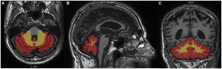
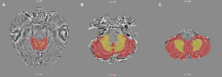
Methods: A total of ALS patients underwent 3-T MRI and a clinical assessment using the ALS Functional Rating Scale-Revised (ALSFRS-R) and the Edinburgh Cognitive and Behavioural ALS Screen (ECAS). Regression models were applied to each subscale of the cognitive section of the ECAS and the ECAS-Carer Interview to examine the effect of QSM-based measures in white and gray matter (WM; GM) of the cerebellum, separately for right, left, and bilateral cerebellar regions of interest (ROIs). These effects were compared to those of cerebellar volumetrics in WM/GM, right and left hemispheres while controlling for demographics, disease status, and total intracranial volume.
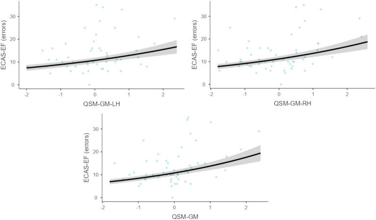
Results: Higher QSM measures of the cerebellar GM on the left, right, and bilateral sides significantly predicted (ps ≤ 0.003) a greater number of errors on the executive functioning (EF) subscale of the ECAS (ECAS-EF). Moreover, higher GM-related, QSM measures of the cerebellum were associated with an increased probability of a below-cut-off performance on the ECAS-EF (ps ≤ 0.024). No significant effects were observed for QSM measures of the cerebellar WM or for volumetric measures on the ECAS-EF. Other ECAS measures showed no significant effects. Bilateral QSM measures of the cerebellar GM also selectively predicted performance on backward digit span and social cognition tasks.
Discussion: Iron accumulation within the cerebellar GM, particularly in the cerebellar cortices, may be associated with executive functioning deficits in non-demented ALS patients. Therefore, QSM-based measures could be useful for identifying the neural correlates of extra-motor cognitive deficits in ALS patients.
Keywords: amyotrophic lateral sclerosis; cerebellum; executive functioning; frontotemporal degeneration; quantitative susceptibility imaging.
Copyright © 2024 Aiello, Contarino, Conte, Solca, Curti, Maranzano, Torre, Casale, Doretti, Colombo, Verde, Silani, Liu, Cinnante, Triulzi, Morelli, Poletti and Ticozzi.
Conflict of interest statement
VS received compensation for consulting services and/or speaking activities from AveXis, Cytokinetics, Italfarmaco, Liquidweb S.r.l., and Novartis Pharma AG, receives or has received research supports from the Italian Ministry of Health, AriSLA, and E-Rare Joint Transnational Call. He is in the Editorial Board of Amyotrophic Lateral Sclerosis and Frontotemporal Degeneration, European Neurology, American Journal of Neurodegenerative Diseases, Frontiers in Neurology. BP received compensation for consulting services and/or speaking activities from Liquidweb S.r.l. She is Associate Editor for Frontier in Neuroscience. NT received compensation for consulting services from Amylyx Pharmaceuticals and Zambon Biotech SA. He is Associate Editor for Frontiers in Aging Neuroscience. ENA serves as an Editorial Board Member for BMC Neurology. FV is Associated Editor for Journal of Alzheimer’s Disease. The remaining authors declare that the research was conducted in the absence of any commercial or financial relationships that could be construed as a potential conflict of interest. The author(s) declared that they were an editorial board member of Frontiers, at the time of submission. This had no impact on the peer review process and the final decision.
Figures
References
LinkOut - more resources
Full Text Sources
Miscellaneous