

Target-based deep learning network surveillance of non-contrast computed tomography for small infarct core of acute ischemic stroke
- PMID: 39364421
- PMCID: PMC11447964
- DOI: 10.3389/fneur.2024.1477811
Target-based deep learning network surveillance of non-contrast computed tomography for small infarct core of acute ischemic stroke
Abstract
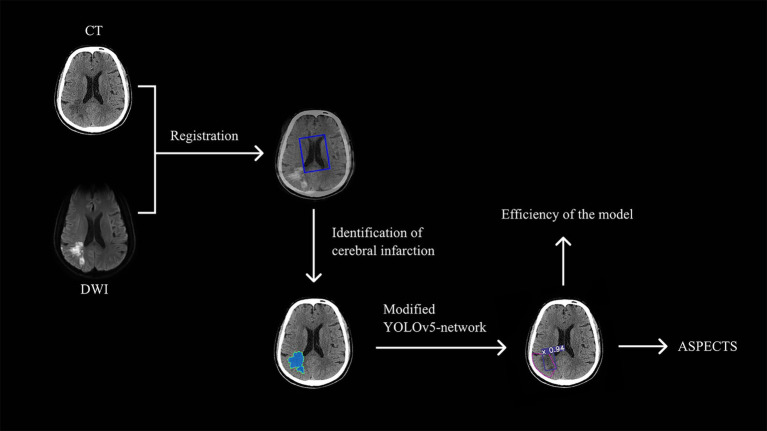
Purpose: Rapid diagnosis of acute ischemic stroke (AIS) is critical to achieve positive outcomes and prognosis. This study aimed to construct a model to automatically identify the infarct core based on non-contrast-enhanced CT images, especially for small infarcts.
Methods: The baseline CT scans of AIS patients, who had DWI scans obtained within less than 2 h apart, were included in this retrospective study. A modified Target-based deep learning model of YOLOv5 was developed to detect infarctions on CT. Randomly selected CT images were used for testing and evaluated by neuroradiologists and the model, using the DWI as a reference standard. Intraclass correlation coefficient (ICC) and weighted kappa were calculated to assess the agreement. The paired chi-square test was used to compare the diagnostic efficacy of physician groups and automated models in subregions. p < 0.05 was considered statistically significant.
Results: Five hundred and eighty four AIS patients were enrolled in total, finally 275 cases were eligible. Modified YOLOv5 perform better with increased precision (0.82), recall (0.81) and mean average precision (0.79) than original YOLOv5. Model showed higher consistency to the DWI-ASPECTS scores (ICC = 0.669, κ = 0.447) than neuroradiologists (ICC = 0.452, κ = 0.247). The sensitivity (75.86% vs. 63.79%), specificity (98.87% vs. 95.02%), and accuracy (96.20% vs. 91.40%) were better than neuroradiologists. Automatic model had better diagnostic efficacy than physician diagnosis in the M6 region (p = 0.039).
Conclusion: The deep learning model was able to detect small infarct core on CT images more accurately. It provided the infarct portion and extent, which is valuable in assessing the severity of disease and guiding treatment procedures.
Keywords: acute ischemic stroke; non-contrast CT; small infarct core; target-based deep learning network; you only look once (YOLO).
Copyright © 2024 Qu, Tang, Gao, Li, Zhao, Ban, Chen, Lu and Wang.
Conflict of interest statement
The authors declare that the research was conducted in the absence of any commercial or financial relationships that could be construed as a potential conflict of interest.
Figures


Similar articles
-
Detecting the Early Infarct Core on Non-Contrast CT Images with a Deep Learning Residual Network.J Stroke Cerebrovasc Dis. 2021 Jun;30(6):105752. doi: 10.1016/j.jstrokecerebrovasdis.2021.105752. Epub 2021 Mar 27. J Stroke Cerebrovasc Dis. 2021. PMID: 33784518
-
Comparison of automated and manual DWI-ASPECTS in acute ischemic stroke: total and region-specific assessment.Eur Radiol. 2021 Jun;31(6):4130-4137. doi: 10.1007/s00330-020-07493-2. Epub 2020 Nov 27. Eur Radiol. 2021. PMID: 33247346
-
Quantification of infarct core signal using CT imaging in acute ischemic stroke.Neuroimage Clin. 2022;34:102998. doi: 10.1016/j.nicl.2022.102998. Epub 2022 Mar 30. Neuroimage Clin. 2022. PMID: 35378498 Free PMC article.
-
ISP-Net: Fusing features to predict ischemic stroke infarct core on CT perfusion maps.Comput Methods Programs Biomed. 2022 Mar;215:106630. doi: 10.1016/j.cmpb.2022.106630. Epub 2022 Jan 12. Comput Methods Programs Biomed. 2022. PMID: 35063712
-
Automated CT Perfusion Detection of the Acute Infarct Core in Ischemic Stroke: A Systematic Review and Meta-Analysis.Cerebrovasc Dis. 2023;52(1):97-109. doi: 10.1159/000524916. Epub 2022 Jun 3. Cerebrovasc Dis. 2023. PMID: 35661075
References
-
- Barber PA, Demchuk AM, Zhang J, Buchan AM. Validity and reliability of a quantitative computed tomography score in predicting outcome of hyperacute stroke before thrombolytic therapy. ASPECTS study group. Alberta stroke Programme early CT score. Lancet (London, England). (2000) 355:1670–4. doi: 10.1016/s0140-6736(00)02237-6, PMID: - DOI - PubMed
LinkOut - more resources
Full Text Sources