. 2024 Oct;12(10):e70008.
doi: 10.1002/iid3.70008.

Exercise preconditioning mitigates brain injury after cerebral ischemia-reperfusion injury in rats by restraining TIMP1

Affiliations

Exercise preconditioning mitigates brain injury after cerebral ischemia-reperfusion injury in rats by restraining TIMP1

Xiangbo Meng et al. Immun Inflamm Dis. 2024 Oct.

Abstract

Background: Cerebral ischemic disease is a common cerebrovascular disease, especially ischemic stroke. Exercise has protective functions on brain tissues following cerebral ischemia-reperfusion injury (CIRI), but its preventive effects and mechanisms in CIRI remain unclear. We aimed to investigate the effects and mechanisms of exercise preconditioning on CIRI.

Methods: The middle cerebral artery occlusion (MCAO) operation was prepared to establish CIRI rats. All rats were randomized into the MCAO, exercise (exercise preconditioning plus MCAO operation), vector (exercise preconditioning, MCAO operation plus intraventricular injection of empty vector), and tissue inhibitor of metalloprotease 1 overexpression (OE-TIMP1, exercise preconditioning, MCAO operation plus intraventricular injection of OE-TIMP1) groups.

Results: The results indicated that exercise preconditioning suppressed approximately 66.67% of neurological deficit scores and 73.79% of TIMP1 mRNA expression in MCAO rats, which were partially offset by OE-TIMP1. The protective effects of exercise against neuron death status and cerebral infarction size in MCAO rats were reversed by OE-TIMP1. It also confirmed that exercise weakened apoptosis and oxidative stress damage, with notable increases of B-cell lymphoma-2, superoxide dismutase, and glutathione peroxidase production, and evident decreases of BCL2-associated X, caspase 3, and malondialdehyde in MCAO rats, while these effects were partially reversed by OE-TIMP1. Additionally, the inhibitory effects of exercise on the protein levels of TIMP1, hypoxia-inducible factor-alpha, vascular endothelial growth factor receptor 2, vascular endothelial growth factor, and neurogenic locus notch homolog protein 1 in MCAO rats were partially reversed by OE-TIMP1.

Conclusion: Altogether, exercise preconditioning had protective effects on CIRI by restraining TIMP1, which provided new therapeutic strategies for preventing CIRI.

Keywords: TIMP1; brain injury; cerebral ischemia‐reperfusion injury; exercise preconditioning.


Conflict of interest statement

The authors declare no conflicts of interest.

Figures

Figure 1
Figure 1
Exercise alleviated neurological deficits in MCAO rats by restraining TIMP1. (A) Relative expression of TIMP1 mRNA in MCAO rats pretreated with or without exercise according to RNA sequencing, n = 5, ## p < .01 versus Sham. (B) The effects of exercise and TIMP1 overexpression on the mRNA level of TIMP1 of the cerebral cortex tissues in MCAO rats were examined by qRT‐PCR. ++ p < .01 versus MCAO; **p < .01 versus Vector. Data were analyzed using One‐way ANOVA with Turkey tests. n = 3. Results were presented as mean ± SD in all figures. Note: MCAO, middle cerebral artery occlusion; TIMP1, tissue inhibitor of matrix metalloprotease 1; qRT‐PCR, real‐time quantitative PCR; ANOVA, analysis of variance; SD, standard deviation.
Figure 2
Figure 2
The effects of exercise and TIMP1 overexpression on the neurological deficit scores in MCAO rats. The neurological deficit score of each rat was assessed 48 h after MCAO. ++ p < .01 versus MCAO; **p < .01 versusVector. Data were analyzed using One‐way ANOVA with Turkey tests. n = 6. Results were presented as mean ± SD.
Figure 3
Figure 3
Exercise reduced cerebral cortex neuronal death in MCAO rats by inhibiting TIMP1. After performing MCAO surgery, the extent of neuronal death in the cerebral cortex of each rat was detected through Nissl staining. (A) Representative images of Nissl staining in the cerebral cortex. Magnification: 200×, scale bar: 100 μm, and magnification: 400×, scale bar: 50 μm. (B) Histograms of the rate of dead neurons obtained via Nissl staining. ++ p < .01 versus MCAO; **p < .01 versus Vector. Data were analyzed using One‐way ANOVA with Turkey tests. n = 3.
Figure 4
Figure 4
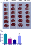
Exercise decreased cerebral infarct size in MCAO rats via weakening TIMP1. Following MCAO surgery, the extent of cerebral infarction from each group was evaluated using TTC staining. (A) Representative images of TTC staining. (B) Histograms of the infarction size obtained via TTC staining. ++ p < .01 versus MCAO; **p < .01 versusVector. Data were analyzed using One‐way ANOVA with Turkey tests. n = 3. TTC, Triphenyl tetrazolium chloride.
Figure 5
Figure 5
Exercise diminished the apoptosis of the cerebral cortex in MCAO rats by suppressing TIMP1. Upon conducting MCAO surgery, TUNEL assay was used to test apoptosis in rat cerebral cortex tissues. (A) Representative images of TUNEL staining in the cerebral cortex. Magnification: 200×, scale bar: 50 μm. (B) Histograms of the TUNEL positive cell rate obtained via TUNEL assay. ++ p < .01 versus MCAO; **p < .01 versus Vector. Data were analyzed using One‐way ANOVA with Turkey tests. n = 3. TUNEL, TdT‐mediated dUTP nick end labeling.
Figure 6
Figure 6
Exercise elevated Bcl‐2 expression but blocked Bax and caspase3 expressions in MCAO rats via impeding TIMP1. Following MCAO, the protein expressions of Bcl‐2, Bax, and caspase 3 in rat cerebral cortex were determined by Western blot assay. (A) Representative images for Western blot assay of Bcl‐2, Bax and caspase 3 levels in the cerebral cortex. Quantifications of Bcl‐2 (B), Bax (C) and caspase3 (D) levels in the cerebral cortex. ++ p < .01 versus MCAO; *p < .05, **p < .01 versus Vector. Data were analyzed using One‐way ANOVA with Turkey tests. n = 3. Bcl‐2, B‐cell lymphoma‐2; Bax, BCL2‐Associated X.
Figure 7
Figure 7
Exercise upregulated GSH‐PX and SOD contents but downregulated MDA content in MCAO rats by inhibiting TIMP1. After MCAO, the contents of GSH‐Px (A), SOD (B) and MDA (C) in the cerebral cortex tissues of MCAO rats were measured using chemical colorimetry. ++ p < .01 versus MCAO; *p < .05, **p < .01 versus Vector. Data were analyzed using One‐way ANOVA with Turkey tests. n = 6. GSH‐Px, glutathione peroxidase; SOD, superoxide dismutase; MDA, malondialdehyde.
Figure 8
Figure 8
Exercise inactivated HIF‐1α/VEGF pathway in MCAO rats by inhibiting TIMP1. After MCAO, Western blot assay was employed to measure TIMP1, HIF‐α, VEGF, VEGFR2, and Notch1 protein expressions in rat cerebral cortex. (A) Representative images for Western blot assay of TIMP1, HIF‐α, VEGF, VEGFR2, and Notch1 levels in the cerebral cortex. Quantifications of TIMP1 (B), HIF‐α (C), VEGF (D), VEGFR2 (E), and Notch1 (F) levels in the cerebral cortex. + p < .05, ++ p < .01 versus MCAO; **p < .01 versus Data were analyzed using One‐way ANOVA with Turkey tests. n = 3. Vector. Note: HIF‐α, hypoxia‐inducible factor‐alpha; VEGF, vascular endothelial growth factor; VEGFR2, vascular endothelial growth factor receptor 2, Notch1, neurogenic locus notch homolog protein 1.

Similar articles

References

    1. Sudheer P, Misra S, Nath M, et al. Micro‐embolic signal monitoring in stroke subtypes: a systematic review and meta‐analysis of 58 studies. Eur Stroke J. 2021;6(4):403–411. - PMC - PubMed
    1. Sun Y, Chen H, Lin Y. Rehabilitation training inhibits neuronal apoptosis by down‐regulation of TLR4/MyD88 signaling pathway in mice with cerebral ischemic stroke. Am J Transl Res. 2021;13(4):2213–2223. - PMC - PubMed
    1. Calabrese B, Jones SL, Shiraishi‐Yamaguchi Y, et al. INF2‐mediated actin filament reorganization confers intrinsic resilience to neuronal ischemic injury. Nat Commun. 2022;13(1):6037. - PMC - PubMed
    1. Lohmann S, Grigoletto J, Bernis ME, et al. Ischemic stroke causes parkinson's disease‐like pathology and symptoms in transgenic mice overexpressing alpha‐synuclein. Acta Neuropathol Commun. 2022;10(1):26. - PMC - PubMed
    1. Sun T, Chen S, Wu K, Sun M, Zhang X, You C. Trends in incidence and mortality of stroke in China from 1990 to 2019. Front Neurol. 2021;12:759221. - PMC - PubMed

Publication types

MeSH terms

Substances