Review
. 2024 Oct 30;44(10):BSR20240139.
doi: 10.1042/BSR20240139.

The role of structure in regulatory RNA elements

Affiliations
Review

The role of structure in regulatory RNA elements

Jan-Niklas Tants et al. Biosci Rep. .

Abstract

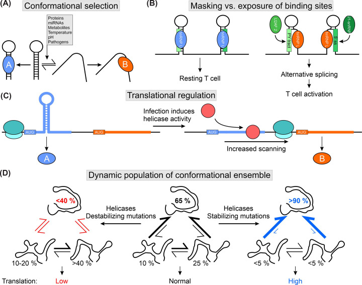
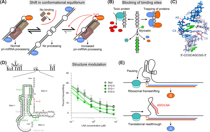
Regulatory RNA elements fulfill functions such as translational regulation, control of transcript levels, and regulation of viral genome replication. Trans-acting factors (i.e., RNA-binding proteins) bind the so-called cis elements and confer functionality to the complex. The specificity during protein-RNA complex (RNP) formation often exploits the structural plasticity of RNA. Functional integrity of cis-trans pairs depends on the availability of properly folded RNA elements, and RNA conformational transitions can cause diseases. Knowledge of RNA structure and the conformational space is needed for understanding complex formation and deducing functional effects. However, structure determination of RNAs under in vivo conditions remains challenging. This review provides an overview of structured eukaryotic and viral RNA cis elements and discusses the effect of RNA structural equilibria on RNP formation. We showcase implications of RNA structural changes for diseases, outline strategies for RNA structure-based drug targeting, and summarize the methodological toolbox for deciphering RNA structures.

Keywords: RNA conformers; RNA structure; RNA-binding proteins; cis-regulatory elements; dynamics; stem-loop.


Conflict of interest statement

The authors declare that there are no competing interests associated with the manuscript.

Figures

Figure 1
Figure 1. Types and function of RNA cis-regulatory elements
(A) Functions of RNA cis elements. (B) Linear and structured RNA elements in a cartoon-model RNA. Sequence-specific recognition of RNA target sites can be mediated by proteins, e.g., to poly-pyrimidine (PY) tracts or AU-rich elements (AREs, green and purple), and miRNAs (red). Specialized protein domains can recognize shapes, e.g., stem-loops (blue) or double-stranded regions (orange), through adapted binding pockets (dark blue and orange). The modular architecture of proteins allows integration of multiple specificities, as exemplified by a protein composed of the purple and blue domains, thereby combining sequence and structure specificity. (C) Examples of cis element structures. Stem-loops (SL) can vary in their stem length and loop size and contain mismatches or bulges that lead to distinct geometries. Complex structures such as branched SLs can be constructed from SLs and bulges. Interaction of two hairpins via their loop regions leads to formation of kissing-loops, often exploited in RNA dimerization. Higher-order structures comprise three-way junctions (3WJ), G-quadruplexes, and pseudoknots (PK). (D) Cis-regulatory cassette composed of multiple cis elements exhibiting additive/cooperative or antagonistic effects for a concerted output. Two hairpin structures can be bound by one or two proteins (blue), leading to a normal or strong output signal, respectively. Binding of a second, antagonistic protein (orange) to a different SL structure counteracts the positive regulatory effect of the other two SLs, leading to a weak output. (E) Complex structure of a regulatory hub for exposure of trans factor binding site. In a properly folded RNA, the green protein-binding site forms a SL that is bound by the blue protein in a shape-specific manner. Point mutations (shown as red spheres) or conformational equilibria can favor the formation of a second conformation, which traps the protein-binding site in a stable stem structure. Through prevention of hairpin formation, the protein is no longer able to engage with the target RNA.
Figure 2
Figure 2. The effect of transient RNA structures on cis-trans pair formation
(A) Schematic of conformational equilibria of an RNA stem-loop (adapted from [25]). Structural changes can be induced by trans factors, environmental stimuli, or by infections. Conformation-specific exposure of trans-factor binding sites leads to distinct formation of cis-trans complexes. Often, this equilibrium is influenced by the abundance of trans factors, e.g., proteins A and B. (B) The competition of hnRNP U and hnRNP L regulates alternative splicing in MALT1 pre-mRNA (adapted from [25]). hnRNP U binds and stabilizes two stem-loop structures, whereas hnRNP L partially unwinds the RNA, exposing two binding sites-the poly-pyrimidine tract (PY tract) and the 5′ splice site (5′ ss)-which are recognized by U2AF2 and U1 snRNP. Binding of both proteins causes exon inclusion and alternate protein production, leading to T cell activation. (C) RNA structure guides start codon selection and modulates translation in Arabidopsis thaliana (adapted from [101]). Hairpin structures slow ribosomal scanning and lead to the translation of protein A (blue ORF). Upon infection, helicases (red) resolve RNA structures, leading to increased scanning through the ribosome and translation of the downstream ORF (orange), i.e., protein B. Downstream ORFs encode immune-relevant proteins. (D) Translation regulation of Csde1 through a dynamic equilibrium of 5′UTR RNA structures (adapted from [6]). In vivo the major conformation (65%) controls translation efficiency. Helicases and point mutations can modulate the conformational ensemble through stabilizing or destabilizing single conformations, thereby shifting the equilibrium and tuning translation efficiency (blue and red equilibria).
Figure 3
Figure 3. RNA structure as a driver of diseases and drug strategies
(A) pri-miRNA-30c binds SRSF3 (blue), hnRNP A1 (orange), and Drosha and DGCR8 for processing (adapted from [22]). The apical loop mediates dimerization, which obscures the protein-binding sites and prevents RNA processing. A cancer-associated G-to-A point mutation in the loop shifts the equilibrium to the monomeric RNA form (red arrow). Furthermore, structural rearrangements in the lower stem facilitate SRSF3 binding (red arrow). Enhanced protein binding leads to an increase in pri-miRNA processing. (B) CAG repeat expansions form repetitive stem-bulge structures, sequester and trap cellular proteins (blue and orange), and cause translation of prionic (toxic) polypeptides (red). The flavonoid myricetin (green) masks stem-bulge structures from protein binding, thereby preventing its toxic effects (adapted from [136]). (C) Structure of a CAG-repeat RNA element in complex with myricetin (adapted from [136]; PDB: 5XI1). Myricetin (green) intercalates in the RNA duplex and pushes A5 from strand 1 (blue) out of the helix. The flavonoid interacts through π-π stacking with the C4-G6 base pair, thereby connecting both RNA strands (blue and light blue). (D) RNA secondary structure scheme of the SARS-CoV-2 genome and frameshifting element (FSE) highlighted in zoom-in. The experimentally determined secondary structure is shown together with the ribosome slippery site and interaction sites of tested ASOs (adapted from [141]). Frameshifting efficiency plotted against LNA concentration from in vitro frameshifting assay (taken from [141] under license 5794170311982). The colors of tested LNAs correspond to binding sites indicated in the secondary structure on the left. A significant reduction in frameshifting is observed at nM-concentrations. (E) Model for mode of action of ASO targeting the FSE (adapted from [141]). The FSE structure causes pausing of the translating ribosome, allowing a frameshift within the slippery site (production of protein B, orange). Binding of ASOs alters the FSE structure and causes a translational read-through to produce protein A (blue).

Similar articles

Cited by

References

    1. Beaudoin J.D., Novoa E.M., Vejnar C.E., Yartseva V., Takacs C.M., Kellis M.et al. . (2018) Analyses of mRNA structure dynamics identify embryonic gene regulatory programs. Nat. Struct. Mol. Biol. 25, 677–686 10.1038/s41594-018-0091-z - DOI - PMC - PubMed
    1. Rouskin S., Zubradt M., Washietl S., Kellis M. and Weissman J.S. (2014) Genome-wide probing of RNA structure reveals active unfolding of mRNA structures in vivo. Nature 505, 701–705 10.1038/nature12894 - DOI - PMC - PubMed
    1. Al'Khafaji A.M., Smith J.T., Garimella K.V., Babadi M., Popic V., Sade-Feldman M.et al. . (2024) High-throughput RNA isoform sequencing using programmed cDNA concatenation. Nat. Biotechnol. 42, 582–586 10.1038/s41587-023-01815-7 - DOI - PubMed
    1. Aw J.G.A., Lim S.W., Wang J.X., Lambert F.R.P., Tan W.T., Shen Y.et al. . (2021) Determination of isoform-specific RNA structure with nanopore long reads. Nat. Biotechnol. 39, 336–346 10.1038/s41587-020-0712-z - DOI - PubMed
    1. Stephenson W., Razaghi R., Busan S., Weeks K.M., Timp W. and Smibert P. (2022) Direct detection of RNA modifications and structure using single-molecule nanopore sequencing. Cell Genom. 2, - PMC - PubMed

Publication types

LinkOut - more resources