. 2024 Oct 29;9(10):e0068624.
doi: 10.1128/msphere.00686-24. Epub 2024 Oct 4.

Pseudomonas aeruginosa kills Staphylococcus aureus in a polyphosphate-dependent manner

Affiliations

Pseudomonas aeruginosa kills Staphylococcus aureus in a polyphosphate-dependent manner

Ritika Shah et al. mSphere. .

Abstract

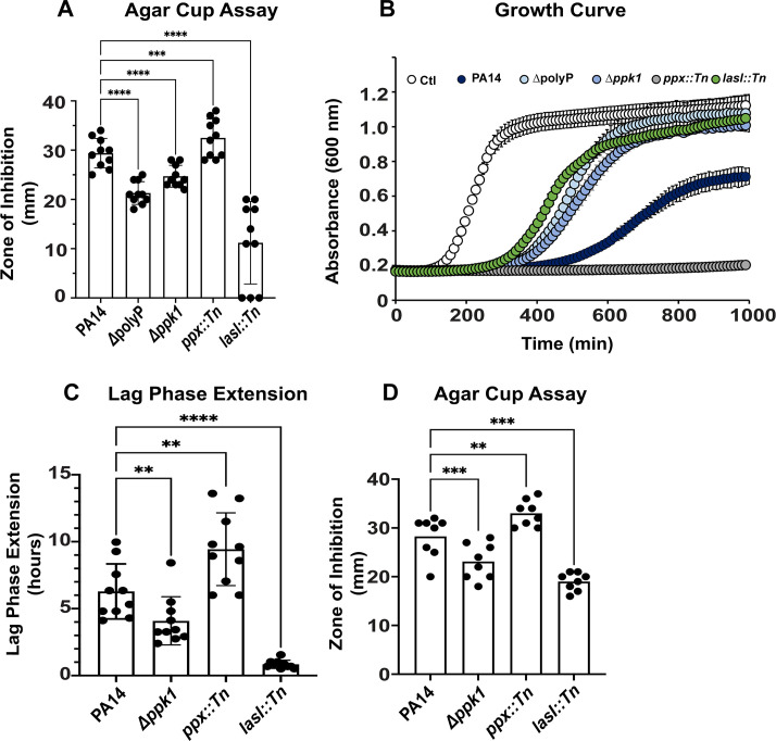
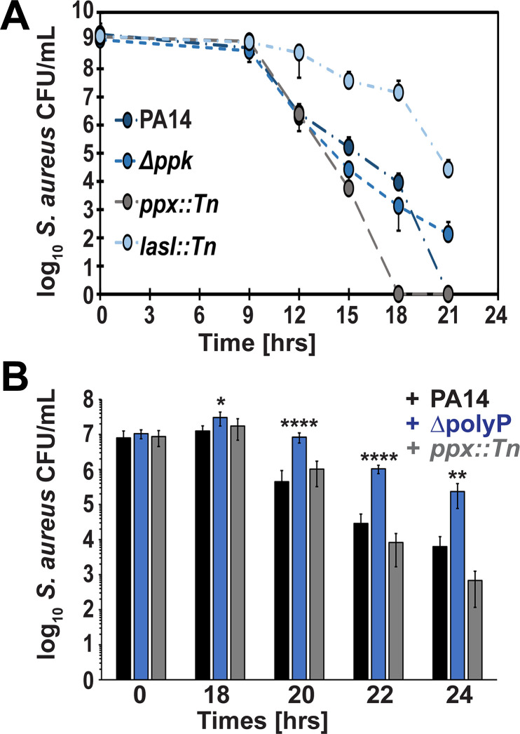
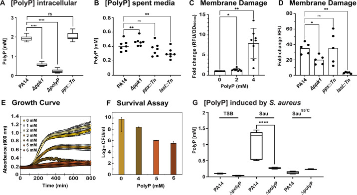
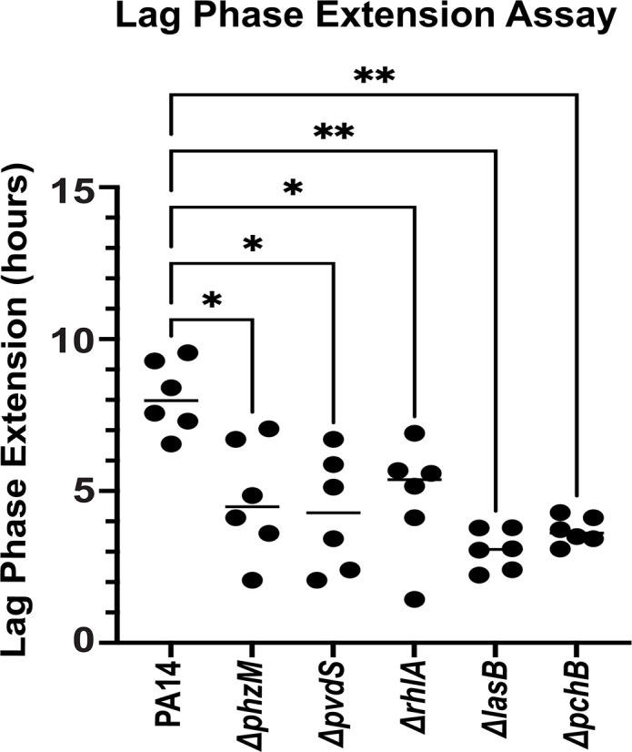
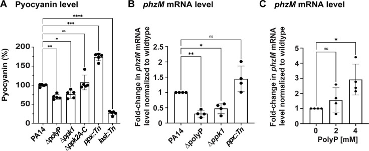
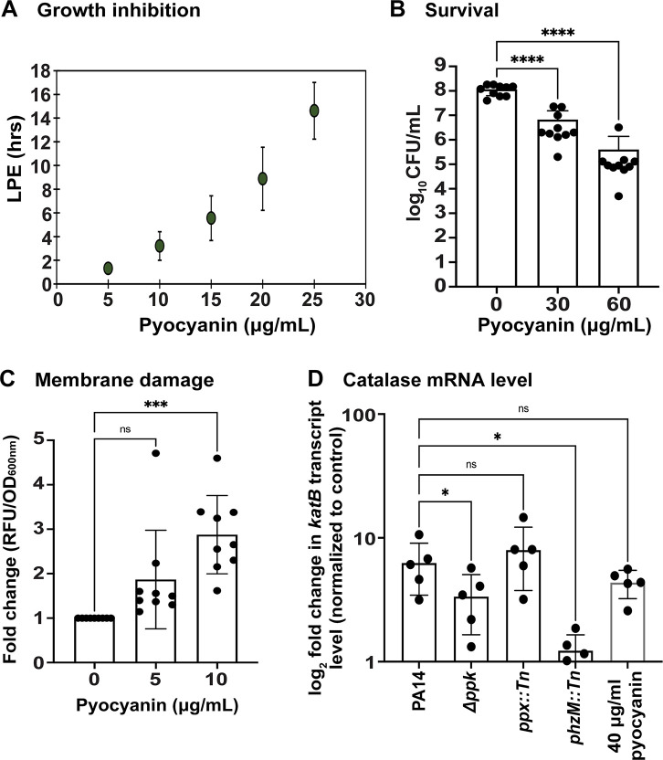
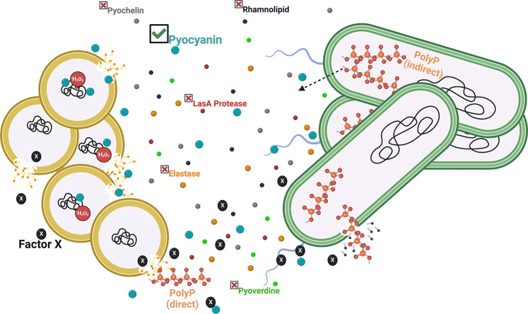
Due to their frequent coexistence in many polymicrobial infections, including in patients with cystic fibrosis or burn/chronic wounds, many studies have investigated the mechanistic details of the interaction between the opportunistic pathogens Pseudomonas aeruginosa and Staphylococcus aureus. P. aeruginosa rapidly outcompetes S. aureus under in vitro cocultivation conditions, which is mediated by several of P. aeruginosa's virulence factors. Here, we report that polyphosphate (polyP), an efficient stress defense system and virulence factor in P. aeruginosa, plays a role in the pathogen's ability to inhibit and kill S. aureus in a contact-independent manner. We show that P. aeruginosa cells characterized by low polyP levels are less detrimental to S. aureus growth and survival while the Gram-positive pathogen is significantly more compromised by the presence of P. aeruginosa cells that produce high levels of polyP. The polyP-dependent phenotype of P. aeruginosa-mediated killing of S. aureus could at least in part be direct, as polyP was detected in the spent media and causes significant damage to the S. aureus cell envelope. However, more likely is that polyP's effects are indirect through modulating the production of one of P. aeruginosa's virulence factors, pyocyanin. We show that pyocyanin production in P. aeruginosa occurs polyP-dependently and harms S. aureus through membrane damage and potentially the generation of reactive oxygen species, resulting in the increased expression of antioxidant enzymes. In summary, our study adds a new component to the list of biomolecules that the Gram-negative pathogen P. aeruginosa generates to compete with S. aureus for resources.IMPORTANCEHow do interactions between microorganisms shape the course of polymicrobial infections? Previous studies have provided evidence that the two opportunistic pathogens Pseudomonas aeruginosa and Staphylococcus aureus generate molecules that modulate their interaction with potentially significant impact on disease outcomes. Our study identified the biopolymer polyphosphate (polyP) as a new effector molecule that impacts P. aeruginosa's interaction with S. aureus. We show that P. aeruginosa kills S. aureus in a polyP-dependent manner, which occurs primarily through the polyP-dependent production of the P. aeruginosa virulence factor pyocyanin. Our findings add a new role for polyP to an already extensive list of functions. A more in-depth understanding of how polyP influences interspecies interactions is critical, as targeting polyP synthesis in bacteria such as P. aeruginosa may have a significant impact on other microorganisms and potentially result in dynamic changes in the microbial composition.

Keywords: Pseudomonas aeruginosa; Staphylococcus aureus; oxidative stress; phenazine; polymicrobial interactions; polyphosphate; virulence factors.


Conflict of interest statement

The authors declare no conflict of interest.

Figures

Fig 1
Fig 1
S. aureus is inhibited by P. aeruginosa spent media in a polyP-dependent manner. (A) S. aureus strain USA300LAC was diluted into Luria Bertani (LB) media to an optical density of 600 nm (OD600) of 0.05 and spread onto LB agar. Wells were cut into the LB agar and filled with 150 µL spent media sterile-filtered from the indicated 24 hrs, stationary phase P. aeruginosa cultures. The zones of inhibition were measured after incubation at 37°C for 16–18 hrs (n = 10, ±S.D.). (B and C) USA300LAC was diluted into tryptic soy broth (TSB) to an OD600 of 0.03 and exposed to the indicated P. aeruginosa spent media in a 15:1 (vol/vol) ratio. A600nm was measured every 10 minutes for 16 hrs using the Tecan Infinite 200 plate reader. White circles, control; dark blue circles, PA14; blue circles, ∆ppk1; bright blue circles, ∆polyP; gray circles, ppx::Tn; green circles, lasI::Tn. Lag phase extensions (LPEs) were calculated as described in Materials and Methods (n = 10, ±S.D.). (D) S. aureus clinical isolate 93 was diluted into LB media to an OD600 of 0.05 and spread onto LB agar. Wells were cut into the LB agar and filled with 150 µL spent media of the indicated 24 hrs, stationary phase P. aeruginosa cultures. The zones of inhibition were measured after incubation at 37°C for 16–18 hrs (n = 8, ±S.D.). Statistical tests: one-way ANOVA, **P ≤ 0.01, ***P ≤ 0.001, and ****P ≤ 0.0001.
Fig 2
Fig 2
P. aeruginosa kills S. aureus in a polyP-dependent manner. (A) Overnight cultures of S. aureus strain USA300LAC and the indicated P. aeruginosa strains were each diluted into LB media to an OD600 of 1, mixed in a ratio of 100:1, and cultivated at 37°C for 21 hrs. Samples were taken at indicated time points and serially diluted for S. aureus CFU counts on mannitol salt agar. Dark blue circles, PA14; blue circles, ∆ppk; bright blue circles, ∆lasI; gray circles, ∆ppx (n = 3, ±S.D.). (B) Overnight cultures of S. aureus strain USA300LAC and the indicated P. aeruginosa strains were each diluted into artificial urine media to an OD600 of 1, mixed in a ratio of 10,000:1, and cultivated at 37°C for 24 hrs. Samples were taken at indicated time points, serially diluted, and plated onto mannitol salt agar for S. aureus CFU counts. Black bars, PA14; blue bars, ∆polyP; gray bars, ppx::Tn (n = 4, ±S.D.). Statistical tests: one-way ANOVA, *P ≤ 0.05, **P ≤ 0.01, and ****P ≤ 0.0001.
Fig 3
Fig 3
PolyP contributes to S. aureus killing by causing membrane damage. (A) Intracellular polyP was extracted from 24 hrs, stationary phase P. aeruginosa cultures and quantified with 25 µM DAPI. All samples were also treated with 1 mg Ppx and deducted from the relative fluorescence unit (RFU) as background. DAPI-polyP fluorescence was measured at excitation/emission wavelengths of 415 and 550 nm, respectively. PolyP concentrations were calculated using a sodium polyP standard curve (n = 4, ±S.D.). (B) PolyP levels were determined in the spent media of 24 hrs, stationary phase P. aeruginosa cultures using 25 µM DAPI. DAPI-polyP fluorescence was measured at excitation/emission wavelengths of 415 and 550 nm, respectively. PolyP concentrations were calculated using a sodium polyP standard curve (n = 7, ±S.D.). (C) Exponentially growing USA300LAC (OD600: 0.2) was exposed to the indicated concentrations of sodium polyP for 1 hr at 37°C, following which membrane damage was analyzed using 0.5 µM propidium iodide (n = 8, ±S.D.). (D) Exponentially growing USA300LAC (OD600: 0.5) was exposed to spent media of the different P. aeruginosa strains for 1 hr at 37°C, following which membrane damage was analyzed using 0.5 µM propidium iodide (n = 5, ±S.D.). (E) USA300LAC was diluted into TSB to an OD600 of 0.03 and exposed to increasing concentrations of sodium polyP. A600nm was measured every 10 minutes for 16 hrs using the Tecan Infinite 200 plate reader [n = 5 (with four technical replicates), ±S.D.]. (F) USA300LAC was diluted into TSB to an OD600 of 0.03 and exposed to increasing concentrations of sodium polyP. USA300LAC survival was determined through CFU counts on TSA plates (n = 3, ±S.D.). (G) Exponentially growing PA14 and ∆polyP cells were exposed to TSB, sterile-filtered spent media of 24-hr USA300LAC cells, and heat-inactivated sterile-filtered spent media of 24-hr USA300LAC cells, respectively. Intracellular polyP was extracted and quantified with 25 µM DAPI. To subtract the background, all samples were also treated with 1 mg Ppx and RFU(+Ppx) deducted from RFU(−Ppx). DAPI-polyP fluorescence was measured at excitation/emission wavelengths of 415 and 550 nm, respectively. PolyP concentrations were calculated using a sodium polyP standard curve (n = 4, ±S.D.). Statistical tests: one-way ANOVA, nsP > 0.05, *P ≤ 0.05, **P ≤ 0.01, and ****P ≤ 0.0001.
Fig 4
Fig 4
S. aureus killing is alleviated in the absence of P. aeruginosa virulence factors. S. aureus strain USA300LAC was diluted into TSB to an OD600 of 0.03 and exposed to the indicated P. aeruginosa spent media in a 15:1 (vol/vol) ratio. A600 nm was measured every 10 minutes for 16 hrs using the Tecan Infinite 200 plate reader. Lag phase extensions were calculated as described in Materials and Methods; n = 6, ± S.D.; one-way ANOVA, *P ≤ 0.05, **P ≤ 0.01.
Fig 5
Fig 5
Production of pyocyanin correlates with the cellular polyP level. (A) Pyocyanin was quantified from the sterile-filtered spent media of the different 24-hr P. aeruginosa cultures (n = 4, ±S.D.). (B) The indicated strains were cultivated in LB for 24 hrs under aerobic conditions. phzM transcript levels were determined using quantitative real-time PCR (qRT-PCR) (n = 3, ±S.D.). (C) PA14∆polyP was cultivated in the presence and absence of the indicated polyP concentrations for 24 hrs under aerobic conditions. phzM transcript levels were determined using qRT-PCR (n = 3, ±S.D.). Statistical tests: one-way ANOVA, nsP > 0.05, *P ≤ 0.05, **P ≤ 0.01, and ***P ≤ 0.001).
Fig 6
Fig 6
PolyP-mediated differences in pyocyanin level may contribute to the killing of S. aureus through the induction of oxidative stress and membrane damage. (A) S. aureus strain USA300LAC was diluted into TSB to an OD600 of 0.03 and cultivated in the presence and absence of the indicated pyocyanin concentrations. A600nm was measured every 10 minutes for 16 hrs using the Tecan Infinite 200 plate reader. Lag phase extensions were calculated as described in Materials and Methods (n = 3, ±S.D.). (B) S. aureus strain USA300LAC was diluted into TSB to an OD600 of 0.05 and exposed to increasing concentrations of pyocyanin. USA300LAC survival was determined through CFU counts on tryptic soy agar (TSA) plates (n = 10, ±S.D.). (C) Exponentially growing USA300LAC (OD600: 0.2) was exposed to the indicated concentrations of pyocyanin for 1 hr at 37°C, following which membrane damage was analyzed using 0.5 µM propidium iodide (n = 9, ±S.D.). (D) Exponentially growing USA300JE2ΔkatAΔahpC were exposed for 10 minutes to sterile-filtered spent media of the indicated PA14 cultures in a 3:1 (vol/vol) ratio or to 40 µg/mL pyocyanin, respectively. Induction of the katB transcript level was determined using qRT-PCR (n = 4, ±S.D.). Statistical tests: one-way ANOVA, nsP > 0.05, *P ≤ 0.05, **P ≤ 0.01, and ***P ≤ 0.001.
Fig 7
Fig 7
Current model of polyP’s role during P. aeruginosa-mediated killing of S. aureus. P. aeruginosa (green cells) sense S. aureus (yellow cells) and generate several virulence factors including pyochelin, rhamnolipid, pyocyanin, pyoverdine, elastase, and LasA, which are released into the extracellular environment. Here, we present evidence that an unknown S. aureus secretion product (Factor X) stimulates polyP formation in P. aeruginosa. The effects of polyP may occur directly upon autolysis of P. aeruginosa, which can cause damage to the S. aureus cell envelope and negatively impact survival when released into the environment. However, more likely is an indirect mechanism, as the production of the P. aeruginosa virulence factor pyocyanin, which causes significant membrane damage and elicits the increased levels of antioxidant enzymes indicating oxidative stress, correlates with the cellular polyP levels, which suggests that the virulence factor production is controlled by polyP.

Update of

Similar articles

Cited by

References

    1. Brogden KA, Guthmiller JM, Taylor CE. 2005. Human polymicrobial infections. Lancet 365:253–255. doi:10.1016/S0140-6736(05)17745-9 - DOI - PMC - PubMed
    1. Stacy A, McNally L, Darch SE, Brown SP, Whiteley M. 2016. The biogeography of polymicrobial infection. Nat Rev Microbiol 14:93–105. doi:10.1038/nrmicro.2015.8 - DOI - PMC - PubMed
    1. Orazi G, O’Toole GA. 2017. Pseudomonas aeruginosa alters Staphylococcus aureus sensitivity to vancomycin in a biofilm model of cystic fibrosis infection. MBio 8:1–17. doi:10.1128/mBio.00873-17 - DOI - PMC - PubMed
    1. Chen Q, Shen Y, Zheng J. 2021. A review of cystic fibrosis: basic and clinical aspects. Animal Model Exp Med 4:220–232. doi:10.1002/ame2.12180 - DOI - PMC - PubMed
    1. Biswas L, Götz F. 2022. Molecular mechanisms of Staphylococcus and Pseudomonas interactions in cystic fibrosis. Front Cell Infect Microbiol 11:824042. doi:10.3389/fcimb.2021.824042 - DOI - PMC - PubMed

MeSH terms

LinkOut - more resources