

Ganglion Cell Complex Thickness and Visual Function in Chronic Leber Hereditary Optic Neuropathy
- PMID: 39365263
- PMCID: PMC11457923
- DOI: 10.1167/iovs.65.12.4
Ganglion Cell Complex Thickness and Visual Function in Chronic Leber Hereditary Optic Neuropathy
Abstract
Purpose: To evaluate the correlation between the macular ganglion cell complex (GCC) thickness measured with manually corrected segmentation and visual function in individuals with chronic Leber hereditary optic neuropathy (LHON).
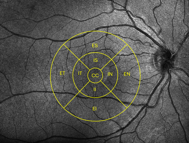
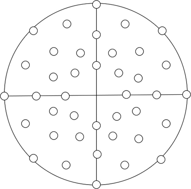
Methods: Twenty-six chronic LHON subjects (60% treated with idebenone or Q10) from the Swedish LHON registry were enrolled. Best-corrected visual acuity (BCVA), visual field tests, and optical coherence tomography (OCT) were performed. Visual field was evaluated with the Haag-Streit Octopus 900 with the Esterman test and a custom 30° test. Canon OCT-HS100 scans were exported to the Iowa Reference Algorithm. GCC thickness was obtained after the segmentation was corrected manually in nine macular sectors.
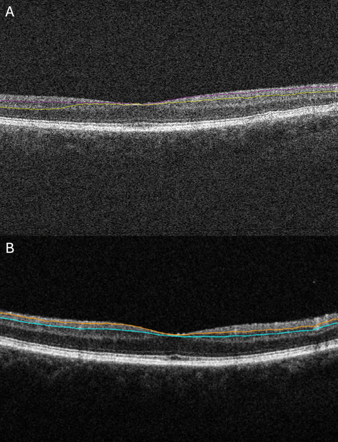
Results: The GCC thickness was overestimated by 16 to 30 µm in different macular sectors with the automated segmentation compared with the corrected (P < 0.001). GCC thickness in all sectors showed significant correlation with all functional parameters. The strongest correlation was seen for the external temporal sector (BCVA: r = 0.604, P < 0.001; mean defect: r = 0.457, P = 0.001; Esterman score: r = 0.421, P = 0.003). No differences were seen between treated and untreated subjects with regard to GCC and visual field scores (P > 0.05), but BCVA was better among treated subjects (P = 0.017).
Conclusions: The corrected GCC thickness showed correlation with visual function in chronic LHON subjects. The frequently occurring segmentation errors in OCT measurements related to chronic LHON can potentially be misleading in monitoring of disease progression and in evaluating the treatment effects. Precise measurements of GCC could serve as a sensitive tool to monitor structural changes in LHON. We therefore emphasize the importance of careful evaluation of the accuracy of OCT segmentation.
Conflict of interest statement
Disclosure:
Figures
References
-
- Rocatcher A, Desquiret-Dumas V, Charif M, et al.. The top 10 most frequently involved genes in hereditary optic neuropathies in 2186 probands. Brain. 2022; 146: 455–460. - PubMed
-
- Poincenot L, Pearson AL, Karanjia R.. Demographics of a large international population of patients affected by Leber's hereditary optic neuropathy. Ophthalmology. 2020; 127(5): 679–688. - PubMed
-
- Puomila A, Hämäläinen P, Kivioja S, et al.. Epidemiology and penetrance of Leber hereditary optic neuropathy in Finland. Eur J Hum Genet. 2007; 15(10): 1079–1089. - PubMed
MeSH terms
Substances
LinkOut - more resources
Full Text Sources
