

Contrast enhanced ultrasound of cystic renal lesions, from diagnosis up to treatment
- PMID: 39365320
- PMCID: PMC11612966
- DOI: 10.3233/CH-248102
Contrast enhanced ultrasound of cystic renal lesions, from diagnosis up to treatment
Abstract
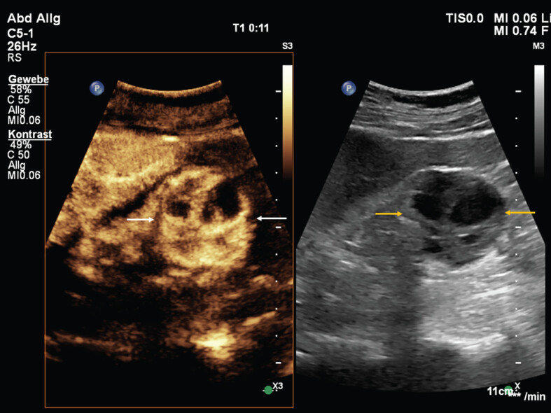
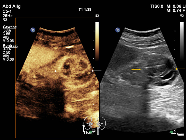
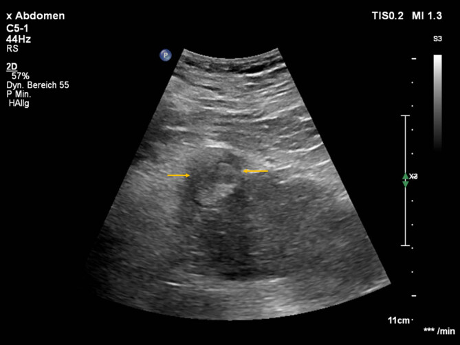
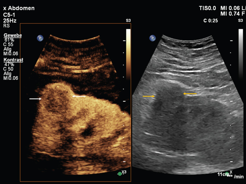
Ultrasound is the most used interdisciplinary imaging technique in clinical routine for assessment of renal pathologies. This includes the monitoring of cystic renal lesions, which can be classified as non-complicated or complicated and by means of occurrence as solitary or multifocal lesions. The Bosniak-classification (I-IV) classifies renal cysts in 5 different categories and is used for decisions of further clinical treatment. This classification was developed for computed tomography and has been adopted for magnetic resonance imaging as well as contrast-enhanced ultrasound. In the following review article, cystic kidney lesions and their differentiation using contrast-enhanced ultrasound are presented and an overview of the therapy options is given. In interventional procedures, CEUS can make a valuable contribution in histological sampling, reduce radiation exposure and, under certain circumstances, the number of interventions for the patient.
Keywords: Contrast-enhanced ultrasound; intervention; kidney lesion; real-time imaging; tissue vascularization.
Figures
Similar articles
-
Contrast-Enhanced Ultrasound (CEUS) of the Kidneys by Using the Bosniak Classification.Ultraschall Med. 2016 Jun;37(3):234-51. doi: 10.1055/s-0042-104646. Epub 2016 Apr 8. Ultraschall Med. 2016. PMID: 27058636 Review. English.
-
Contrast-enhanced ultrasound (CEUS) of cystic renal lesions in comparison to CT and MRI in a multicenter setting.Clin Hemorheol Microcirc. 2020;75(4):419-429. doi: 10.3233/CH-190764. Clin Hemorheol Microcirc. 2020. PMID: 32039837
-
Multislice computed tomography versus contrast-enhanced ultrasound in evaluation of complex cystic renal masses using the Bosniak classification system.Clin Hemorheol Microcirc. 2008;39(1-4):171-8. Clin Hemorheol Microcirc. 2008. PMID: 18503122
-
Contrast-enhanced ultrasound in renal cystic lesions: an update.J Med Ultrason (2001). 2024 Oct;51(4):635-647. doi: 10.1007/s10396-024-01489-x. Epub 2024 Aug 20. J Med Ultrason (2001). 2024. PMID: 39164480 Free PMC article. Review.
-
Ultrasound-based "CEUS-Bosniak"classification for cystic renal lesions: an 8-year clinical experience.World J Urol. 2023 Mar;41(3):679-685. doi: 10.1007/s00345-022-04094-0. Epub 2022 Aug 20. World J Urol. 2023. PMID: 35986781 Free PMC article.
Cited by
-
Possibility of modern ultrasound imaging of portal venous system.Clin Hemorheol Microcirc. 2025 May;90(1):3-13. doi: 10.1177/13860291251324086. Epub 2025 Jun 2. Clin Hemorheol Microcirc. 2025. PMID: 40452393 Free PMC article. Review.
References
-
- Cantisani V, et al.. EFSUMB 2020 Proposal for a Contrast-Enhanced Ultrasound-Adapted Bosniak Cyst Categorization – Position Statement. Ultraschall Med. 2021;42(2):154–66. - PubMed
-
- Schwarze V, et al.. Single-center study: the diagnostic performance of contrast-enhanced ultrasound (CEUS) for assessing renal oncocytoma. Scand J Urol. 2020;54(2):135–40. - PubMed
Publication types
MeSH terms
Substances
LinkOut - more resources
Full Text Sources
Medical
