

Effect of primary copper metabolism disturbance on elemental, protein, and lipid composition of the organs in Jackson toxic milk mouse
- PMID: 39365499
- PMCID: PMC11754380
- DOI: 10.1007/s10534-024-00640-y
Effect of primary copper metabolism disturbance on elemental, protein, and lipid composition of the organs in Jackson toxic milk mouse
Abstract
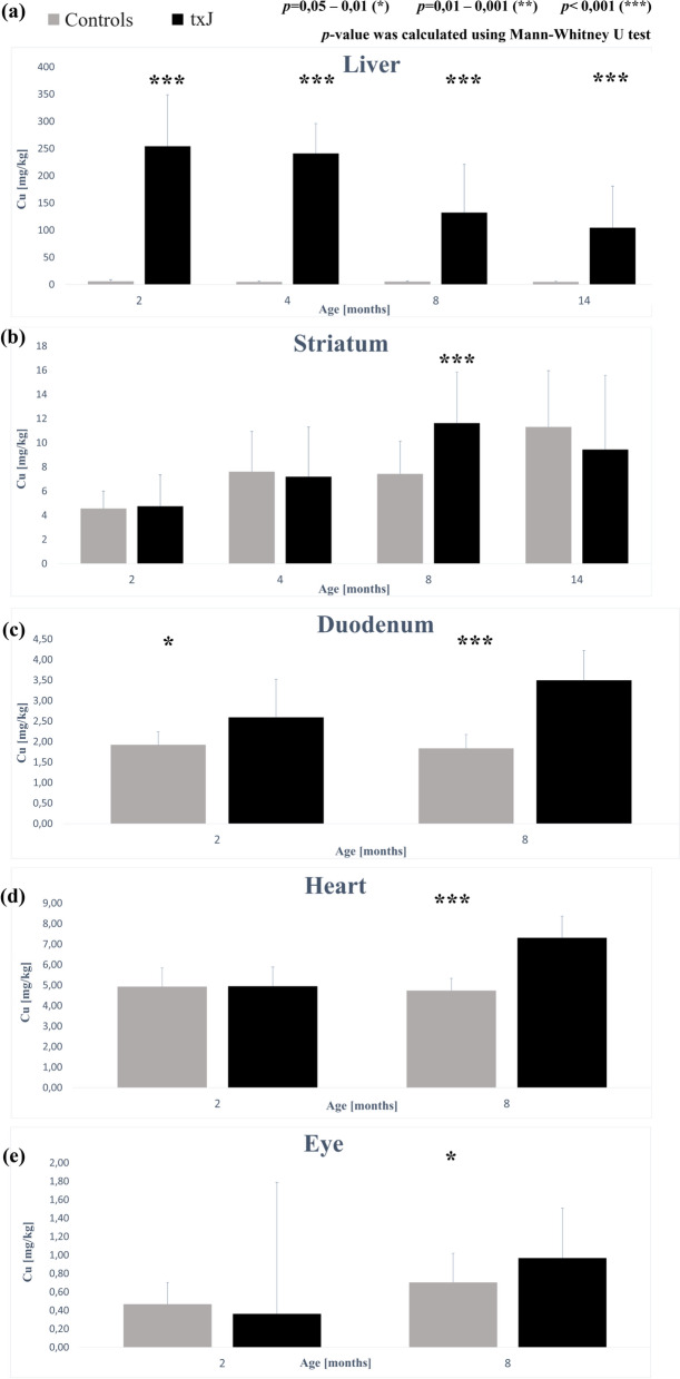
Toxic milk (txJ) is an autosomal recessive mutation in the Atp7b gene in the C3H/HeJ strain, observed at The Jackson Laboratory in Maine, USA. TxJ mice exhibit symptoms similar to those of human Wilson's disease (WD). The study aimed to verify organ involvement in a mouse model of WD. TxJ mice and control animals were sacrificed at 2, 4, 8, and 14 months of age. Total X-ray Fluorescence Spectroscopy (TXRF) was used to determine the elemental concentration in organs. Tissue chemical composition was measured by Fourier Transform Infrared Spectroscopy (FTIR). Additionally, hybrid mapping of FTIR and microXRF was performed. Elevated concentrations of Cu were observed in the liver, striatum, eye, heart, and duodenum of txJ mice across age groups. In the striatum of the oldest txJ mice, there was lower lipid content and a higher fraction of saturated fats. The secondary structure of striatum proteins was disturbed in txJ mice. In the livers of txJ mice, higher concentrations of saturated fats and disturbances in the secondary structure of proteins were observed. The concentration of neurofilaments was significantly higher in txJ serum. The distribution of Cu deposits in brains was uniform with no prevalence in any anatomic structure in either group, but significant protein structure changes were observed exclusively in the striatum of txJ. In this txJ animal model of WD, pathologic copper accumulation occurs in the duodenum, heart, and eye tissues. Increased copper concentration in the liver and brain results in increased saturated fat content and disturbances in secondary protein structure, leading to hepatic injury and neurodegeneration.
Keywords: Copper metabolism; Neurodegeneration; Neurofilament; Toxic milk mouse; Wilson’s disease.
© 2024. The Author(s).
Conflict of interest statement
Declarations. Conflict of interest: The authors have no relevant financial or nonfinancial interests to disclose. Ethical approval: This is an observational study. The first Local Bioethics Committee for Animal Research in Warsaw, Poland has confirmed that no ethical approval is required.
Figures


References
-
- Bronson RTSH, Davisson MT (1995) Acute cerebral neuronal necrosis in copper deficient offspring of female mice with the toxic milk mutation. Mouse Genome 93(1):152–154
MeSH terms
Substances
LinkOut - more resources
Full Text Sources
Medical
Molecular Biology Databases
