. 2024 Oct 8;121(41):e2401071121.
doi: 10.1073/pnas.2401071121. Epub 2024 Oct 4.

Dysregulated miR-124-3p in endometrial epithelial cells reduces endometrial receptivity by altering polarity and adhesion

Affiliations

Dysregulated miR-124-3p in endometrial epithelial cells reduces endometrial receptivity by altering polarity and adhesion

Wei Zhou et al. Proc Natl Acad Sci U S A. .

Abstract

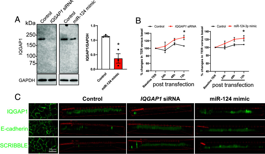
The endometrium undergoes substantial remodeling in each menstrual cycle to become receptive to an implanting embryo. Abnormal endometrial receptivity is one of the major causes of embryo implantation failure and infertility. MicroRNA-124-3p is elevated in both the serum and endometrial tissue of women with chronic endometritis, a condition associated with infertility. MicroRNA-124-3p also has a role in cell adhesion, a key function during receptivity to allow blastocysts to adhere and implant. In this study, we aimed to determine the function of microRNA-124-3p on endometrial epithelial adhesive capacity during receptivity and effect on embryo implantation. Using a unique inducible, uterine epithelial-specific microRNA overexpression mouse model, we demonstrated that elevated uterine epithelial microRNA-124-3p impaired endometrial receptivity by altering genes associated with cell adhesion and polarity. This resulted in embryo implantation failure. Similarly in a second mouse model, increasing microRNA-124-3p expression only in mouse uterine surface (luminal) epithelium impaired receptivity and led to implantation failure. In humans, we demonstrated that microRNA-124-3p was abnormally increased in the endometrial epithelium of women with unexplained infertility during the receptive window. MicroRNA-124-3p overexpression in primary human endometrial epithelial cells (HEECs) impaired primary human embryo trophectoderm attachment in a 3-dimensional culture model of endometrium. Reduction of microRNA-124-3p in HEECs from infertile women normalized HEEC adhesive capacity. Overexpression of microRNA-124-3p or knockdown of its direct target IQGAP1 reduced fertile HEEC adhesion and its ability to lose polarity. Collectively, our data highlight that microRNA-124-3p and its protein targets contribute to endometrial receptivity by altering cell polarity and adhesion.

Keywords: embryo implantation; endometrial epithelium; endometrial receptivity; miR-124-3p; microRNA.


Conflict of interest statement

Competing interests statement:The authors declare no competing interest.

Figures

Fig. 1.
Fig. 1.
Overexpression of miR-124-3p in the murine uterine epithelium during endometrial receptivity impaired implantation. (A) Endogenous miR-124-3p levels in uterine luminal epithelium from pregnant wildtype mouse uterine luminal epithelium at E3 and E4 (qPCR; N = 3 to 4). (B–F) Uterine epithelium specific miR-124-3p overexpression significantly reduced implantation success in mice. (B) Timeline of doxycycline treatment to induce miR-124-3p overexpression during endometrial receptivity. (C) miR-124-3p overexpression in the uterus was verified at E4.5 by qPCR (N = 5 to 6). (D) miR-124-3p overexpression significantly reduced implantation site number (IS, arrowhead) at E4.5 (N = 5 to 6). (E) miR-124-3p overexpression was localized to the luminal and glandular epithelium by in situ hybridization. (F) miR-124-3p did not affect embryo viability, IS found in transgenic miR-124-3p mice stained with the cell apoptosis marker Cleaved Caspase-3. Wildtype mouse spleen was used as a positive control for staining. All data are presented as mean ± SEM. *P < 0.05, ****P < 0.0001.
Fig. 2.
Fig. 2.
miR-124-3p overexpression in mouse uterine epithelium impairs receptivity. (A) Representative receptivity and decidualization genes in E4.5 transgenic control and miR-124-3p uteri (qPCR; N = 4 to 6). All data are presented as mean ± SEM. (B) Heatmap of results from customized PCR gene array on E4.5 uteri from transgenic miR-124-3p and control mice. Data presented as dCt value in heat map and ranked by expression level (high to low) (N = 5 to 6). Genes having miR-124-3p binding site(s) highlighted in bold. Original data provided in Dataset S1. *P < 0.05, **P < 0.01, ***P < 0.001, ****P < 0.0001. (C) Immunohistochemistry staining at E4.5 of cell polarity markers (E-cadherin, SCRIBBLE), proliferation marker (KI-67), receptivity marker (P-STAT3), and decidualization marker (DESMIN). As no implantation sites were recorded in some miR-124-3p transgenic mice, uteri at E4.5 were also stained. Arrow: implanting blastocyst. The tissue sections are oriented so that the antimesometrial (AM) sides of the uterus is at the top and the mesometrial (M) is at the bottom.
Fig. 3.
Fig. 3.
Luminal epithelium only miR-124-3p overexpression impaired implantation. (A) Schematic diagram of the intrauterine injection to overexpress miR-124-3p in the mouse luminal epithelium. For each individual mouse, AgomiR-124-3p or scrambled mimic was injected into one side of the uterine horn at E3. O: Ovary, F: Fallopian tube, U: Uterus. (B) miR-124-3p was significantly elevated in the luminal epithelium after intrauterine injection compared to the remaining uterus and control uterine horn (qPCR; N = 3). (C) miR-124-3p overexpression localized to the luminal epithelium as determined by in situ hybridization. (D) Intrauterine injection of miR124-3p significantly reduced implantation site number in the treated horn compared to untreated control at E6 (N = 8). (E) Exposure of unhatched wildtype mouse embryos at E4 to miR-124-3p showed no effect on outgrowth compared to control. (F and G) The effect of miR-124-3p overexpression on receptivity and decidualization markers was determined by immunohistochemistry (F) and qPCR (G). Arrow: implanting blastocyst (N = 3 to 4). The tissue sections are oriented so that the AM sides of the uterus is at the top and the M is at the bottom. All data are presented as mean ± SEM. *P < 0.05, **P < 0.01, ***P < 0.001, IS: implantation site.
Fig. 4.
Fig. 4.
miR-124-3p was overexpressed in infertile human endometrial luminal epithelium during receptive phase. (A) miR-124-3p expression in endometrial tissue (qPCR; N = 6 to 11) from fertile and infertile endometrium. Expression levels were normalized to U44. The unpaired t test was used to compare miR-124-3p levels between each phase and between fertile and infertile groups. Data are presented as mean ± SEM. *P < 0.05, **P < 0.01. (B) In situ hybridization of miR-124-3p on mid-secretory phase endometrial sections revealed strong fluorescence in the luminal epithelial cells of the infertile endometrium, compared to fertile endometrium (green arrows, N = 3). Sections were counterstained with DAPI to reveal the cell nuclei (blue). As controls, endometrial sections were either incubated with U6 probe (positive control) or scrambled sequence probe (scrambled control). L: Luminal epithelium; G: Glandular epithelium; S: Stroma.
Fig. 5.
Fig. 5.
miR-124-3p impaired HEEC receptivity. (A–C) Outgrowth of primary human trophectoderm spheroids on a 3D primary human endometrial cell construct. Primary HEECs in the construct were transfected with miR-124-3p mimic or scrambled control (N = 3). (A) Before culture, inner cell mass (arrow) was removed from day 6 human blastocysts. (B) The remaining blastocyst readily re-formed into a spheroid. (C) Human trophectoderm attachment and outgrowth on the 3D endometrial construct (outlined, cultured up to 120 h) (N = 3). (D and E) Adhesion of HTR8/SVneo spheroid to (D) 3D endometrial construct (N = 3) or (E) HEEC monolayer (N = 3 to 5). (F) miR-124-3p levels in fertile and infertile HEECs (N = 3 to 5). (G) HTR8/SVneo spheroid adhesion to infertile HEECs transfected with miR-124-3p inhibitor or control (N = 5). Data are presented as mean ± SEM. *P < 0.05, **P < 0.01.
Fig. 6.
Fig. 6.
miR-124-3p targets adhesion genes in human and mouse uterus. (A) Fluidigm BioMark HD System was applied to quantify the expression of 88 genes in HEECs transfected with scrambled control or miR-124-3p mimic. These genes are predicted miR-124-3p targets and fall into broad categories of cell adhesion and attachment. Data were presented as dCt value in heat map (N = 5). Original data provided in Dataset S2. (B) Expression of miR-124-3p gene targets identified in A in E4.5 uterus from transgenic miR-124-3p or control mice (N = 4 to 6). Data are presented as mean ± SEM. *P < 0.05, **P < 0.01, ***P < 0.001, **** P < 0.0001.
Fig. 7.
Fig. 7.
miR-124-3p target IQGAP1 alters HEEC adhesion. (A and B) IQGAP1 immunolocalization in fertile and infertile secretory phase human endometrium. Higher magnification images of the luminal epithelium (L), glandular epithelium (G), and stromal cells (S) are depicted on the right of panels with their original locations indicated by outlines on the left. Sections were counterstained with hematoxylin to indicate cell nuclei (blue). IQGAP1 staining intensity (N = 4) was semiquantitated by scoring. Endometrial tissues were blinded to fertility status. (C) Immunoblot showing reduction in IQGAP1 protein following HEEC transfection with miR-124-3p mimic (10 nM, N = 4 to 6). (D and E) HTR8/SVneo spheroid attachment (D) to HEECs following IQGAP1 knockdown (E, qPCR) (N = 4). (F and G) Target site blocker (TSB) was used to confirm the direct interaction between miR-124-3p and IQGAP1. (F) miR-124-3p and IQGAP1 TSB binding sequences of IQGAP1 mRNA. (G) HEECs were transfected with miR-124-3p mimic (30 nM) in combination with either IQGAP1 or control TSB (30 nM) and subjected to immunoblotting to determine expression of IQGAP1 (N = 6). (H) Protection of IQGAP1 via TSB in miR-124-3p overexpressed HEECs were able to improve their adhesion to HTR8/SVneo spheroids (N = 6). (I) Iqgap1 expression in mouse E4 luminal epithelium (intrauterine injection model). Data are presented as mean ± SEM. *P < 0.05, **P < 0.01, ***P < 0.001.
Fig. 8.
Fig. 8.
miR-124-3p overexpression and IQGAP1 knockdown increased endometrial epithelial cell polarity. (A) Immunoblotting identified reduction of IQGAP1 in Ishikawa cells treated with either IQGAP1 siRNA or miR-124-3p, compared to control. IQGAP1 was undetectable in cells treated with IQGAP1 siRNA therefore densitometric analysis was not conducted (N = 3). (B) To determine cell polarity, Ishikawa cells transfected with IQGAP1 siRNA, miR-124-3p, or respective controls were subjected to transepithelial electrical resistance assay to monitor percentage changes of cell barrier integrity (N = 5). Data are presented as mean ± SEM. *P < 0.05. (C) 3D lateral scanning of transfected Ishikawa monolayers stained with IQGAP1, E-cadherin, or cell polarity marker SCRIBBLE.

References

    1. Wilcox A. J., Baird D. D., Weinberg C. R., Time of implantation of the conceptus and loss of pregnancy. N. Engl. J. Med. 340, 1796–1799 (1999). - PubMed
    1. Kao L., et al. , Global gene profiling in human endometrium during the window of implantation. Endocrinology 143, 2119–2138 (2002). - PubMed
    1. Strowitzki T., Germeyer A., Popovici R., Von Wolff M., The human endometrium as a fertility-determining factor. Hum. Reprod. Update 12, 617–630 (2006). - PubMed
    1. Psychoyos A., Uterine receptivity for nidation. Ann. N. Y. Acad. Sci. 476, 36–42 (1986). - PubMed
    1. Paria B., Huet-Hudson Y., Dey S., Blastocyst’s state of activity determines the “window” of implantation in the receptive mouse uterus. Proc. Natl. Acad. Sci. U.S.A. 90, 10159–10162 (1993). - PMC - PubMed

LinkOut - more resources