Review
. 2025 Feb 4;24(2):188-199.
doi: 10.1158/1535-7163.MCT-23-0726.

Inflammation and Immunity in Liver Neoplasms: Implications for Future Therapeutic Strategies

Affiliations
Review

Inflammation and Immunity in Liver Neoplasms: Implications for Future Therapeutic Strategies

Jieun Kim et al. Mol Cancer Ther. .

Abstract

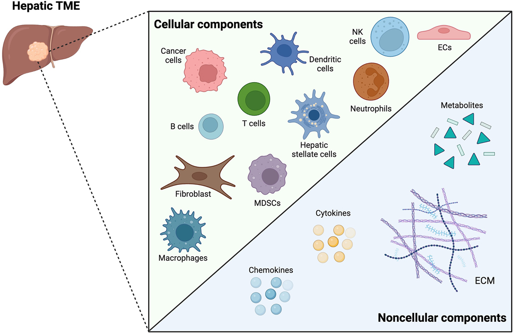
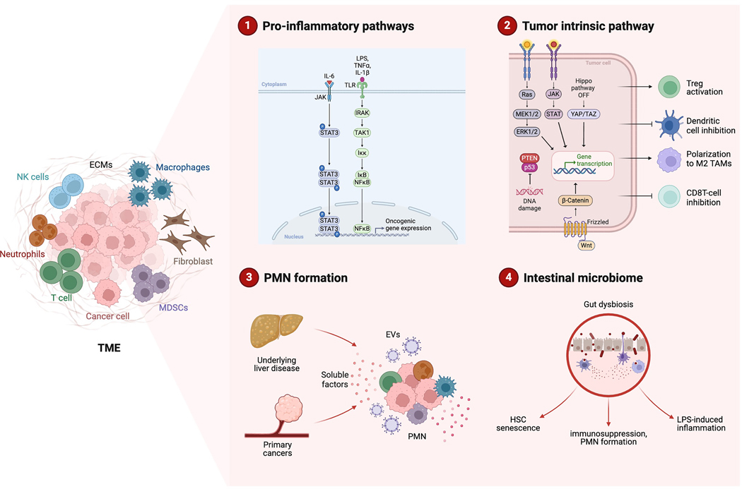
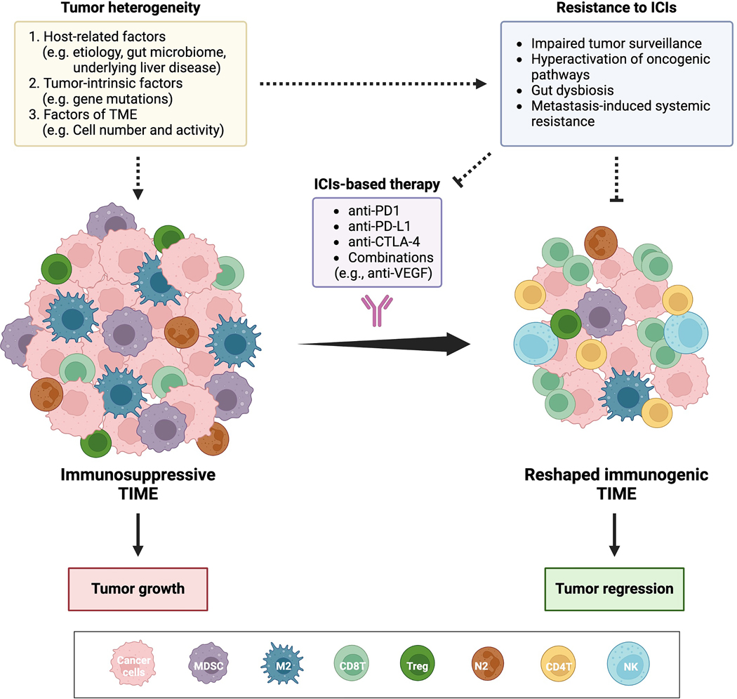
Over the past two decades, the "hallmarks of cancer" have revolutionized cancer research and highlighted the crucial roles of inflammation and immunity. Protumorigenic inflammation promotes cancer development along with inhibition of antitumor immunity, shaping the tumor microenvironment (TME) toward a tumor-permissive state and further enhancing the malignant potential of cancer cells. This immunosuppressive TME allows tumors to evade immunosurveillance. Thus, understanding the complex interplay between tumors and the immune system within the TME has become pivotal, especially with the advent of immunotherapy. Although immunotherapy has achieved notable success in many malignancies, primary liver cancer, particularly hepatocellular carcinoma, presents unique challenges. The hepatic immunosuppressive environment poses obstacles to the effectiveness of immunotherapy, along with high mortality rates and limited treatment options for patients with liver cancer. In this review, we discuss current understanding of the complex immune-mediated mechanisms underlying liver neoplasms, focusing on hepatocellular carcinoma and liver metastases. We describe the molecular and cellular heterogeneity within the TME, highlighting how this presents unique challenges and opportunities for immunotherapy in liver cancers. By unraveling the immune landscape of liver neoplasms, this review aims to contribute to the development of more effective therapeutic interventions, ultimately improving clinical outcomes for patients with liver cancer.


Conflict of interest statement

The authors declare they have nothing to disclose.

Figures

Figure 1.
Figure 1.. Components of tumor microenvironment in the liver.
The hepatic tumor microenvironment (TME) is heterogeneous, composed of both cellular and noncellular components. Major cellular components include cancer cells, immune cells (such as macrophages, natural killer [NK] cells, dendritic cells, T and B lymphocytes, myeloid-derived suppressor cells (MDSCs), or neutrophils), as well as stromal cells (such as endothelial cells [ECs], hepatic stellate cells [HSCs], or fibroblasts). Noncellular components consist of the extracellular matrix (ECM), chemokines, cytokines, and metabolites. These diverse cell populations interact directly with each other or with noncellular components to regulate cancer growth.
Figure 2.
Figure 2.. The dynamic interactions of inflammation and cancers in the tumor microenvironment.
The tumor microenvironment (TME) is regulated by inflammation and immunity during liver cancer development. (1) Inflammatory pathways connect inflammation with hepatocarcinogenesis. Pro-inflammatory cytokines, like IL-6, TNFɑ, and IL-1β, or pathogen-derived molecules such as LPS, drive inflammation-associated hepatocarcinogenesis. The IL6/JAK/STAT3 pathway is oncogenic and promote hepatocellular carcinoma (HCC) development. Additionally, the NF-κB pathway regulates hepatocarcinogenesis and contributes to drug resistance. (2) Tumor intrinsic pathway plays a critical role in reshaping the tumor immune microenvironment (TIME) during liver cancer progression. Genetic mutations, including loss of TP53 or PTEN, and activation of oncogenic pathways such as Wnt/β-Catenin, Ras-MAPK-ERK, or YAP, contribute to inflammation, immunosuppression, or suppression of anti-tumor immunity. These reprogramming events collectively create a microenvironment that supports tumor growth and progression. (3) Premetastatic niche (PMN) in the liver is established by factors released from primary cancers. These factors include TIMP-1, VEGFA, IL-6 and extracellular vesicles (EVs). Underlying liver disease is an important factor that modifies PMN. Hepatic steatosis resulting from metabolic dysfunction-associated liver disease or alcohol-associated liver disease predisposes the liver to develop a PMN by creating immunosuppressive TIME. (4) Gut dysbiosis is linked to HCC development. The gut microbiome contributes to hepatic stellate cell (HSC) senescence, immunosuppressive TIME formation, tumor-favorable PMN formation, and pro-tumorigenic inflammation (e.g. LPS-induced), enhancing tumor growth.
Figure 3.
Figure 3.. Reshaping the TIME in liver cancer with immune checkpoint inhibitors reduces tumor burden.
The tumor immune microenvironment (TIME) in both primary and secondary liver cancer development is characterized as immunosuppressive or poorly immunogenic, marked by an abundance of immunosuppressive cell types such as myeloid-derived suppressor cells (MDSCs), M2-polarized tumor-associated macrophages, N2-polarized neutrophils, and regulatory T (Treg) cells. These cell types collectively promote tumor growth and progression. Immunotherapeutic strategies, particularly immune checkpoint inhibitors (ICIs), either alone or in combination therapies, have been extensively investigated in preclinical settings. These strategies aim to reshape the liver cancer TIME into a more immunogenic environment characterized by increased presence of CD8 T cells, CD4 T cells, and natural killer (NK) cells, and reduced numbers of immunosuppressive cells, thereby creating a less favorable environment for tumor growth. However, the response and resistance to ICIs are influenced by the heterogeneous nature of the TME. (1) Host-related factors such as cancer etiology, alterations in the gut microbiome, and underlying liver disease contribute to TIME heterogeneity and influence ICI response. The distinct immunosuppressive TIME, shaped by host-related factors, impacts therapy outcomes. (2) Tumor-intrinsic factors including genetic mutations modulate the TIME and affect ICI response. Genetic variations contribute to individual differences in the impact of ICIs on immune responses and resistance. (3) Variability in the composition of TIME among individuals can limit the effectiveness of ICIs, with differences in immune cell numbers and activity, such as the absence of CD8 T cells, impacting therapy outcomes. Understanding these complexities is crucial for optimizing ICI therapies and enhancing treatment outcomes in liver cancer.

Similar articles

Cited by

References

    1. Hanahan D Hallmarks of Cancer: New Dimensions. Cancer Discov 2022;12(1):31–46 doi 10.1158/2159-8290.CD-21-1059. - DOI - PubMed
    1. de Visser KE, Joyce JA. The evolving tumor microenvironment: From cancer initiation to metastatic outgrowth. Cancer Cell 2023;41(3):374–403 doi 10.1016/j.ccell.2023.02.016. - DOI - PubMed
    1. Wasik AA, Schiller HB. Functional proteomics of cellular mechanosensing mechanisms. Semin Cell Dev Biol 2017;71:118–28 doi 10.1016/j.semcdb.2017.06.019. - DOI - PubMed
    1. Binnewies M, Roberts EW, Kersten K, Chan V, Fearon DF, Merad M, et al. Understanding the tumor immune microenvironment (TIME) for effective therapy. Nat Med 2018;24(5):541–50 doi 10.1038/s41591-018-0014-x. - DOI - PMC - PubMed
    1. Hodi FS, O’Day SJ, McDermott DF, Weber RW, Sosman JA, Haanen JB, et al. Improved survival with ipilimumab in patients with metastatic melanoma. N Engl J Med 2010;363(8):711–23 doi 10.1056/NEJMoa1003466. - DOI - PMC - PubMed

MeSH terms