. 2024 Oct 4;10(40):eadn8760.
doi: 10.1126/sciadv.adn8760. Epub 2024 Oct 4.

The generation of stable microvessels in ischemia is mediated by endothelial cell derived TRAIL

Affiliations

The generation of stable microvessels in ischemia is mediated by endothelial cell derived TRAIL

Siân P Cartland et al. Sci Adv. .

Abstract

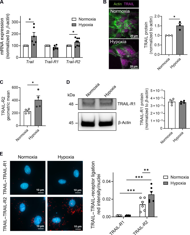
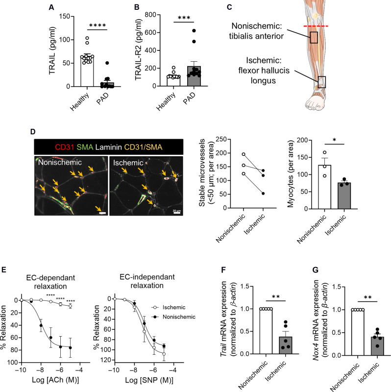
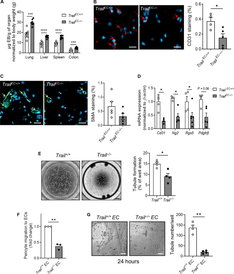
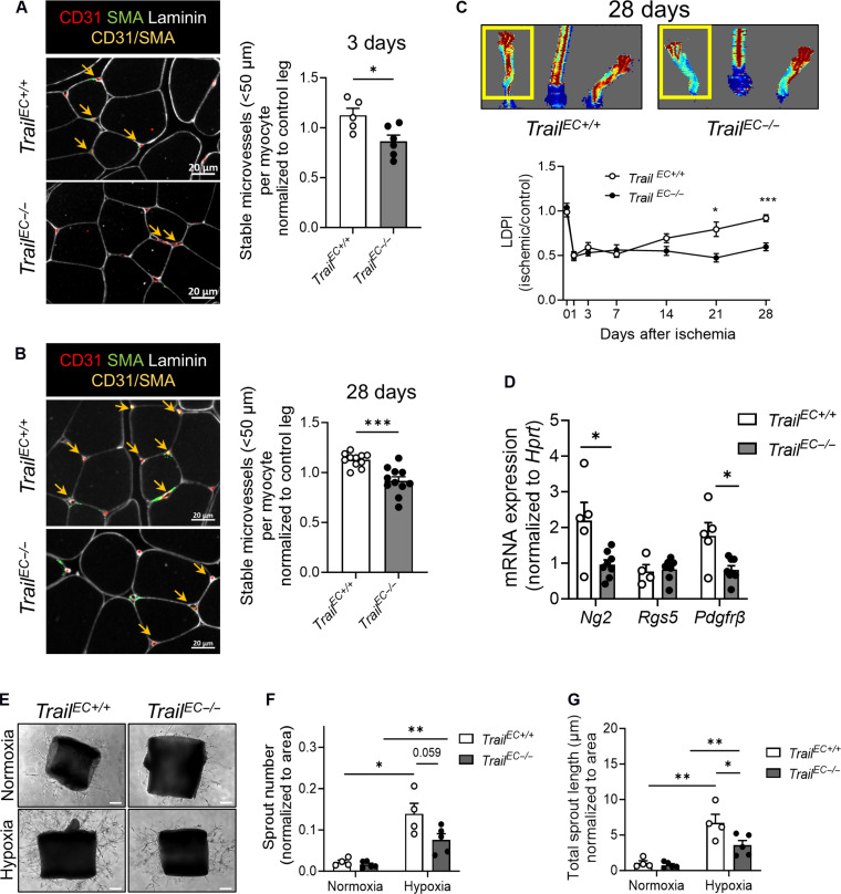
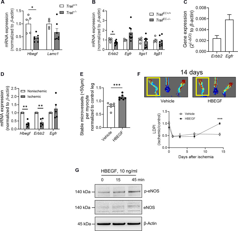
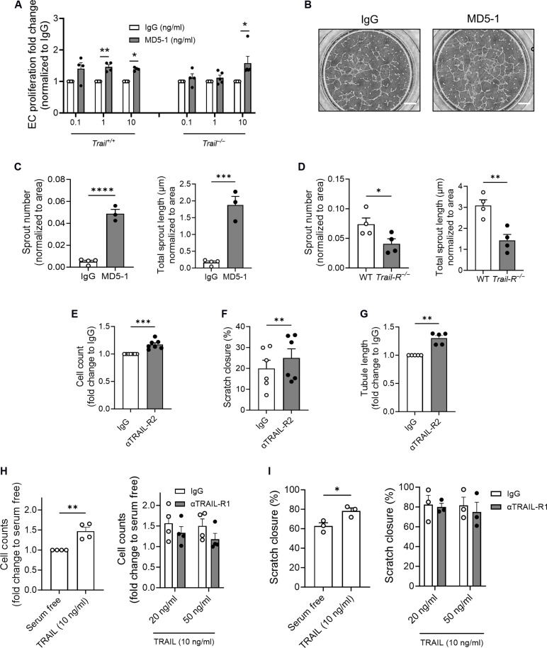
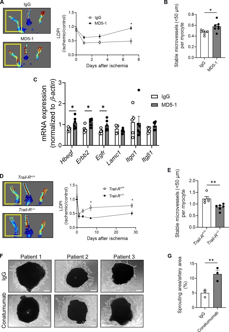
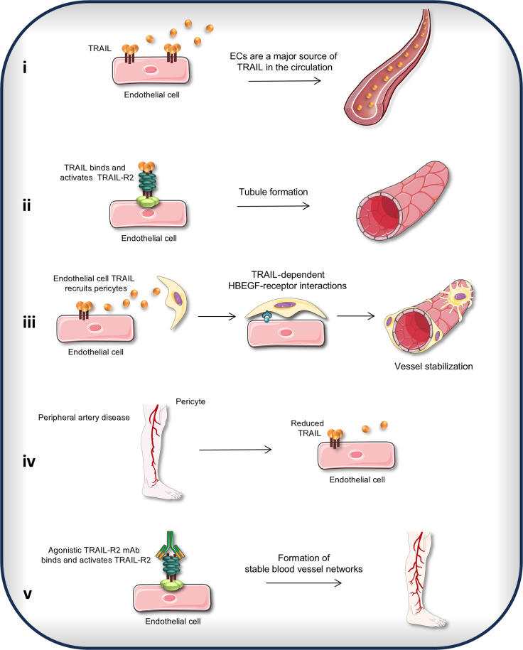
Reversal of ischemia is mediated by neo-angiogenesis requiring endothelial cell (EC) and pericyte interactions to form stable microvascular networks. We describe an unrecognized role for tumor necrosis factor-related apoptosis-inducing ligand (TRAIL) in potentiating neo-angiogenesis and vessel stabilization. We show that the endothelium is a major source of TRAIL in the healthy circulation compromised in peripheral artery disease (PAD). EC deletion of TRAIL in vivo or in vitro inhibited neo-angiogenesis, pericyte recruitment, and vessel stabilization, resulting in reduced lower-limb blood perfusion with ischemia. Activation of the TRAIL receptor (TRAIL-R) restored blood perfusion and stable blood vessel networks in mice. Proof-of-concept studies showed that Conatumumab, an agonistic TRAIL-R2 antibody, promoted vascular sprouts from explanted patient arteries. Single-cell RNA sequencing revealed heparin-binding EGF-like growth factor in mediating EC-pericyte communications dependent on TRAIL. These studies highlight unique TRAIL-dependent mechanisms mediating neo-angiogenesis and vessel stabilization and the potential of repurposing TRAIL-R2 agonists to stimulate stable and functional microvessel networks to treat ischemia in PAD.


Figures

Fig. 1.
Fig. 1.. TRAIL and TRAIL-R2 expression and interaction are augmented in hypoxia.
(A) Trail, Trail-R1, and Trail-R2 mRNA expression in HMEC-1 after exposure to hypoxia (2% O2) or normoxia (21% O2) for 24 hours, qPCR normalized to β-actin (n = 6 to 8 per treatment). (B) Total internal reflection fluorescence microscopy images (left) and quantification (right) of TRAIL cell surface expression (n = 3 per treatment). (C) TRAIL-R2 cell surface expression by flow cytometry (n = 4 per treatment). (D) Left: Western blot of TRAIL-R1 and β-actin. Right: TRAIL-R1 protein expression normalized to β-actin (n = 4 per group). (E) Protein-protein interactions between TRAIL–TRAIL-R1 and TRAIL–TRAIL-R2, measured using the Duolink Proximity Ligation Assay. Interactions are indicated by red staining. Left: Representative image. Right: Quantification (n = 6 per treatment). Results are means ± SEM; Students t test or two-way analysis of variance (ANOVA); *P < 0.05, **P < 0.01, and ***P < 0.001.
Fig. 2.
Fig. 2.. Plasma TRAIL levels are reduced in PAD, associating with reduced microvessel numbers with increasing ischemia.
Plasma (A) TRAIL and (B) TRAIL-R2 levels in PAD versus healthy individuals (n = 11 to 12 per group). (C) Schematic illustrating tissue harvest locations from below-knee amputations. (D) Stable microvessel numbers in nonischemic and ischemic regions of amputated tissues. Left: Representative image of stable microvessels (CD31+SMA+, yellow arrows). Laminin (myocytes, white), CD31 (ECs, red), and SMA (pericytes, green); scale bars, 20 μm. Middle: Microvessel quantification. Right: Myocyte number is reduced with ischemia (n = 3 per group). (E) Myography showing impaired endothelial function in arteries isolated from nonischemic versus ischemic regions. Left: Arterial relaxation in response to increasing doses of acetylcholine (ACh). Right: No change in sodium nitroprusside (SNP)–mediated relaxation (n = 5 per group). (F) Trail and (G) Nox4 mRNA expression in patient tissues (n = 5 per group). mRNA normalized to β-actin. Results are means ± SEM; Mann-Whitney U test, paired t test, or two-way ANOVA; *P < 0.05, **P < 0.01, ***P < 0.001, and ****P < 0.0001.
Fig. 3.
Fig. 3.. EC-derived TRAIL regulates neo-angiogenesis, pericyte recruitment, and vessel stabilization.
(A) Permeability of Evans Blue (EB) into highly vascularized organs from TrailEC+/+ and TrailEC−/− mice (n = 7 to 8 per group). TrailEC+/+ and TrailEC−/− mice were injected with Matrigel; plugs were collected 28 days later and (B) capillary density (CD31, red) and (C) perivascular cell (SMA, green) content determined by microscopy; scale bars, 20 μm (n = 4 to 5 per group). (D) EC and pericyte mRNA marker expression in Matrigel plugs was measured by qPCR, normalized to β-actin (n = 4 to 5 per group). (E) Tubule formation in Trail+/+ and Trail−/− ECs at 6 hours. Left: Representative images; scale bars, 500 μm. Right: Quantification (n = 4 per group). (F) Pericyte migration to Trail+/+ and Trail−/− ECs by Transwell assay at 8 hours (n = 3 per group). (G) Increased tubule numbers at 24 hours in Trail+/+ but not Trail−/− ECs cocultured with pericytes. Left: Representative images; scale bars, 100 μm. Right: Quantification. ECs and pericytes isolated from mouse brain. Results are means ± SEM; Student’s t test, paired t test, or Mann-Whitney U test; *P < 0.05, **P < 0.01, ***P < 0.001, and ****P < 0.01.
Fig. 4.
Fig. 4.. Stable microvessel numbers are reduced in ischemic limb tissues of TrailEC−/− mice.
(A) Stable microvessels (CD31+SMA+; yellow arrows) in gastrocnemius muscle of TrailEC+/+ and TrailEC−/− mice 3 days after HLI. Left: Representative image of ischemic muscle stained for laminin, CD31, and SMA; scale bars, 20 μm. Right: Quantification (n = 5 to 6 per genotype). (B) Stable microvessels (CD31+SMA+; yellow arrows) in gastrocnemius muscle of TrailEC+/+ and TrailEC−/− 28 days after HLI. Left: Representative image of ischemic muscle stained for laminin, CD31, and SMA; scale bars, 20 μm. Right: Quantification (n = 11 per genotype). (C) Laser Doppler imaging showing blood perfusion over time in TrailEC+/+ and TrailEC−/− mice after HLI. Top: Representative image of blood flow at 28 days. Bottom: Quantification over time (n = 9 to 11 mice per genotype). (D) Pericyte marker mRNA expression in ischemic limbs of TrailEC+/+ and TrailEC−/− mice 3 days after HLI, normalized to hypoxanthine-guanine phosphoribosyl transferase, Hprt (n = 4 to 8 per group). All data from (A) to (D) were normalized to the respective nonischemic control limb. (E) Representative sprouting images. Aortae from TrailEC+/+ and TrailEC−/− mice in response to 8-day hypoxia (2% O2) ex vivo; scale bars, 100 μm. Quantification of (F) sprout number and (G) total sprout length (n = 4 to 5 per genotype). Results are means ± SEM; Mann-Whitney U test or two-way ANOVA; *P < 0.05, **P < 0.01, and ***P < 0.001.
Fig. 5.
Fig. 5.. EC-pericyte interactions are altered in ischemic limbs of TrailEC−/− mice.
(A) Hbegf and Lamc1 mRNA levels in ECs isolated from Trail+/+ and Trail−/− mice (n = 4 to 7 per genotype). (B) mRNA expression of Hbegf receptors Erbb2 and Egfr, and Lamc1 receptors Itgα1 and Itgb1 in gastrocnemius muscle from TrailEC+/+ and TrailEC−/− mice (n = 5 to 8 per genotype). (C) Erbb2 and Egfr expression in primary murine pericytes. (D) mRNA expression of Hbegf receptors Erbb2 and Egfr in nonischemic and ischemic skeletal muscle from patients with PAD (n = 5). (E) Stable microvessel numbers in gastrocnemius muscle of TrailEC−/− mice treated with vehicle or recombinant murine HBEGF and 14 days after HLI (n = 6 to 7 per treatment). (F) Laser Doppler imaging showing blood perfusion over time in TrailEC−/− mice treated with vehicle or recombinant HBEGF (1 μg I.M., 2 days before surgery, day of surgery, and then daily). Top: Representative image of blood flow at 14 days. Bottom: Quantification (n = 6 to 7 per treatment). (G) Recombinant human HBEGF increases eNOS expression and phosphorylation (p-eNOS) in HMEC-1. β-Actin demonstrates unbiased loading. All mRNA expression was normalized to β-actin. Student’s t test, paired t test, or two-way ANOVA; *P < 0.05 and ***P < 0.001.
Fig. 6.
Fig. 6.. TRAIL-receptor activation stimulates features of neo-angiogenesis.
(A) Trail+/+ and Trail−/− EC proliferation is increased with MD5-1 at 24 hours, normalized to immunoglobulin G (IgG; n = 4 to 5 per group). (B) Representative images showing increased tubule formation of Trail+/+ ECs with MD5-1 versus IgG at 6 hours (10 ng/ml); scale bars, 500 μm. (C) Aortic rings from wild-type mice were exposed to MD5-1 or IgG for 4 days, and sprout number and total sprout length were assessed (n = 3 to 4 per group). (D) Aortic rings from Trail-R−/− mice exposed to hypoxia for 6 days have reduced sprout number and reduced total sprout length (n = 4 per group). Human TRAIL-R2 agonistic antibody (αTRAIL-R2, 10 ng/ml) stimulates HMEC-1 (E) proliferation, (F) migration, and (G) tubule formation (n = 5 to 7 per group). Conversely, neutralization by αTRAIL-R1 has no effect on TRAIL-inducible HMEC-1 (H) proliferation and (I) migration (n = 3 to 4 per group). Results are means ± SEM; two-way ANOVA, Mann-Whitney U test, or Students t test; *P < 0.05, **P < 0.01, ***P < 0.001, and ****P < 0.0001.
Fig. 7.
Fig. 7.. TRAIL-R2 activation stimulates angiogenesis.
(A) Laser Doppler imaging showing blood perfusion in HLI TrailEC−/− mice treated with MD5-1 or IgG over 7 days (1 μg I.M., 2 days before, 2 days after, and 6 days after surgery). Left: Representative image at 7 days. Right: Quantification over time and (B) stable microvessel numbers (CD31+SMA+) in gastrocnemius from ischemic limbs (n = 6 to 7 per group). (C) mRNA expression of Hbegf and its receptors Erbb2 and Egfr, and Lamc1 and its receptors Itgα1 and Itgb1 in gastrocnemius muscle of mice, normalized to β-actin (n = 5 to 6 per genotype). (D) Laser Doppler imaging showing blood perfusion in HLI Trail-R+/+ and Trail-R−/− mice over 28 days. Left: Representative image at 28 days. Right: Quantification over time and (E) stable microvessel numbers (CD31+SMA+) in gastrocnemius from ischemic limbs 28 days after HLI (n = 5 to 6 per group). (F) Sprouting in blood vessels harvested from ischemic tissues of patients with PAD undergoing below-knee amputation. Vessels were exposed to Conatumumab or IgG (500 ng/ml) for 7 days; scale bars, 200 μm. (G) Quantification of sprout area (white dotted lines), normalized to vessel segment area using ImageJ (n = 3 per treatment). Results are means ± SEM; Students t test; *P < 0.05 and **P < 0.01.
Fig. 8.
Fig. 8.. TRAIL-dependent EC-pericyte interactions generate stable microvessel formation.
(i) ECs are a major source of TRAIL in the circulation. (ii) TRAIL binds and activates TRAIL-R2 to stimulate the formation of immature microvessels. (iii) EC-derived TRAIL recruits pericytes to the endothelium to form stable blood vessel networks. EC-pericyte communication is mediated by HBEGF and HBEGF receptor interactions. (iv) EC-derived TRAIL is suppressed in PAD. (v) Agonistic TRAIL-R2 mAb stimulates the formation of stable microvessels in PAD.

References

    1. Song P., Rudan D., Zhu Y., Fowkes F. J. I., Rahimi K., Fowkes F. G. R., Rudan I., Global, regional, and national prevalence and risk factors for peripheral artery disease in 2015: An updated systematic review and analysis. Lancet Glob. Health 7, e1020–e1030 (2019). - PubMed
    1. Ziegler-Graham K., MacKenzie E. J., Ephraim P. L., Travison T. G., Brookmeyer R., Estimating the prevalence of limb loss in the United States: 2005 to 2050. Arch. Phys. Med. Rehabil. 89, 422–429 (2008). - PubMed
    1. Emanueli C., Madeddu P., Angiogenesis gene therapy to rescue ischaemic tissues: Achievements and future directions. Br. J. Pharmacol. 133, 951–958 (2001). - PMC - PubMed
    1. Cao R., Xue Y., Hedlund E. M., Zhong Z., Tritsaris K., Tondelli B., Lucchini F., Zhu Z., Dissing S., Cao Y., VEGFR1-mediated pericyte ablation links VEGF and PlGF to cancer-associated retinopathy. Proc. Natl. Acad. Sci. U.S.A. 107, 856–861 (2010). - PMC - PubMed
    1. Azahri N. S., Kavurma M. M., Transcriptional regulation of tumour necrosis factor-related apoptosis-inducing ligand. Cell. Mol. Life Sci. 70, 3617–3629 (2013). - PMC - PubMed

Publication types

MeSH terms

Substances