. 2024 Oct 4;10(40):eado8540.
doi: 10.1126/sciadv.ado8540. Epub 2024 Oct 4.

Site-specific sulfations regulate the physicochemical properties of papillomavirus-heparan sulfate interactions for entry

Affiliations

Site-specific sulfations regulate the physicochemical properties of papillomavirus-heparan sulfate interactions for entry

Fouzia Bano et al. Sci Adv. .

Abstract

Certain human papillomaviruses (HPVs) are etiological agents for several anogenital and oropharyngeal cancers. During initial infection, HPV16, the most prevalent cancer-causing type, specifically interacts with heparan sulfates (HSs), not only enabling initial cell attachment but also triggering a crucial conformational change in viral capsids termed structural activation. It is unknown, whether these HPV16-HS interactions depend on HS sulfation patterns. Thus, we probed potential roles of HS sulfations using cell-based functional and physicochemical assays, including single-molecule force spectroscopy. Our results demonstrate that N-sulfation of HS is crucial for virus binding and structural activation by providing high-affinity sites, and that additional 6O-sulfation is required to mechanically stabilize the interaction, whereas 2O-sulfation and 3O-sulfation are mostly dispensable. Together, our findings identify the contribution of HS sulfation patterns to HPV16 binding and structural activation and reveal how distinct sulfation groups of HS synergize to facilitate HPV16 entry, which, in turn, likely influences the tropism of HPVs.


Figures
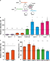
Fig. 1.
Fig. 1.. Changes in HS modifications by RNAi or overexpression of modifying enzymes affect HPV16 infectivity.
(A) Representation of a HS disaccharide with its modifications and the modifying enzymes, with OST, NDST, GLCE, GlcN, GlcA, and IdoA; circles represent sulfations. (B) HeLa cells were transfected with siRNAs against NDST1 and NDST2 (10 nM), 6OST-2 (10 nM), 3OST-3a (15 nM, pool), 2OST-1 (10 nM), and GLCE (10 nM). Two days post-transfection (p.t.), cells were infected with HPV16 PsV and fixed 48 hours post-infection (p.i.). Shown are average infection levels relative to control siRNA (10 nM, dotted line, 100%) ± SD. (C) HeLa cells were transfected with expression constructs of myc-tagged Sulf 1 and 2. Cells were infected 24 hours p.t. and fixed 48 hours p.i. Subsequently, cells were stained against myc and analyzed by flow cytometry, where transfected cells were gated and infection was scored as compared to the transfected control (dotted line, 100%). Displayed are averages of three independent experiments ± S.D. (D) Hela cells were transfected with eGFP-tagged constructs of 6OST-1, 6OST-2, 6OST-3, 3OST-2, 3OST-3a, and 2OST-1. Cells were infected 24 hours p.t. and fixed 48 hours p.i. Infection of transfected cells was scored by microscopy and is shown as the average of the relative infection (normalized to an eGFP transfection control) of three independent experiments. For all quantifications, a two-tailed Student’s t test was performed with *P < 0.05, **P < 0.01, ***P < 0.005, ****P < 0.0001, or nonsignificant (n.s.).
Fig. 2.
Fig. 2.. N-sulfation and 6O-sulfation are required for stable interaction with HPV16 capsids.
(A) To test for competition of desulfated and parental heparins for binding to cells (add-on assay), HPV16 PsV were incubated with polysaccharides for 1 hour at RT. This inoculum was added to cells and allowed to bind for 2 hours at 37°C. After washing off unbound virus, infection was allowed to proceed until 48 hours p.i., when cells were fixed and infection was scored by automated microscopy. Shown are infection levels relative to an untreated HPV16 control (dotted line, 100%) from three independent experiments ± SD. (B) Desulfated/parental heparins were preincubated at a concentration of 1 mg/ml with fluorescently labeled HPV16. Two hours after addition of the inoculum to cells, cells were fixed and analyzed by confocal microscopy. Shown are maximum intensity projections of confocal stacks with cell outlines in yellow and virus particles in white. Scale bars, 10 μm. (C) Virus binding on cells was quantified using the Fiji three-dimensional object counter plugin. The signal of cell-associated virus is depicted normalized to an untreated HPV16 control (dotted line). Depicted are averages of three independent experiments ± SD. For all quantifications, a two-tailed Student’s t test was performed with *P < 0.05, **P < 0.01, ***P < 0.005, or ****P < 0.0001.
Fig. 3.
Fig. 3.. N- and 6O-sulfation contribute to structural activation of HPV16.
(A) Schematic representation of the “seed-over assay” to test for structural activation of HPV16. (B) HPV16 PsV were pre-incubated with desulfated and parental heparins for 1 hour at RT and allowed to bind to undersulfated ECM derived from HaCaT cells for another hour. Infection of overseeded, undersulfated HeLa cells was scored 48 hours p.i. as a readout for structural activation. Shown are infection levels relative to untreated, add-on infection of HPV16 (full line), whereas background infection levels of the undersulfated, NaClO3-treated cells in seed-over condition are indicated by a dotted line. Depicted are averages of three independent experiments ± SD. (C and D) Exclusively N-sulfated heparin and the parental fully sulfated heparin were used in add-on (C) and seed-over (D) assays. Infection levels are shown relative to untreated HPV16 (full line) ± SD for three independent experiments. In (C), the dotted line represents background infection of NaClO3-treated cells as in (B). Please note that the supplier/batches of heparins are distinct for (B) versus (C)/(D). For all quantifications, a two-tailed Student’s t test was performed with *P < 0.05, **P < 0.01, or ***P < 0.005.
Fig. 4.
Fig. 4.. Quantifying HPV16-heparin bonds by AFM-based SMFS.
(A) Schematic illustration (created with BioRender.com) of the experimental setup of AFM-based SMFS. A single HPV16 virion is attached to an AFM probe via a PEG linker and brought into contact with a heparin-presenting surface for the repeated recording of extend and retract curves. (B) Representative retract curves recorded at 1 μm/s with a maximum applied load of 600 pN for specific unbinding events between HPV16 or fcHPV16 and indicated heparin surfaces. (C) Dynamic force spectra covering a range between 1 × 10³ and 5 × 10⁵ pN/s for HPV16 interactions with four heparin surfaces and fcHPV16 interaction with heparin, as indicated. Data are representative from at least two independently prepared AFM probes and heparin surfaces. Solid symbols represent mean rupture force obtained from fig. S7, and error bars represent SDs. The solid lines are best fits to the Bell-Evans model. (D) koff values extracted from the fits.(E) Plots of BP as a function of contact time are shown for the indicated HPV16-heparin interactions. Solid lines are the best fits to an exponential function (Eq. 2) to extract the interaction time (τ) needed to reach half maximum BP. Error bars represent the standard error of the mean (SEM) from three independent experiments. (F) kon values derived from BP plots. (G) Kd values derived from koff and kon from (D) and (F), respectively. (H) Box and whisker plot with 10 to 90 percentiles of unbinding force data for four heparin surfaces for data points = 1204 (heparin), 971 (2O-deS), 1088 (6O-deS), and 1217 (N-deS) from three (heparin and N-deS) and two (2O-deS and 6O-deS) independent experiments. P values were determined by one-way analysis of variance (ANOVA) test in GraphPad Prism (Brown-Frosythe and Welch ANOVA test). ****P < 0.0001, **P = 0.0033, and *P = 0.0119.
Fig. 5.
Fig. 5.. Proposed model of the contribution of HS sulfation moieties to virus binding kinetics and structural activation.
(A) Schematic illustration of four binding sites (inset) on the HPV16 capsomer. (B) N-, 6O-, and 2O-sulfation moieties contribute to the association of HS. (C) Predominantly, N-sulfation controls unbinding.(D) 6O-sulfation contributes to structural activation by providing mechanical stability against HS bending strain with N-sulfation contributing low koff rates. The individual contributions of specific sulfations are depicted separately but are likely to occur close to instantaneously upon engagement.

References

    1. J. D. Esko, K. Kimata, U. Lindahl, “Proteoglycans and sulfated glycosaminoglycans” in Essentials of Glycobiology, A. Varki, R. D. Cummings, J. D. Esko, H. H. Freeze, P. Stanley,C. R. Bertozzi, G. W. Hart, M. E. Etzler, Eds. (Cold Spring Harbor, ed. 2, 2009). - PubMed
    1. Turnbull J., Powell A., Guimond S., Heparan sulfate: Decoding a dynamic multifunctional cell regulator. Trends Cell Biol. 11, 75–82 (2001). - PubMed
    1. Liu J., Thorp S. C., Cell surface heparan sulfate and its roles in assisting viral infections. Med. Res. Rev. 22, 1–25 (2002). - PubMed
    1. Ströh L. J., Stehle T., Glycan engagement by viruses: Receptor switches and specificity. Annu. Rev. Virol. 1, 285 (2014). - PubMed
    1. Esko J. D., Selleck S. B., Order out of chaos: Assembly of ligand binding sites in heparan sulfate. Annu. Rev. Biochem. 71, 435–471 (2002). - PubMed

MeSH terms

Substances

LinkOut - more resources