. 2024 Oct 4;10(40):eado7120.
doi: 10.1126/sciadv.ado7120. Epub 2024 Oct 4.

BCL-XL-targeting antibody-drug conjugates are active in preclinical models and mitigate on-mechanism toxicity of small-molecule inhibitors

Affiliations

BCL-XL-targeting antibody-drug conjugates are active in preclinical models and mitigate on-mechanism toxicity of small-molecule inhibitors

Andrew S Judd et al. Sci Adv. .

Abstract

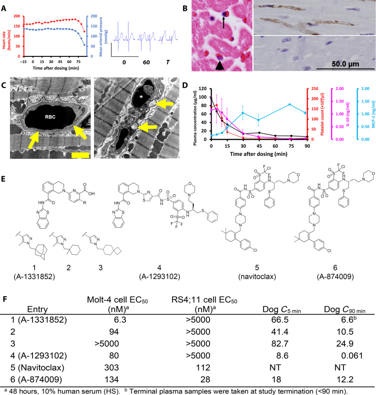
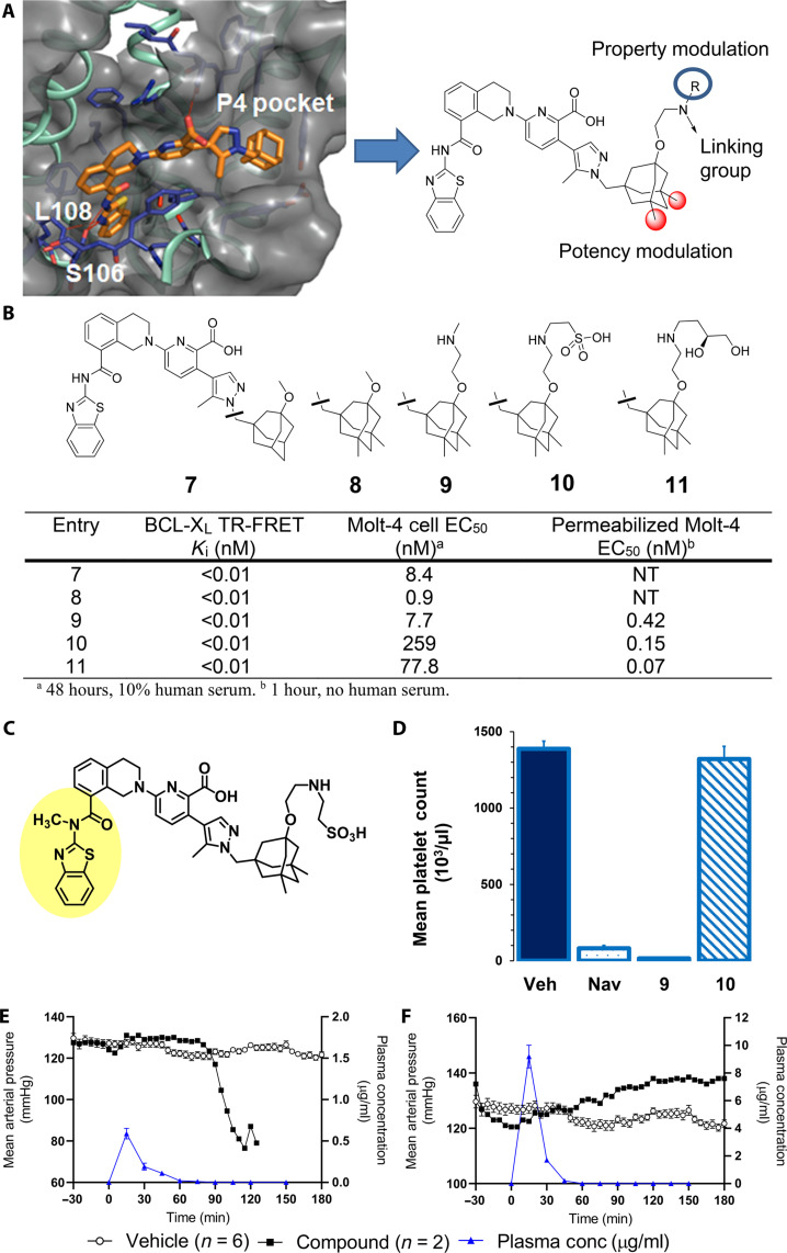
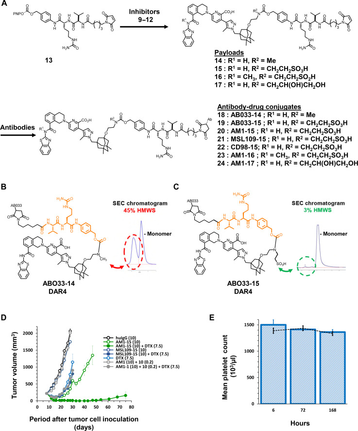
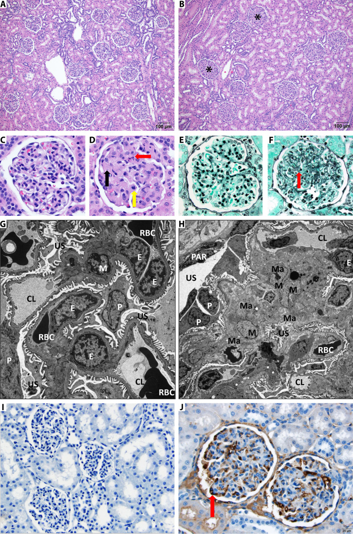
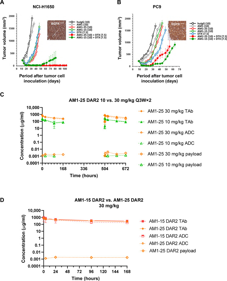
Overexpression of the antiapoptotic protein B-cell lymphoma-extra large (BCL-XL) is associated with drug resistance and disease progression in numerous cancers. The compelling nature of this protein as a therapeutic target prompted efforts to develop selective small-molecule BCL-XL inhibitors. Although efficacious in preclinical models, we report herein that selective BCL-XL inhibitors cause severe mechanism-based cardiovascular toxicity in higher preclinical species. To overcome this liability, antibody-drug conjugates were constructed using altered BCL-XL-targeting warheads, unique linker technologies, and therapeutic antibodies. The epidermal growth factor receptor-targeting antibody-drug conjugate AM1-15 inhibited growth of tumor xenografts and did not cause cardiovascular toxicity nor dose-limiting thrombocytopenia in monkeys. While an unprecedented BCL-XL-mediated toxicity was uncovered in monkey kidneys upon repeat dosing of AM1-15, this toxicity was mitigated via further drug-linker modification to afford AM1-AAA (AM1-25). The AAA drug-linker has since been incorporated into mirzotamab clezutoclax, the first selective BCL-XL-targeting agent to enter human clinical trials.


Figures

Fig. 1.
Fig. 1.. BCL-XL–selective inhibitors cause on-target cardiovascular toxicity in dogs.
(A) In instrumented, anesthetized beagle dogs’ heart rate and mean arterial blood pressure drop precipitously approximately 60 min following an intravenous bolus dose (2.5 mg/kg) of A-1331852 (left; n = 2). Marked reduction in R-wave amplitude in electrocardiogram tracings at 0 and 60 min after dosing (right). (B) Left: Hematoxylin and eosin–stained heart demonstrates nuclear pyknosis of an endothelial cell (arrow) and a comparatively normal nucleus (arrowhead). Top right: Cardiac endothelial cells are immunoreactive (brown) for activated caspase-3 in an A-1331852 (2.5 mg/kg)–treated dog 90 min after dosing, whereas no activated caspase-3 is detected in a dog treated with structurally related inactive compound 3 (2.5 mg/kg) that does not cause cardiovascular collapse (bottom right). (C) Electron photomicrograph of a myocardial capillary from dog treated intravenously with active compound 2 (left), approximately 90 min after an intravenous dose (2.5 mg/kg). An erythrocyte [red blood cell (RBC)] is present within a well-defined capillary (arrows). Right: The endothelium is collapsed with condensed nuclear material (*) and intracytoplasmic vesicles (arrows) approximately 90 min following an intravenous dose (2.5 mg/kg) of a potent SMI 2. Scale bar, 2 μm (on electron micrograph). (D) Plasma A-1331852 is rapidly cleared following an intravenous dose (2.5 mg/kg), shown as mean from the same two dogs. Platelet count and serum IL-10 rapidly fall from baseline levels following dosing, whereas serum MCP-1 increases and remains elevated from 30 min after dosing. Error bars are SEM. (E and F) Structure, binding affinity, and cellular potency of molecules used in anesthetized dog studies and navitoclax, as well as the plasma concentration (in micrograms per milliliter) of each molecule evaluated in the dog studies at 5 and 90 min after initiation of study. The RS4;11 and Molt-4 cell lines are dependent on BCL-2 and BCL-XL, respectively. NT, not tested.
Fig. 2.
Fig. 2.. Discovery of linkable BCL-XL inhibitors with different solubility and permeability properties affords putative ADC payload component with high target affinity yet minimal systemic toxicity in mice and dogs.
(A) X-ray cocrystal structure of BCL-XL bound to A-1331852 (1.93 Å; Protein Data Bank code 9AQZ; see the Supplementary Materials) and key regions of the BCL-XL inhibitor pharmacophore for modification as derived from the x-ray structure. (B) Binding, cellular potency in Molt-4 cells, and functional potency in digitonin-permeabilized Molt-4 cells of BCL-XL inhibitors. (C) Structurally related inactive inhibitor 11; methylation of P2-binding amide bond nitrogen disrupts key hydrogen binding network and leads to full ablation of cellular activity. (D) Platelet count 6 hours after treatment with BCL-XL inhibitors in heparinized whole mouse blood. Navitoclax (50 mg/kg) was given orally, and 9 (0.2 mg/kg) and 10 (0.6 mg/kg) were intravenously administered. Each bar represents the average platelet count in five SCID/bg mice. Error bars depict the SEM. (E and F) MAP of dogs administered vehicle (black filled circles) or BCL-XL inhibitor (open circles) and plasma concentration of BCL-XL inhibitors (blue triangles) following an intravenous bolus dose of either 9 or 10.
Fig. 3.
Fig. 3.. Modification of payload leads to ADC (AM1-15) with robust in vivo efficacy and no thrombocytopenia in mice.
(A) General synthetic scheme for coupling inhibitors 9 to 12 to linker 13 to afford drug-linkers 14 to 17 and subsequent conjugation of drug-linkers 14 to 17 with antibodies targeting EGFR (AB033 and AM1), CD98 or a nontargeted control (MSL109) immunoglobulin G1 (IgG1) antibody to generate ADCs 18 to 24. (B and C) Aggregation profiles of ADCs AB033-14 and AB033-15 in pH 7.4 buffer as assessed by size exclusion chromatography (SEC) and presence of HMWS. (D) The volume of subcutaneous H1650 tumors was plotted as a function of the time after tumor inoculation. SCID/bg mice with a subcutaneous tumor of approximately 200 mm3 were treated with AM1-15 or MSL109-15 at dose levels of 10 mg/kg. The conjugates were given as either single agent (open symbol) or in combination with docetaxel (DTX) (closed symbol). Treatment started at day 9 after tumor inoculation. An unconjugated mixture of AM1 and free inhibitor 10 was administered with (closed symbol) or without docetaxel (open symbol). Conjugates, antibody, and inhibitor 10 were intraperitoneally administered at a QDx1 (every day for one dose) regimen. Docetaxel was administered intravenously at 7.5 mg/kg (QDx1). Dose levels of each treatment (in milligrams per kilogram) are specified between brackets in the legend. Each point of a curve represents the arithmetic mean of the volumes of eight tumors. Error bars depict the SEM. (E) Platelet count after treatment with AM1-15 (bars) compared to treatment with vehicle (dashed line) in heparinized whole mouse blood. AM1-15 was administered intravenously at 30 mg/kg. Each bar and data point represent the average platelet count in five mice. Error bars depict the SEM.
Fig. 4.
Fig. 4.. Multiple dose administration of AM1-15 to cynomolgus monkeys causes unique renal toxicity.
Hematoxylin and eosin–stained kidney from control (A) and treated cynomolgus monkey (B). There is diffuse thickening of glomerular matrix in the AM1-15–treated kidney (asterisk). Scale bar, 100 μm. At high magnification as compared to control monkey glomeruli (C), the AM1-15–treated glomeruli (D) has global thickening of glomerular matrix by eosinophilic material (black arrow), and occasional degenerate cells (red arrow) and mitotic figures (yellow arrow) are also present. Periodic acid methenamine stain revealed an increased mesangial matrix component (red arrow) in the AM1-15–treated glomeruli (F) as compared to control monkey glomeruli (E). Electron microscopy revealed enlarged mesangial cell with increased cytoplasm (hypertrophy) along with moderate increase in extracellular mesangial matrix deposition in the AM1-15–treated glomeruli (H). The rest of the glomerular components, namely, endothelial cells, podocytes, and filtration apparatus, was within normal limit as compared to control glomeruli (G). M, mesangial cell; Ma, mesangial matrix; E, endothelial cells; P, podocyte; PAR, parietal epithelium; US, urinary space; CL, capillary space; RBC, red blood cell. Magnification, ×6000 (G) and ×3000 (H). (I and J) Anti–BCL-XL drug-linker–specific monoclonal antibody was synthesized, and IHC was performed to visualize the distribution of drug-linker. Moderate-to-strong immunoreactivity was observed in glomerular endothelial cells (red arrow) in a cynomolgus monkey administered AM1-15 (J) (scale bar, 20 μm) as compared to control (I).
Fig. 5.
Fig. 5.. Monkey pharmacokinetics of AM1-15 DAR4 and purified DAR2 and modification of drug-linker for solubility enable the utility of semipermeable inhibitor 11 in the context of drug-like ADC AM1-25.
(A) AM1-15 (broad DAR4) and AM1-15 DAR2 show linear pharmacokinetics in monkeys, and DAR purification to 2 reduces free payload in circulation. (B) Components of intact drug-linker 15 and aggregation profile of corresponding DAR4 AM1-15 ADC in pH 7.4 buffer as determined by SEC. VC, valine-citrulline; MC, maleimidocaproyl. (C) Components of enabling drug-linker 25 including solubilized para-aminobenzyloxy (p-ABO) spacer, valine-alanine (VA) dipeptide trigger unit and solubilized antibody attachment unit, and aggregation profile of corresponding DAR4 AM1-15 ADC in pH 7.4 buffer as determined by SEC.
Fig. 6.
Fig. 6.. AM1-25 inhibits tumor growth in vivo and exhibits linear pharmacokinetics in monkeys.
(A and B) The volume of subcutaneous tumors was plotted as a function of the time after tumor inoculation. SCID/bg mice with a subcutaneous tumor of approximately 200 mm3 were treated with docetaxel, huIgG, AM1, AM1-25, or AM1-15 (open symbols) or a combination of AM1-25 or AM1-15 with docetaxel (closed symbols). The numbers in the legend between parentheses indicates the dose levels (in milligrams per kilogram). Treatment started at day 10 after tumor inoculation. Conjugates and antibody were intraperitoneally administered at a Q7D×2 regimen. Docetaxel was administered intravenously at 7.5 mg/kg (QDx1). Each point of a curve represents the arithmetic mean of the volumes of eight tumors. Error bars depict the SEM. The inset shows EGFR expression of a xenograft as revealed by IHC for H1650 (A) and EBC-1 (B). Concentration-time profiles of TAb, conjugated antibody (ADC), and unconjugated payload following two intravenous doses (10 or 30 mg/kg) of AM1-25 (C), or AM1-15 (D) 3 weeks apart in cynomolgus monkeys. Data are presented as means ± SD (N = 3).

References

    1. Hanahan D., Weinberg R. A., Hallmarks of cancer: The next generation. Cell 144, 646–674 (2011). - PubMed
    1. Frenzel A., Grespi F., Chmelewskij W., Villunger A., Bcl2 family proteins in carcinogenesis and the treatment of cancer. Apoptosis 14, 584–596 (2009). - PMC - PubMed
    1. Khan N., Kahl B., Targeting BCL-2 in hematologic malignancies. Target. Oncol. 13, 257–267 (2018). - PubMed
    1. Amundson S. A., Myers T. G., Scudiero D., Kitada S., Reed J. C., Fornace A. J. Jr., An informatics approach identifying markers of chemosensitivity in human cancer cell lines. Cancer Res. 60, 6101–6110 (2000). - PubMed
    1. Vogler M., Targeting BCL2-proteins for the treatment of solid tumours. J. Adv. Med. 2014, 1–14 (2014). - PMC - PubMed