. 2024 Nov:77:103383.
doi: 10.1016/j.redox.2024.103383. Epub 2024 Oct 2.

Mechanism of action and impact of thiol homeostasis on efficacy of an enzyme replacement therapy for classical homocystinuria

Affiliations

Mechanism of action and impact of thiol homeostasis on efficacy of an enzyme replacement therapy for classical homocystinuria

Thilo Magnus Philipp et al. Redox Biol. 2024 Nov.

Abstract

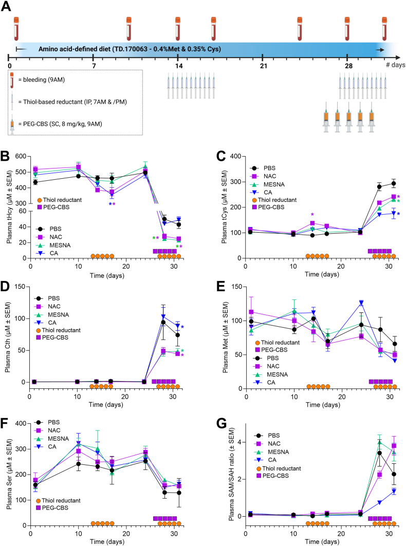
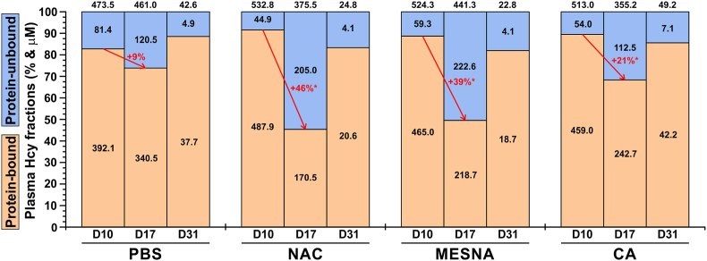
Homocystinuria (HCU) due to cystathionine beta-synthase (CBS) deficiency is characterized by elevated plasma and tissue homocysteine levels. There is no cure, but HCU is typically managed by methionine/protein restriction and vitamin B6 supplementation. Enzyme replacement therapy (ERT) based on human CBS has been developed and has shown significant efficacy correcting HCU phenotype in several mouse models by bringing plasma total homocysteine below the clinically relevant 100 μM threshold. As the reactive nature of homocysteine promotes disulfide formation and protein binding, and ERT is unable to normalize plasma total homocysteine levels, the mechanism of action of ERT in HCU remains to be further characterized. Here we showed that only a reduced homocysteine serves as a substrate for CBS and its availability restricts the homocysteine-degrading capacity of CBS. We also demonstrated that cells export homocysteine in its reduced form, which is efficiently metabolized by CBS in the culture medium. Availability of serine, a CBS co-substrate, was not a limiting factor in our cell-based model. Biological reductants, such as N-acetylcysteine, MESNA or cysteamine, increased the availability of the reduced homocysteine and thus promoted its subsequent CBS-based elimination. In a transgenic I278T mouse model of HCU, administration of biological reductants significantly increased the proportion of protein-unbound homocysteine in plasma, which improved the efficacy of the co-administered CBS-based ERT, as evidenced by significantly lower plasma total homocysteine levels. These results clarify the mechanism of action of CBS-based ERT and unveil novel pharmacological approaches to further increase its efficacy.

Keywords: Cystathionine beta-synthase; Disulfide isomerization; Enzyme replacement therapy; Homocysteine; Homocystinuria; N-acetylcysteine; Plasma redox status.


Conflict of interest statement

Declaration of competing interest TM is an inventor on patents related to pegtibatinase, provides ad-hoc consulting to Travere Therapeutics and receives research support from Travere Therapeutics. TB receives compensation for metabolomic analyses from Travere Therapeutics. WC, KL and SR are current or former employees and stockholders of Travere Therapeutics, which clinically develops pegtibatinase as an enzyme replacement therapy for classical homocystinuria. TMP and CS declare no conflicting interests.

Figures

Image 1
Graphical abstract
Fig. 1
Fig. 1
Validation of Probe 1 for quantification of homocysteine and cysteine in biological samples. The tHcy (A, D) and tCys (B, E) were quantified in plasma of I278T mice fed an amino acid-defined diet with different amount of Met using either the fluorescent Probe 1 (10 μM final concentration in the assay; A, B) or LC-MS/MS (D, E) as a current standard. Samples were measured as single values (LC-MS/MS) or technical triplicates (Probe 1) and presented as means ± SD (n = 5–6). Panels C and F show correlation between plasma tHcy and tCys as determined by Probe 1 and LC-MS/MS, respectively. Panels G and H show correlation between Probe 1 and LC-MS/MS quantification of tHcy and tCys, respectively. Inset in the bottom right corner shows structure and basic properties of Probe 1.
Fig. 2
Fig. 2
Impact of reductant on Hcy availability and efficacy of CBS to degrade it in vitro. (A) To mimic concentrations found in HCU plasma, PBS was supplemented with 120 μM Ser and either reduced thiols (360 μM Hcy, 130 μM Cys; grey) or their equimolar oxidized disulfide forms (180 μM homocystine, 65 μM cystine; purple). Additional 500 μM Ser was added to one set of samples containing the reduced thiols (black). Subsequently, 25 ng/μl CBS45 was added and Hcy elimination was monitored using Probe 1 (10 μM). (B) Pooled plasma from I278T mice (black) was supplemented with 25 ng/μl CBS45 alone (purple) or in combination with 400 μM NAC (lime) or mixture containing 400 μM NAC and 400 μM Ser (blue). NAC or NAC + Ser combination was repeatedly added as indicated by the arrows. Data are displayed as relative means ± SD (n = 4). (C) Similarly, pooled plasma from WT mice (black) was supplemented with 25 ng/μl CBS45 alone (purple) or in combination with 400 μM NAC (lime) or 400 μM Ser (blue). Addition of 400 μM Hcy in 10 min increments is indicated by the arrows. Concentration of tHcy were determined by using Probe 1 (10 μM). Data are displayed as relative means ± SD (n = 4). (D) HepG2 cells were seeded at the density of 300,000 cells per well into a 6-well plate. Standard media or media containing additional 2 mM Met were supplemented with PBS for controls (black, grey), 25 ng/μl CBS45 (blue, purple) or 25 ng/μl CBS45 in combination with 1 mM Ser (light blue, pink). The tHcy in media was quantified by using Probe 1 (10 μM) at designated timepoints with the inset zooming in at the last time point after 96 h. (E) HepG2 cells were seeded at the density of 600,000 cells per well into a 6-well plate and were incubated untreated for 48 h to generate a substantial fraction of oxidized Hcy (homo-/hetero-disulfides, protein-bound). Thereafter (indicated by the arrow), media were supplemented with equal volumes of PBS (black), 25 ng/μl CBS45 (grey) or combination of 25 ng/μl CBS with 2 mM NAC (purple), MESNA (lime) or CA (blue). The tHcy in media was quantified by using Probe 1 (10 μM) at designated timepoints with the inset zooming in at the last time point after 96 h (48 h after the treatment). Data are displayed as mean ± SD (n = 4) and were statistically analyzed by ordinary one-way ANOVA with Tukey's multiple comparison test against PBS controls. (F) Pooled I278T plasma was treated with different concentrations (0–3 mM) of NAC (purple), MESNA (lime), CA (blue), Vitamin C (black) and Trolox (grey). Subsequently, samples (n = 4 each group) were precipitated and protein-unbound Hcy was quantified in the clarified supernatants by using Probe 1 (10 μM). (G) Reductants NAC (purple), MESNA (lime) and CA (blue) were tested for their ability to compete with Cys as the first substrate forming PLP-aminoacrylate adduct in CBS catalytic site. The CBS specific activity was determined by methylene blue assay quantifying generated H2S as a common by-product of CBS-catalyzed condensations. Data represent means ± SDs (n = 4 each group). (H) Similarly, reductants NAC (purple), MESNA (lime) and CA (blue) were tested for their ability to compete with Hcy as the second substrate attacking PLP-aminoacrylate adduct using Cys as the first substrate. The CBS specific activity was determined by methylene blue assay quantifying generated H2S as a common by-product of CBS-catalyzed condensations (n = 4 each group). Data are presented as mean ± SD (n = 4 each group).
Fig. 3
Fig. 3
Preparation of PEG-CBS. Three linear 5, 10, and 20 kDa NHS ester PEGs (5NHS, 10NHS, and 20NHS) were used to modify human CBS40 and yeast tyCBS in a variable molar excess of PEG to CBS subunit as indicated in the figure. Newly generated functional analogs of pegtibatinase were compared to a previously evaluated unmodified CBS45 and its PEGylated version OT-58 (also known as pegtibatinase). (A) Unmodified enzymes and their PEGylated conjugates were resolved on a 10 % Miniprotein TGX SDS-PAGE (10 μg/lane) and stained with Safe Stain (Invitrogen) according to manufacturer's recommendation. The arrow points to subunits of unmodified enzymes, while the bracket encompasses the region where their PEGylated conjugates migrated. (B) Unmodified enzymes and their PEGylated conjugates were assayed using radiometric CBS activity assay and compared. Bars represent average from four separate measurements and error bars designate SEMs. Asterisks (∗ = p < 0.05) designate a significantly increased CBS specific activity of tyCBS compared to CBS45.
Fig. 4
Fig. 4
Evaluation of the thiol-based reductants in I278T mice. Four I278T mouse cohorts (n = 6–8 each) were acclimated to the amino acid-defined diet containing 0.4 % Met and received vehicle (PBS, black), NAC (purple), MESNA (lime) or CA (blue) alone and after a washout period received the same in combination with PEG-CBS as described in detail in the main text. The orange circles and purple squares denote administration days of PBS or reductants via intraperitoneal injections and PEG-CBS via subcutaneous injections, respectively. (A) Schematics of a mouse study. Plasma tHcy (B), tCys (C), Cth (D), methionine (E), serine (F) and SAM/SAH ratio (G). Color-coded asterisks denote significance (∗ = p < 0.05).
Fig. 5
Fig. 5
Distribution of plasma Hcy fractions during evaluation of thiol-based reductants in I278T mice. Plots display relative proportions of protein-bound (light brown) and protein-unbound (light blue) Hcy of plasma tHcy pool (representing 100 %) at the baseline (D10), after the treatment with thiol-based reductants (D17) and after the co-administration of PEG-CBS and thiol-based reductants (D31) in I278T cohorts designated as PBS (control), NAC, MESNA and CA. Black numbers within and above the bars correspond to average concentrations in protein-bound/unbound Hcy and plasma tHcy, respectively. Red color and arrows designate a change in relative proportion of protein-bound versus protein-unbound plasma Hcy with a significance of the change (∗ = p < 0.05).

References

    1. Mudd S.H., Levy H.L., Kraus J.P. In: The Metabolic and Molecular Bases of Inherited Disease. Scriver C.R., Beaudet A.L., Sly W.S., Valle D., Childs B., Kinzler K., Vogelstein B., editors. McGraw-Hill; New York: 2001. Disorders of transsulfuration; pp. 2007–2056.
    1. Brosnan J.T., Brosnan M.E., Bertolo R.F.P., Brunton J.A. Methionine: a metabolically unique amino acid. Livest. Sci. 2007;112:2–7.
    1. Zuhra K., Augsburger F., Majtan T., Szabo C. Cystathionine-beta-Synthase: molecular regulation and pharmacological inhibition. Biomolecules. 2020;10:697. - PMC - PubMed
    1. Jakubowski H. Pathophysiological consequences of homocysteine excess. J. Nutr. 2006;136:1741S–1749S. - PubMed
    1. Barber G.W., Spaeth G.L. The successful treatment of homocystinuria with pyridoxine. J. Pediatr. 1969;75:463–478. - PubMed

Publication types

LinkOut - more resources