Review
. 2024 Sep 18;48(5):fuae024.
doi: 10.1093/femsre/fuae024.

Biodiversity of microorganisms in the Baltic Sea: the power of novel methods in the identification of marine microbes

Affiliations
Review

Biodiversity of microorganisms in the Baltic Sea: the power of novel methods in the identification of marine microbes

Hanna Mazur-Marzec et al. FEMS Microbiol Rev. .

Erratum in

Abstract

Until recently, the data on the diversity of the entire microbial community from the Baltic Sea were relatively rare and very scarce. However, modern molecular methods have provided new insights into this field with interesting results. They can be summarized as follows. (i) Although low salinity causes a reduction in the biodiversity of multicellular species relative to the populations of the North-East Atlantic, no such reduction occurs in bacterial diversity. (ii) Among cyanobacteria, the picocyanobacterial group dominates when considering gene abundance, while filamentous cyanobacteria dominate in means of biomass. (iii) The diversity of diatoms and dinoflagellates is significantly larger than described a few decades ago; however, molecular studies on these groups are still scarce. (iv) Knowledge gaps in other protistan communities are evident. (v) Salinity is the main limiting parameter of pelagic fungal community composition, while the benthic fungal diversity is shaped by water depth, salinity, and sediment C and N availability. (vi) Bacteriophages are the predominant group of viruses, while among viruses infecting eukaryotic hosts, Phycodnaviridae are the most abundant; the Baltic Sea virome is contaminated with viruses originating from urban and/or industrial habitats. These features make the Baltic Sea microbiome specific and unique among other marine environments.

Keywords: Baltic Sea; diversity of microorganisms; marine ecosystem; marine viruses; molecular methods; prokaryotic and eukaryotic microorganisms.


Conflict of interest statement

The authors declare no conflict of interest.

Figures

Figure 1.
Figure 1.
Map of the Baltic Sea with the surface salinity and catchment area, and indication of major deeps. Salinity levels are indicated, with a scale representing regions of the sea with corresponding concentrations of NaCl. The catchment is shown as an area around the sea region. Vertical distributions of salinity at Bornholm (IDEAL), Gdansk (P1), and Gotland (BY15) Deeps within 2020–2023 (Szymczycha et al. 2024) are indicated.
Figure 2.
Figure 2.
Epifluorescent microphotographs of bacterial cells from surface coastal waters of the Baltic Sea. DAPI (4',6-diamidino-2-phenylindole dye) staining is shown in panels A and B. The live/dead staining (with SYTO 9 and propidium iodide dyes), where alive cells and dead cells are marked by fluorescence, is shown in panels C and D, respectively. Photographs taken by Ewa Kotlarska.
Figure 3.
Figure 3.
Microphotographs of cyanobacteria from the Baltic Sea. Cells of the following species are shown: Aphanizomenon flos-aquae (A), N. spumigena (B), and Dolichospermum lemmermannii (C). Photographs taken by Justyna Kobos.
Figure 4.
Figure 4.
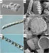
Microphotographs of diatoms from the Baltic Sea. Cells of the following species are shown in images obtained using light (A, C, and E) and electron (B, D, and F) microscopic techniques: Gedaniella mutabilis (A and B), P. trainorii (C and D), and Gedaniella flavoviren (E and F). Photographs taken by Przemysław Dąbek.
Figure 5.
Figure 5.
Microphotographs of dinoflagellates from the Baltic Sea. Cells of the following species are shown in images obtained using light microscopy (A and C), Imaging Flow Cytobot (B), and electron microscopy (D): Tripos mulleri (A and B), Peridiniella catenata (C), and Dinophyis acuminata (D). Photographs taken by Maria Karlberg and Bengt Karlson.
Figure 6.
Figure 6.
Microphotograph of a ciliate from the Baltic Sea. Cells of Mesodinium major are shown in the microphotograph. Photograph taken by Krzysztof Rychert.
Figure 7.
Figure 7.
Photographs of fungi from the Baltic Sea. Ascomycota perithecium on F. vesiculosus blade (A and B), and colonies of Rhodotorula sp. (C and D) and Fusarium sp. (E) are shown. Photographs taken by Marija Kataržytė.
Figure 8.
Figure 8.
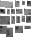
Electron microphotographs of bacteriophages from Baltic Sea samples. Several morphotypes are shown, including myoviruses (B, D, E, G, H, K, L, and N), podoviruses (M), and siphoviruses with either short (C, F, M, and Q) or extremely long tails (A, J, O, and P), and prolate heads (I, M, and Q). The bar corresponds to 200 nm. Reproduced from Jakubowska-Deredas et al. (2012), with permission of the publisher (Elsevier Masson SAS).
Figure 9.
Figure 9.
The key elements and features of the unique diversity of Baltic microorganisms. A Cyske rosette chart showing major groups of Baltic microorganisms, indicating key elements and/or features of microbial diversity among each group.

References

    1. Aevarsson A, Kaczorowska AK, Adalsteinsson BT et al. Going to extremes—a metagenomic journey into the dark matter of life. FEMS Microbiol Lett. 2021;368:fnab067. - PubMed
    1. Ahmed MN, Wahlsten M, Jokela J et al. Potent inhibitor of human trypsin from the aeruginosin family of natural products. ACS Chem Biol. 2021;16:2537–46. 10.1021/acschembio.1c00611. - DOI - PMC - PubMed
    1. Ahrens R, Moll G. Ein neues knospendes bakterium aus der Ostsee. (A new budding bacterium from the Baltic Sea). Archiv Mikrobiol. 1970;70:243–65. - PubMed
    1. Akinbowale OL, Peng H, Barton MD. Antimicrobial resistance in bacteria isolated from aquaculture sources in Australia. J Appl Microbiol. 2006;100:1103–13. 10.1111/j.1365-2672.2006.02812.x. - DOI - PubMed
    1. Alacid E, Reñé A, Gallisai R et al. Description of two new coexisting parasitoids of blooming dinoflagellates in the Baltic Sea: Parvilucifera catillosa sp. nov. and Parvilucifera sp. (Perkinsea, Alveolata). Harmful Algae. 2020;100:101944. 10.1016/j.hal.2020.101944. - DOI - PubMed