. 2024 Oct 4;15(1):8606.
doi: 10.1038/s41467-024-52573-2.

Reversible covalent c-Jun N-terminal kinase inhibitors targeting a specific cysteine by precision-guided Michael-acceptor warheads

Affiliations

Reversible covalent c-Jun N-terminal kinase inhibitors targeting a specific cysteine by precision-guided Michael-acceptor warheads

Dániel Bálint et al. Nat Commun. .

Abstract

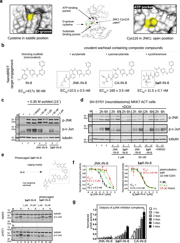
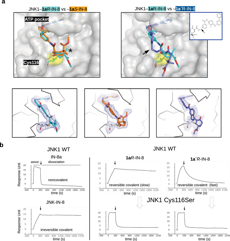
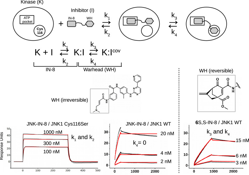
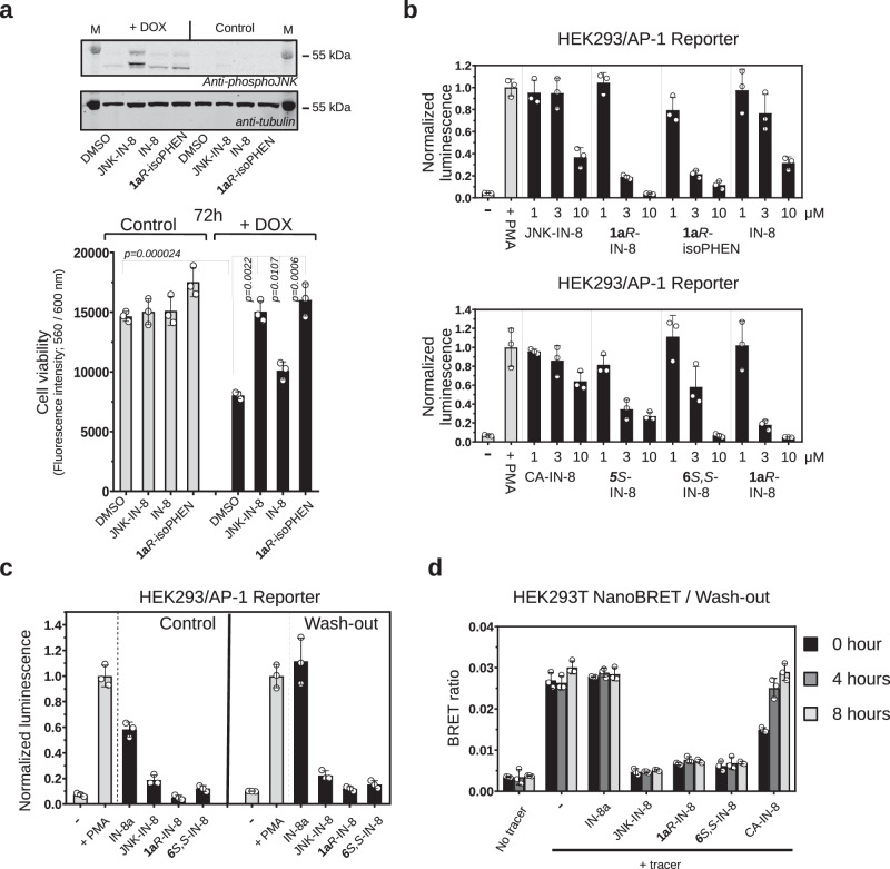
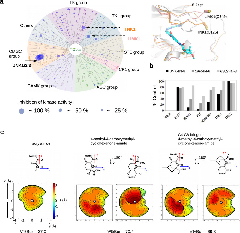
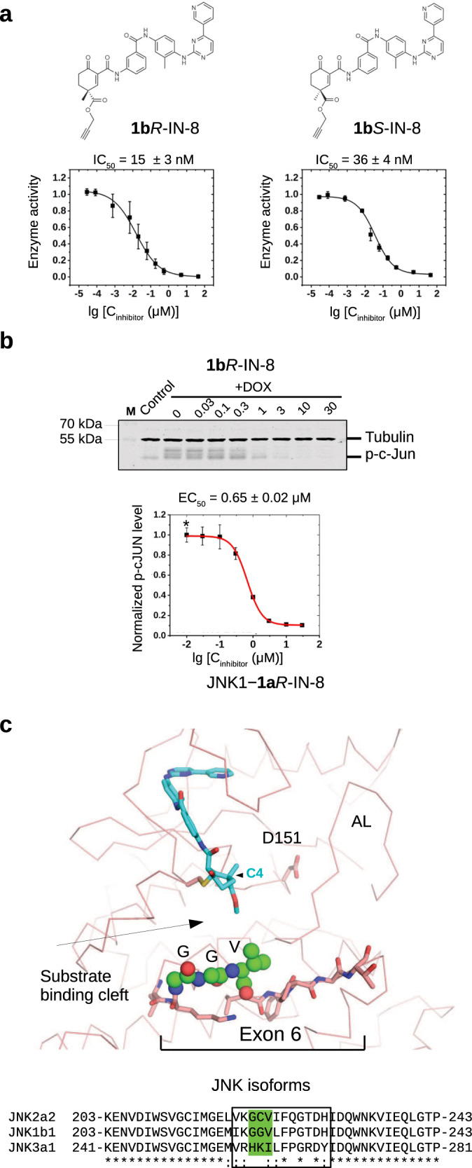
There has been a surge of interest in covalent inhibitors for protein kinases in recent years. Despite success in oncology, the off-target reactivity of these molecules is still hampering the use of covalent warhead-based strategies. Herein, we disclose the development of precision-guided warheads to mitigate the off-target challenge. These reversible warheads have a complex and cyclic structure with optional chirality center and tailored steric and electronic properties. To validate our proof-of-concept, we modified acrylamide-based covalent inhibitors of c-Jun N-terminal kinases (JNKs). We show that the cyclic warheads have high resilience against off-target thiols. Additionally, the binding affinity, residence time, and even JNK isoform specificity can be fine-tuned by adjusting the substitution pattern or using divergent and orthogonal synthetic elaboration of the warhead. Taken together, the cyclic warheads presented in this study will be a useful tool for medicinal chemists for the deliberate design of safer and functionally fine-tuned covalent inhibitors.


Conflict of interest statement

A.R., T.S., Á.L.P., D.B., A.A., P.S., L.T., K.A., T.I., E.SZ. and R.P. are inventors in a patent application pending approval (PCT/HU2023/050079) on the use of cyclic designer scaffolds for the covalent targeting of proteins via Michael addition. The remaining authors declare no competing interests.

Figures

Fig. 1
Fig. 1. JNK inhibitors containing different chemical warheads.
a Position of Cys116 in JNK1 compared to the MAPK D-groove cysteine. Left panels show the ERK2 D-groove in surface representation with Cys161 highlighted in yellow (PDB ID: 2ERK). This cysteine, similarly to other MAPK D-groove cysteines from p38α and JNK1, is more buried since it is located in a less accessible (saddle) position compared to the JNK unique cysteine (Cys116 in JNK1) located next to the ATP-pocket, which is more open. The middle panel shows the relative position of these two cysteines on JNK1 (cysteine side-chain atoms are shown with spheres), while the panel on the right shows the JNK1 surface area around Cys116 (colored in yellow). b Comparison of three covalent JNK inhibitors comprised of the IN-8 ATP-pocket binding moiety and a Michael acceptor based warhead (acrylamide – irreversible, cyanoacrylamide – reversible, and cyclohexenone – reversible; * indicates the Michael acceptor carbon (β or C3) forming the C-S bond; the numbering of carbon atoms in the cyclohexenone ring is indicated: C2 and C4 contain electron-withdrawing amide or ester groups increasing the reactivity of C3; C4 is an asymmetric center and the IN-8 directing group is linked to C2 via an amide). The EC50 values of JNK1 binding were measured in live HEK293 cells using the NanoBRET target engagement assay (the values show the mean and parameter error estimates are from weighted least squares method; n = 2, independent experiments, see Supplementary Fig. 1). c The impact of JNK-IN-8 and 1aR-IN-8 on sorbitol stimulated JNK and c-Jun phosphorylation in HEK293T cells. Cells were stimulated with sorbitol for 15 min which gives robust JNK activation. ‘-‘ indicates no treatment with inhibitors and +DMSO indicates treatment with DMSO used in the same amount (0.1%) as an organic solvent for the inhibitors. Inhibitors were added 2 h before stimulation and blots are representative of two independent experiments. Note the changed electrophoretic mobility of JNK in the presence of JNK-IN-8, indicating irreversible JNK–JNK-IN-8 binding (compare lane 4-5 with the other lanes). (p-JNK: western blot, WB, signal with phosphoJNK antibody; p-c-Jun: WB with phospho-c-Jun (Ser73) antibody where MAPK mediated c-Jun phosphorylation results in differently phosphorylated phospho-c-Jun (p-c-Jun) species with different electrophoretic mobility; the fastest migrating band is the least phosphorylated; Tubulin: WB with anti-tubulin antibody which serves as the load control; M: molecular marker). d Effects of JNK-IN-8 and 1aR-IN-8 on c-Jun phosphorylation in an engineered neuroblastoma cell line (SH-SY5Y MKK7 ACT). Phosphorylation of JNK (p-JNK) was stimulated by 2 or 6 h of doxycycline (DOX) treatment and blots are representative of two independent experiments. e Photocaged JNK inhibitor for light-controlled regulation of JNK activity. 1aR-IN-8 was linked to a photolysable moiety via its pyridine nitrogen, which blocked its capacity to bind into the JNK ATP-pocket. HEK293T cells were unstimulated (-) or stimulated with 10 μg/mL anisomycin (A) to turn JNK on. One set of experiment was carried out in the dark (DARK), while in the parallel experiment cells were illuminated briefly with 450 nm blue LED light (LIGHT) but otherwise also kept in the dark. The original inhibitor and its photocaged version were added in 1 or 10 μM concentrations and JNK-mediated c-Jun phosphorylation was monitored by western blots (and the panel shows a representative set of two independent experiments). Notice that the photocaged inhibitor blocks c-Jun phosphorylation only in LIGHT but not in DARK (compare Lane 7–10 between the upper and the lower panels). (tubulin was used as the load control; lower and upper bands show the position of hypophosphorylated or hyperphosphorylated c-Jun, respectively; p-c-Jun.). f Stability in a nucleophile rich environment. The inhibitory potential of JNK-IN-8 and 1aR-IN-8 were tested using the in vitro PhALC assay after pre-incubating the compounds for different amounts of time in 10 mM GSH (see Methods). In contrast to JNK-IN-8, the effect of 1aR-IN-8 on JNK is apparently intact since it forms only a weak and transient reversible covalent adduct with GSH. Data show the mean value and error bars show SD (n = 3, independent experiments). g Results of in vitro dialysis experiments. 50 nM phosphorylated JNK1 (pJNK1) was incubated with 10 μM inhibitor for 1.5 h, then samples were loaded into dialysis tubes and dialyzed in 50 mM HEPES pH = 7.4, 100 mM NaCl, 5 mM MgCl2, 2 mM DTT. The samples before the start of the dialysis experiment (0 h) and after the indicated times (1, 2, 3,4, 5 days) were tested for enzymatic activity using the PhALC assay (n = 3; independent experiments; data show the mean and error bars show SD). The control sample (used for normalization) was treated the same way but no inhibitor was added in the incubation step before the dialysis experiment. Source data are provided as a Source Data file.
Fig. 2
Fig. 2. Crystal structures of covalent inhibitors bound to JNK1.
a Structural comparisons of JNK1–1aR-IN-8, –1aS-IN-8, and –1a’R-IN-8 crystallographic complexes (with the following newly deposited PDB IDs: 8PTA, 8PT9, 8PT8, respectively). These show that the C4 stereogenic center could be used to direct the carboxymethyl group towards different directions in the substrate binding pocket next to Cys116 (left panel), or an additional methylene between the ATP-pocket binding moiety and the warhead necessitates a dramatically different conformational solution to form the cysteine covalent adduct (right panel). Lower panels display the Fo-Fc omit map for the cysteine-small molecule covalent adduct contoured at 1.5ϭ. b Validation of the cysteine mediated covalent bond by SPR experiments. Mutation of Cys116 to serine increases the kinetic dissociation rate validating the importance of covalent bond formation upon binding. Panels show the results of surface plasmon resonance (SPR) experiments with IN-8a (acetylated IN-8; noncovalent), JNK-IN-8 (irreversible covalent), 1aR-IN-8 (reversible covalent, slow dissociation) and 1a’R-IN-8 (reversible covalent, fast dissociation) binding to JNK1. Note that the association phase (assoc.) of the SPR curve is determined by the kon, while the dissociation by the koff. Compounds were injected over the JNK surface for 5 min at a concentration corresponding to their ~KD and the dissociation of the JNK-compound complex or adduct was monitored in time (the start of the dissociation phase is shown with an arrow). Note that the kinetic binding profiles of 1aR-IN-8 or 1a’R-IN-8 on the JNK1 C116S surface resembles that of IN-8a (the ATP-pocket binding moiety without the warhead) on the wild-type (WT) JNK1 surface, suggesting that Cys116 is indispensable for the decreased koff.
Fig. 3
Fig. 3. Inhibitor potency, binding energetics and covalent residence based on SPR data.
Scheme of the 2-step reversible kinetic binding model showing the relevant kinetic binding constants for noncovalent binding via the IN-8 ATP-binding moiety (k1 and k2) and for warhead-mediated covalent bond formation (k3 and k4). The panels at the bottom show the kinetic binding plots for JNK-IN-8 on the JNK1(Cys116Ser) mutant surface injected at 100, 300, and 1000 nM concentrations (KD ~ 200-300 nM) as well as the response curves on the wild-type JNK1 surface (WT) at three different concentrations. The analysis on the JNK1(Cys116Ser) mutant surface gives the value of k1 and k2 based on a one-site noncovalent binding model; these values were independently determined for each inhibitor this way (see Supplementary Table 1). In the case of the irreversible JNK-IN-8 inhibitor k4 = 0 (and thus only k3 was fit), while for the reversible inhibitors k3 and k4 were both numerically fit to the experimental kinetic curves obtained on the JNK1 WT surface (and the panel on the right with 6S,S-IN-8 shows an example of this, for further data see Supplementary Fig. 5). The experimental binding curves are shown in black and the fitted curves in red.
Fig. 4
Fig. 4. Characterization of covalent JNK inhibitors in cell-based tests.
a Effect of inhibitors on JNK mediated cell death in SH-SY5Y MKK7 ACT cells. Endogenous JNK activation was initiated artificially by the addition of 2 μg/mL doxycycline (DOX) for 72 h in engineered SH-SY5Y neuroblastoma cell line (in which the expression of an active MLK3-MKK7 chimera is controlled via the DOX dependent Tet-ON system). The top panel shows the results of a phospho-JNK western blot confirming JNK activation upon doxycycline treatment ( + DOX; the two different bands on the phospho-JNK western blot correspond to different JNK isoforms). The panel below shows the results of the experiment with 1 μM JNK-IN-8 (irreversible), IN-8 (the ATP-pocket binding moiety without any warhead) or the reversible covalent 1aR-isoPHEN. (Data show the mean value and error bars show SD, n = 3, independent experiments; p-values were calculated based on two-sided, unpaired t-test.). b Effect of inhibitors on JNK mediated AP-1 transcription factor promoter activity. Reporter AP-1 – HEK293 Recombinant Cell Line was unstimulated (-) or stimulated with phorbol 12-myristate 13-acetate ( + PMA) and AP-1 promoter driven transcription of the luciferase reporter gene was monitored by measuring luminescence after 6 h. Inhibitors were co-administered with PMA and were used in 1, 3, or 10 μM concentrations. (Data show the mean and error bars show SD, n = 3, independent experiments.). c Results of wash-out experiments with noncovalent (IN-8a), irreversible covalent (JNK-IN-8) and reversible covalent (1aR-IN-8 or 6S,S-IN-8) inhibitors in the AP-1 promoter assay. Inhibitors (10 μM) were incubated with Reporter AP-1 – HEK293 cells for 4 h then were left untreated (-) or stimulated with phorbol 12-myristate 13-acetate ( + PMA). AP-1 promoter activity was monitored by luminescence measurements after 6 h. Wash-out samples were washed by PBS twice before adding fresh media with PMA (but without any inhibitor), while for control cells the media, in addition to PMA, contained the respective inhibitor in the same concentration as before the wash-out. (Data show the mean and error bars show SD, n = 3, independent experiments.). d Results of wash-out experiments monitoring long-term target engagement by NanoBRET. HEK293T cells were transiently transfected with a NanoLuc-JNK1 fusion expression plasmid and the binding of a fluorescent JNK ATP-pocket binding compound (tracer) was monitored in the absence or presence of different JNK inhibitors used in 10 μM concentration. The BRET ratio is low when there was no tracer added (No tracer), while the maximum BRET signal is expected upon addition of the tracer with no inhibitor added (-). Cells were preincubated with the inhibitors for 2 h and washed three times with media followed by the addition of fresh media supplemented with the tracer. The remaining amount of the respective JNK-inhibitor complex was monitored by the BRET signal right after of the wash-out (0 h) or 4 and 8 h later. (Data show the mean and error bars show SD, n = 3, independent experiments.) Source data are provided as a Source Data file.
Fig. 5
Fig. 5. Specificity of 1aR-IN-8 in the human kinome panel and analysis of steric congestion near the reactive center of open-chain versus cyclic warheads.
a Results of the Wild Type Kinase Panel (Reaction Biology Corp, USA; 340 human kinases) with 1aR-IN-8 used at 1 μM concentration. Inhibition of a specific kinase is depicted with a circle on the human kinome tree, where circle size correlates with the amount of inhibition. The panel on the right shows the structural models of LIMK1 (PDB ID: 3S95) and TNK1 (homology model created by AlphaFold 2) superimposed with the JNK1–1aR-IN-8 crystallographic model. LIMK1 and TNK1 are the only two human off-target kinases whose activity were inhibited more than 50% (see Supplementary Table 2). b Off-target kinase panel (1 target + 6 off-target kinases) and compound binding tested by the DiscoverX platform. Compounds were used in 1 μM concentration. Note that JNK3 is the target kinase and interacted strongly with all three inhibitors (its %Control value is 0 for all three compounds which is invisible in the bar graph). For %Control lower numbers indicate stronger binding of the compound to the tested kinase (where 0 means complete binding and 100 means no binding of the test compound to the kinase; the value shows the mean of duplicate measurements). (INSR: insulin receptor, IRAK1: interleukin 1 receptor associated kinase 1, KIT: c-KIT receptor tyrosine kinase, PDGFRB: platelet derived growth factor receptor beta, TNK1: tyrosine kinase non-receptor 1, TNK2: tyrosine kinase non-receptor 2, also known as ACK1). c Topographic steric maps of selected simplified warheads and buried volume calculation (V%Bur). Steric maps are derived from the DFT-optimized structures (see Supplementary Note 1). The isocontour scheme is in Å and a coloring scheme from dark red to deep blue is used to display sterically encumbered regions around the reactive center. The gray dot shown at the center of the xy plane represents the reactive carbon atom in the Michael acceptor and the steric map is viewed down the z-axis. Comparison of the steric maps of the simplified warheads indicates that the establishment of the cyclic form notably increases steric congestion and/or reshapes the encumbered region near the reactive center. Source data are provided as a Source Data file.
Fig. 6
Fig. 6. JNK isoform specificity of cyclohexenone warhead containing inhibitors.
a PhALC assay results with 1bR-IN-8 and 1bS-IN-8. These compounds contain a propargyl ester moiety allowing further C4 extensions by either CuAAc click chemistry or by Sonogashira coupling (see Supplementary Fig. 14). Error bars show SD (n = 3, independent experiments). b Results of the in-cell c-Jun phosphorylation assay with 1bR-IN-8. The Western-blot panel shows the results of one experiment. Data show the mean value and error bars show SD on the graph (n = 3, three independent experiments). Note that the EC50 of this modestly C4-extended compound displays similar inhibitory capacity compared to 1aR-IN-8 (see Table 1). c The structural panel shows the crystal structure of the JNK1–1aR-IN-8 complex highlighting residues corresponding to exon 6 (shown with stick representation). This short region varies among JNK isoforms, and it forms the base of the substrate binding cleft next to the active site (D151). The three residues (GGV) displaying the greatest variation among the examined JNK isoforms are shown with spheres on the structural panel. Note that the substrate binding cleft is remodeled upon JNK activation loop (AL) phosphorylation, but the region corresponding to exon 6, especially the residues shown with spheres, will stay in the proximity of the cyclohexenone warhead. Inhibitor contacts will likely be affected by the stereochemistry of the warhead and/or the length of the substituent groups at C4. Source data are provided as a Source Data file.

Similar articles

Cited by

References

    1. Boike, L., Henning, N. J. & Nomura, D. K. Advances in covalent drug discovery. Nat. Rev. Drug Discov.21, 881–898 (2022). - PMC - PubMed
    1. Roskoski, R. Orally effective FDA-approved protein kinase targeted covalent inhibitors (TCIs). Pharmacol. Res.165, 105422 (2021). - PubMed
    1. Krishnan, S. et al. Design of reversible, cysteine-targeted michael acceptors guided by kinetic and computational analysis. J. Am. Chem. Soc.136, 12624–12630 (2014). - PMC - PubMed
    1. Gehringer, M. & Laufer, S. A. Emerging and Re-Emerging Warheads for Targeted Covalent Inhibitors: Applications in Medicinal Chemistry and Chemical Biology. J. Med. Chem.62, 5673–5724 (2019). - PubMed
    1. Resnick, E. et al. Rapid Covalent-Probe Discovery by Electrophile-Fragment Screening. J. Am. Chem. Soc.141, 8951–8968 (2019). - PMC - PubMed

Publication types

MeSH terms

Substances

LinkOut - more resources