. 2024 Oct 4;15(1):8627.
doi: 10.1038/s41467-024-52995-y.

Evolution of the conformational dynamics of the molecular chaperone Hsp90

Affiliations

Evolution of the conformational dynamics of the molecular chaperone Hsp90

Stefan Riedl et al. Nat Commun. .

Abstract

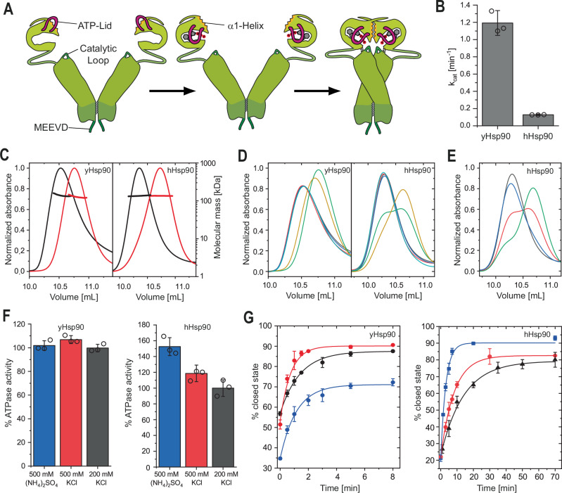
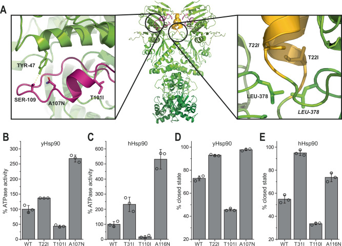
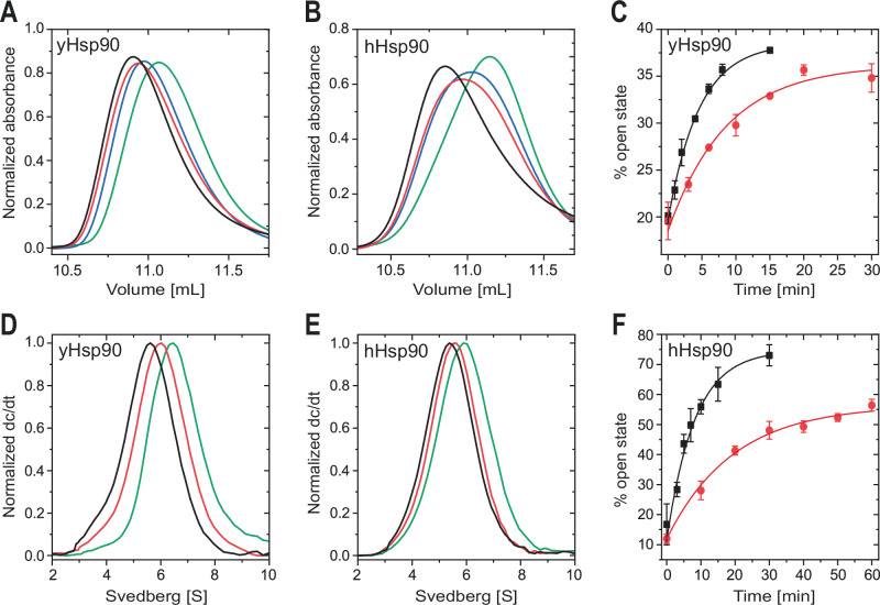
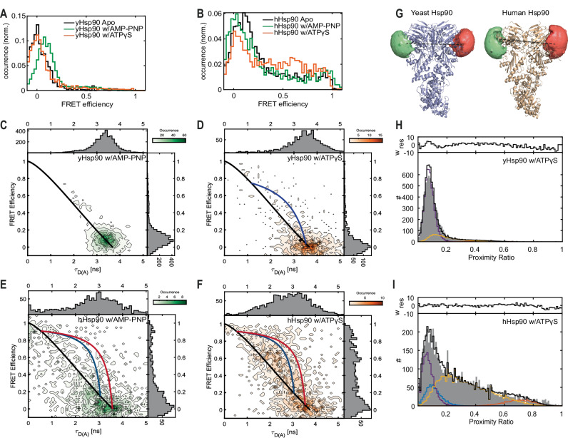
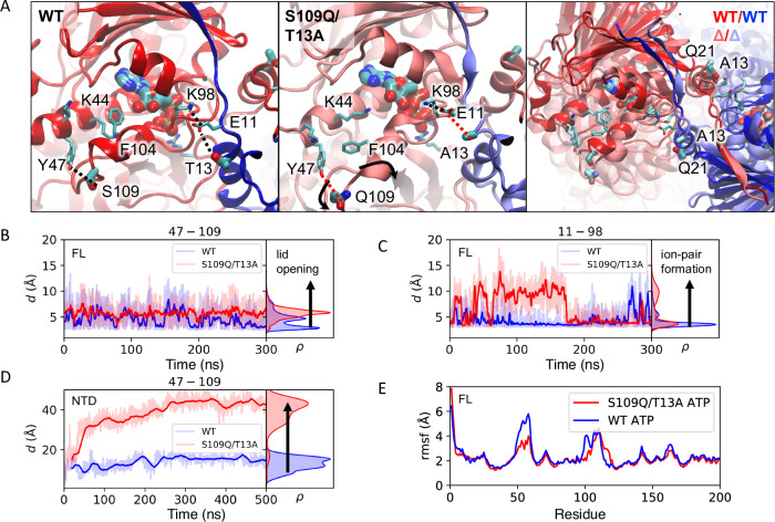
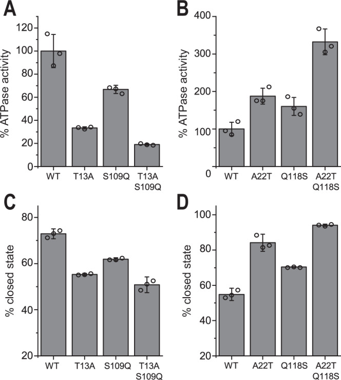
Hsp90 is a molecular chaperone of central importance for protein homeostasis in the cytosol of eukaryotic cells, with key functional and structural traits conserved from yeast to man. During evolution, Hsp90 has gained additional functional importance, leading to an increased number of interacting co-chaperones and client proteins. Here, we show that the overall conformational transitions coupled to the ATPase cycle of Hsp90 are conserved from yeast to humans, but cycle timing as well as the dynamics are significantly altered. In contrast to yeast Hsp90, the human Hsp90 is characterized by broad ensembles of conformational states, irrespective of the absence or presence of ATP. The differences in the ATPase rate and conformational transitions between yeast and human Hsp90 are based on two residues in otherwise conserved structural elements that are involved in triggering structural changes in response to ATP binding. The exchange of these two mutations allows swapping of the ATPase rate and of the conformational transitions between human and yeast Hsp90. Our combined results show that Hsp90 evolved to a protein with increased conformational dynamics that populates ensembles of different states with strong preferences for the N-terminally open, client-accepting states.


Conflict of interest statement

The authors declare no competing interests.

Figures

Fig. 1
Fig. 1. Nucleotide induced closing of yHsp90 and hHsp90.
A Schematic illustration of Hsp90 closing upon nucleotide binding. The N-terminal lid (magenta), the α1-helix (yellow) and the catalytic loop (dark green) are highlighted. B ATPase activity of yHsp90 and hHsp90 measured via a regenerative ATPase assay. Data is presented as mean values +/− SD from technical replicates (n = 3). C Size exclusion chromatography-multi-angle light scattering (SEC-MALS) elution profiles of yHsp90 (left) and hHsp90 (right). Proteins were pre-incubated without (black) or with ATPγS (red) to induce closing. Shifts to higher elution volumes indicate conformational rearrangements to a more compact state. D yHsp90 (left) and hHsp90 (right) elution profiles from SEC after incubation (yHsp90: 30 °C, 30 min, hHsp90: 37 °C, 4 h) with varying nucleotides (black: w/o, red: ADP, blue: ATP, green: AMP-PNP, yellow: ATPγS, teal: ADP-AlF4). E SEC elution profiles of hHsp90 after different time points of incubation with AMP-PNP at 37 °C (black: 0 min, blue: 1 h, red: 4 h, green: 16 h). F Correlation of yHsp90 (left) and hHsp90 (right) ATPase activity with varying buffer salts and concentrations. Measurements were normalized to the activity of the respective protein in 200 mM KCl. Data is presented as mean values +/− SD from technical replicates (n = 3). G The obtained closing kinetics of yHsp90 (left) and hHsp90 (right) with 2 mM ATPγS for different buffer conditions. Kinetics were calculated by fitting SEC elution profiles at different time points of nucleotide incubation with a bi-Gaussian fit to determine the open- and closed-state fractions. Obtained kinetics are color coded according to F. (yHsp90: 200 mM KCl, kclosing = 1.03 min−1; 500 mM KCl, kclosing = 1.94 min−1; 500 mM (NH4)2SO4, kclosing = 1.26 min−1; hHsp90: 200 mM KCl, kclosing = 0.103 min−1; 500 mM KCl, kclosing = 0.177 min−1; 500 mM (NH4)2SO4, kclosing = 0.439 min−1). All measurements were performed as technical replicates (n = 3) to allow calculation of the mean and standard deviation.
Fig. 2
Fig. 2. Mutational analysis in the NTD reveals important interactions for Hsp90 ATPase activity and closing.
A Modified crystal structure of yHsp90 (PDB ID: 2CG9) highlighting mutations affecting ATPase activity. ATP lid (magenta) and α1-helix (yellow) are highlighted according to Fig. 1A. B Comparison of wt yHsp90 and mutant ATPase activities. The obtained activity was normalized to wt yHsp90. C Comparison of analog hHsp90 mutants. D wt yHsp90 closed-state fraction after 1 min of incubation at 30 °C with 2 mM ATPγS compared to mutants. Data were obtained by fitting SEC elution profiles with a bi-Gaussian fit. E Wt hHsp90 compared to analog mutants after 10 min of incubation at 37 °C. All measurements were performed as technical replicates (n = 3) to allow calculation of the mean and standard deviation.
Fig. 3
Fig. 3. Nucleotide exchange induced dimer opening.
A Size exclusion chromatography elution profiles of ATPγS closed yHsp90 (green) after additional incubation with 2 mM ATP (blue), ADP (red) or 500 µM radicicol (black) for 1 h at 30 °C. Shift to lower elution volumes indicate conformational rearrangements to a more open state. B Analog hHsp90 SEC elution profiles after incubation for 1 h at 37 °C. Peaks are normalized by peak area. C Obtained opening kinetics of yHsp90 and (F) hHsp90 with 500 µM radicicol (black) or 2 mM ADP (red). Kinetics were calculated by fitting SEC elution profiles with a bi-Gaussian fit to determine the open- and closed-state fractions (yHsp90 ADP kopening = 0.167 min−1, radicicol kopening = 0.312 min−1; hHsp90 ADP kopening = 0.0742 min−1, radicicol kopening = 0.176 min−1). All measurements were performed as technical replicates (n = 3) to allow calculation of the mean and standard deviation. D Analytical ultracentrifugation sedimentation profiles of Atto488 labeled yHsp90 and (E) hHsp90 re-opened with ADP (red) or radicicol (black).
Fig. 4
Fig. 4. Nucleotide dependent conformations and their dynamics for yHsp90 and hHsp90 measured by smFRET.
A SmFRET efficiency histograms of yHsp90 in the apo state (black), in the presence of AMP-PNP (green) and in the presence of ATPγS (orange). B SmFRET efficiency histograms of hHsp90 in the apo state (black), in the presence of AMP-PNP (green) and in the presence of ATPγS (orange). FRET histograms are of representatives from at least two independent measurements unless otherwise mentioned. CF 2D histograms of FRET efficiency vs. donor fluorescence lifetime in the presence of an acceptor (τD(A)) for C yHsp90 w/AMP-PNP, D yHsp90 w/ATPγS, E hHsp90 w/AMP-PNP and F hHsp90 w/ATPγS. Black lines indicate the static FRET line whereas both the red and blue curved lines depict dynamic FRET lines. G Accessible volume (AV) calculations of residues C61 for yHsp90 (left panel) and residue C70 for hHsp90 (right panel) labeled with Atto532 as a donor and Atto643 as an acceptor dye. Distances of 88.8 Å (AV) versus 89.6 Å (measured) for yHsp90 and 89.9 Å (AV) and 82–86 Å (measured) for hHsp90 were determined. H, I Dynamic photon distribution analysis (PDA) for yHsp90 and hHsp90, respectively, in the presence of ATPγS. The open conformation is shown in violet, the closed conformation in blue, a compact conformation with high FRET efficiency in red and the contribution of dynamically interconverting populations are shown in yellow. For experiments with yHsp90, nucleotides were preincubated with the protein for 2 h. In the case of hHsp90, the preincubation time was 4 h.
Fig. 5
Fig. 5. HDX comparison of the open and closed state.
A Change (Δ) in fractional uptake between the open and closed state of yHsp90 plotted on its crystal structure in the closed state (PDB ID: 2CG9). Red indicates decreased exchange after closing, blue areas with increased exchange. B Change (Δ) in fractional uptake between the open and closed state of hHsp90 plotted on its crystal structure (PDB ID: 5FWK). C Fractional uptake (Δ) of yHsp90 and (D) hHsp90 plotted by residue. Blue highlights crucial elements exceeding the significant threshold (a: α1-helix, b: ATP-lid, c: catalytic loop, d: M1/M2-hinge, e: MD/CTD-hinge, f: dimerization interface); gray areas indicate the standard deviation of the respective residues. All measurements were performed as independent technical replicates (n = 2) to allow calculation of the mean and standard deviation.
Fig. 6
Fig. 6. Molecular dynamics (MD) simulations of WT and S109Q/T13A yHsp90.
A MD simulations of the full-length yHsp90 suggest that the S109Q/T13A mutations result in conformational changes in the NTD, particularly around the ATP lid, and residues surrounding the ATP binding site. B Dynamic changes in the ATP lid (Tyr47-Ser/Gln109 distance) and C ion-pairs affecting Hsp90 dimerization (Glu11-Lys98 distance). D Introduction of the S109Q/T13A mutation results in an opening of the ATP binding site in simulations of the NTD construct (see Methods). E Conformational dynamics of the NTD (from full length yHsp90 simulations) indicated by the changes in root-mean-square-fluctuation (rmsf). See also Supplementary Figs. 14–16 for further analysis.
Fig. 7
Fig. 7. Inherent evolutionary differences of yHsp90 and hHsp90.
A ATPase activity of wt yHsp90 and yeast-to-human mutants measured via a regenerative ATPase assay. B ATPase activity of wt hHsp90 and human-to-yeast mutants. C Calculated yHsp90 closed-state fraction after 1 min of incubation at 30 °C with 2 mM ATPγS compared to the mutants. Data were obtained by fitting SEC elution profiles with a bi-Gaussian fit. D Closed-state percentage of wt hHsp90 compared to mutants after 10 min of incubation at 37 °C. The ATPase activity was normalized to that of the respective wt protein. All measurements were performed as technical replicates (n = 3) to allow calculation of the mean and standard deviation.

Similar articles

Cited by

References

    1. Richter, K., Haslbeck, M. & Buchner, J. The heat shock response: life on the verge of death. Mol. Cell40, 253–266 (2010). - PubMed
    1. Biebl, M.M. & Buchner, J. Structure, function, and regulation of the Hsp90 machinery. Cold Spring Harb. Perspect Biol.11, a034017 (2019). - PMC - PubMed
    1. Karagoz, G. E. & Rudiger, S. G. Hsp90 interaction with clients. Trends Biochem. Sci.40, 117–125 (2015). - PubMed
    1. Zhao, R. et al. Navigating the chaperone network: an integrative map of physical and genetic interactions mediated by the hsp90 chaperone. Cell120, 715–727 (2005). - PubMed
    1. Echeverria, P. C., Bernthaler, A., Dupuis, P., Mayer, B. & Picard, D. An interaction network predicted from public data as a discovery tool: application to the Hsp90 molecular chaperone machine. PLoS One6, e26044 (2011). - PMC - PubMed

Publication types

MeSH terms

Substances

LinkOut - more resources