. 2024 Oct 4;14(1):23078.
doi: 10.1038/s41598-024-73950-3.

Biofilm infections of endobronchial valves in COPD patients after endoscopic lung volume reduction: a pilot study with FISHseq

Affiliations

Biofilm infections of endobronchial valves in COPD patients after endoscopic lung volume reduction: a pilot study with FISHseq

Eva Pappe et al. Sci Rep. .

Abstract

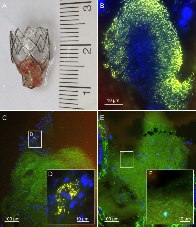
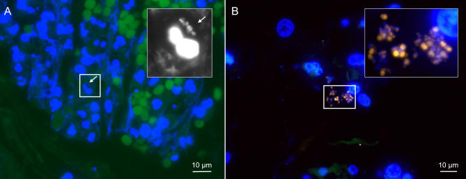
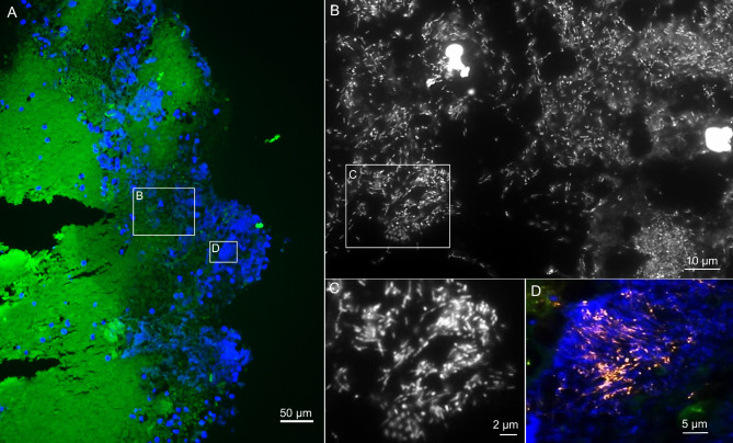
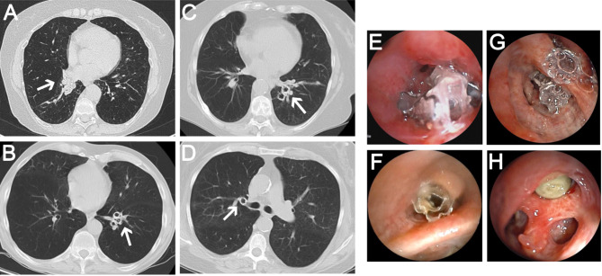
Endoscopic lung volume reduction (ELVR) using endobronchial valves (EBV) is a treatment option for a subset of patients with severe chronic obstructive pulmonary disease (COPD), suffering from emphysema and hyperinflation. In this pilot study, we aimed to determine the presence of bacterial biofilm infections on EBV and investigate their involvement in lack of clinical benefits, worsening symptomatology, and increased exacerbations that lead to the decision to remove EBVs. We analyzed ten COPD patients with ELVR who underwent EBV removal. Clinical data were compared to the microbiological findings from conventional EBV culture. In addition, EBV were analyzed by FISHseq, a combination of Fluorescence in situ hybridization (FISH) with PCR and sequencing, for visualization and identification of microorganisms and biofilms. All ten patients presented with clinical symptoms, including pneumonia and recurrent exacerbations. Microbiological cultures from EBV detected several microorganisms in all ten patients. FISHseq showed either mixed or monospecies colonization on the EBV, including oropharyngeal bacterial flora, Staphylococcus aureus, Pseudomonas aeruginosa, Streptococcus spp., and Fusobacterium sp. On 5/10 EBV, FISHseq visualized biofilms, on 1/10 microbial microcolonies, on 3/10 single microorganisms, and on 1/10 no microorganisms. The results of the study demonstrate the presence of biofilms on EBV for the first time and its potential involvement in increased exacerbations and clinical worsening in patients with ELVR. However, further prospective studies are needed to evaluate the clinical relevance of biofilm formation on EBV and appropriate treatment options to avoid infections in patients with ELVR.

Keywords: Bacterial biofilms; COPD; Endoscopic lung volume reduction; Exacerbations.


Conflict of interest statement

The authors declare no competing interests.

Figures

Fig. 1
Fig. 1
FISHseq analysis of patient 1 showing a mature Pseudomonas aeruginosa biofilm on endobronchial valve. Fluorescence in situ hybridization (FISH) of the endobronchial valve (EBV) from patient no. 1 fixed in the operating room. Histological sections show in green the autofluorescent tissue background, in yellow the Pseudomonas. aeruginosa-specific FISH probe PSMG-Cy3 and in blue the nucleic acid stain 4’,6-diamidino-2-phenylindole (DAPI). (A) – Macroscopic impression of the explanted EBV. (B) – FISH visualizing rod-shaped Pseudomonas sp. positive for PSMG-Cy3. (C) – Tissue overview showing host cell nuclei in blue and the green tissue background. (D) – Higher magnification of the inset marked in C shows a different position with rod-shaped Pseudomonas sp. in PSMG-Cy3 and host cell nuclei in blue. (E) – Tissue overview showing host cell nuclei in blue and the green tissue background. (F) – Higher magnification of the inset marked in E shows autofluorescent erythrocytes and tissue background in green with host cell nuclei in blue, no bacteria are detectable.
Fig. 2
Fig. 2
Examples of single bacteria and thin biofilms in tissue attached to the EBVs as visualized by FISH. (A) – Single bacteria (patient 6) and host cell nuclei shown with the nucleic acid stain DAPI (bacteria beside a host cell nucleus shown enlarged in the inset in black and white, marked with an arrow). Note the erythrocytes with strong background autofluorescence in green. (B) – Thin Staphylococcus aures biofilms (patient 10) as visualized by FISH. Shown is the overlay of the microscopic channels for the nucleic acid stain DAPI (blue), and the Staphylococcus aures specific FISH probe SAU in Cy3 (orange). The inset marks the region that is shown enlarged in the inset. Note the varying FISH signal intensity of bacteria due to their different ribosome content.
Fig. 3
Fig. 3
FISHseq analysis of patient 2 showing oral biofilm on EBV. Fluorescence in situ hybridization (FISH) of the endobronchial valve (EBV) from patient 2 fixated in the operating room. Histological sections show in green the autofluorescent tissue background, in orange the Fusobacterium nucleatum-specific FISH probe FUNU-Cy3 (sequence deposited in Probebase, where details on the probe are available) and in blue the nucleic acid stain 4’,6-diamidino-2-phenylindole (DAPI). (A) – Overview of the sample showing the tissue background in green and the nucleic acid stain DAPI in blue. Extensive biofilms are visible as blue clouds. (B) - Higher magnification of the inset A shows that the biofilm consists of rod shaped bacteria and cocci, in line with oral flora (DAPI filter set in black and white). (C) – Higher magnification of the inset in B showing rods and cocci. (D) - Higher magnification of the inset A shows that the biofilm contains Fusobacterium nucleatum (orange). Nucleic acids in blue and tissue background in green.
Fig. 4
Fig. 4
Radiological and bronchoscopic findings in patients after endoscopic lung volume reduction: (A) – CT scans of patient no. 1 with biofilm presented mucus layers on endoscopic valves (EBV) with almost complete obstruction of the right middle lobe. The White arrow points the mucus layer. (B) CT scans of patient no. 2 with biofilm showed signs of mucus layers on EBV in the left lower lobe, indicated by the white arrow. (C) No mucus layers were observed in CT scans of patient no. 5 and patient no. 6 (D), both without bacterial biofilm on EBV. (E-H) Bronchoscopy visualized mucus layers on EBV in patient no. 1, 2, 5 and 6.

References

    1. Adeloye, D. et al. Global, regional, and national prevalence of, and risk factors for, chronic obstructive pulmonary disease (COPD) in 2019: A systematic review and modelling analysis. Lancet Respir. Med.10, 447–458. 10.1016/S2213-2600(21)00511-7 (2022). - PMC - PubMed
    1. Lin, C. R., Bahmed, K. & Kosmider, B. Dysregulated cell signaling in pulmonary emphysema. Front. Med. (Lausanne)8, 762878. 10.3389/fmed.2021.762878 (2021). - PMC - PubMed
    1. Slebos, D. J., Shah, P. L., Herth, F. J. & Valipour, A. Endobronchial valves for endoscopic lung volume reduction: Best practice recommendations from expert panel on endoscopic lung volume reduction. Respiration93, 138–150. 10.1159/000453588 (2017). - PMC - PubMed
    1. Davey, C. et al. Bronchoscopic lung volume reduction with endobronchial valves for patients with heterogeneous emphysema and intact interlobar fissures (the BeLieVeR-HIFi study): A randomised controlled trial. Lancet386, 1066–1073. 10.1016/S0140-6736(15)60001-0 (2015). - PubMed
    1. Herth, F. J. et al. Efficacy predictors of lung volume reduction with Zephyr valves in a European cohort. Eur. Respir. J.39, 1334–1342. 10.1183/09031936.00161611 (2012). - PubMed

LinkOut - more resources