

Structural elucidation of recombinant Trichomonas vaginalis 20S proteasome bound to covalent inhibitors
- PMID: 39366995
- PMCID: PMC11452676
- DOI: 10.1038/s41467-024-53022-w
Structural elucidation of recombinant Trichomonas vaginalis 20S proteasome bound to covalent inhibitors
Abstract
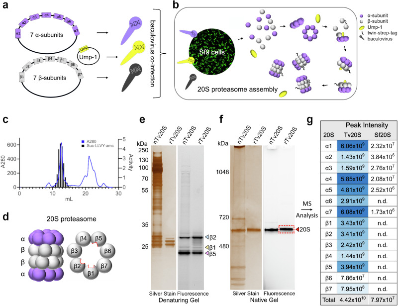
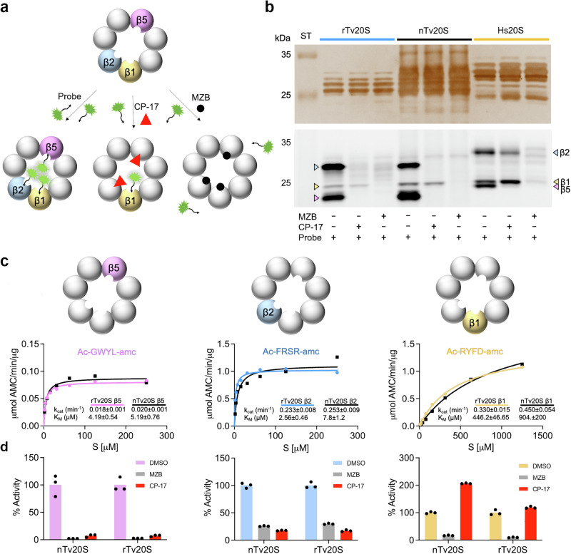
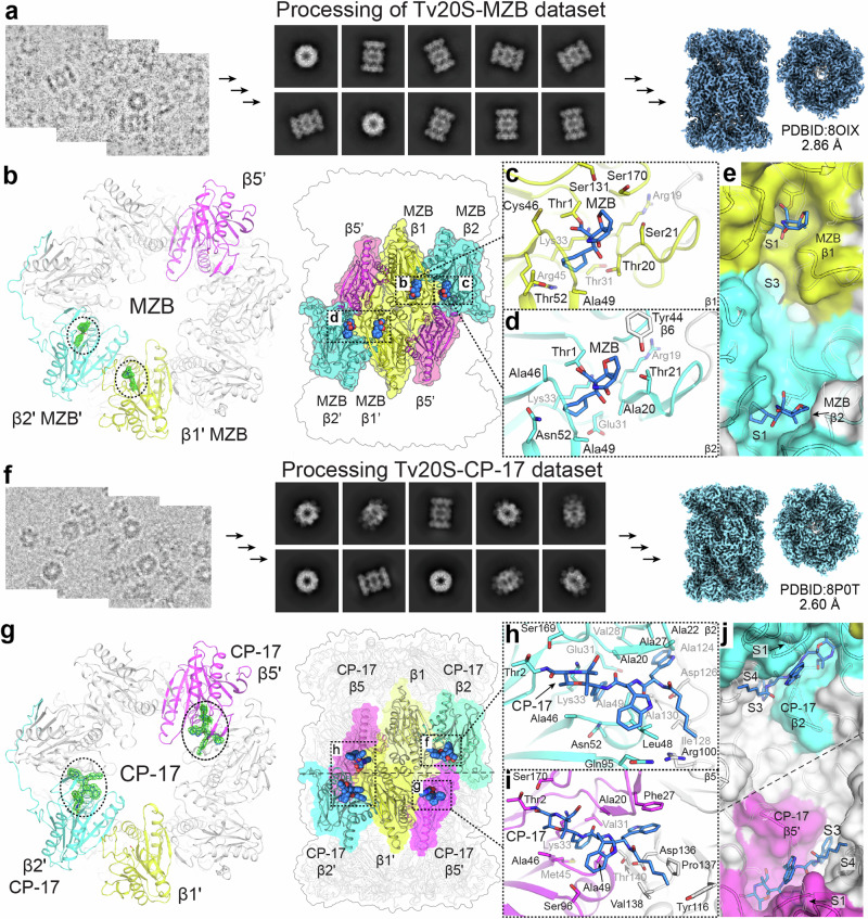
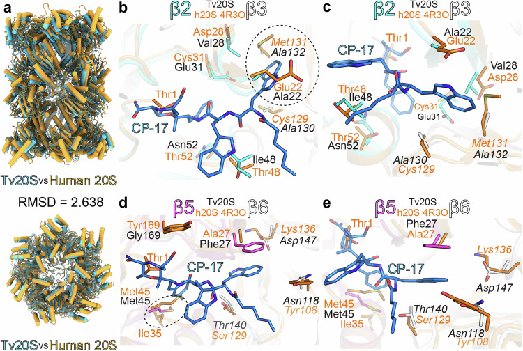
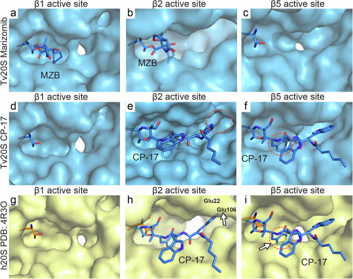
The proteasome is a proteolytic enzyme complex essential for protein homeostasis in mammalian cells and protozoan parasites like Trichomonas vaginalis (Tv), the cause of the most common, non-viral sexually transmitted disease. Tv and other protozoan 20S proteasomes have been validated as druggable targets for antimicrobials. However, low yields and purity of the native proteasome have hindered studies of the Tv 20S proteasome (Tv20S). We address this challenge by creating a recombinant protozoan proteasome by expressing all seven α and seven β subunits of Tv20S alongside the Ump-1 chaperone in insect cells. The recombinant Tv20S displays biochemical equivalence to its native counterpart, confirmed by various assays. Notably, the marizomib (MZB) inhibits all catalytic subunits of Tv20S, while the peptide inhibitor carmaphycin-17 (CP-17) specifically targets β2 and β5. Cryo-electron microscopy (cryo-EM) unveils the structures of Tv20S bound to MZB and CP-17 at 2.8 Å. These findings explain MZB's low specificity for Tv20S compared to the human proteasome and demonstrate CP-17's higher specificity. Overall, these data provide a structure-based strategy for the development of specific Tv20S inhibitors to treat trichomoniasis.
© 2024. The Author(s).
Conflict of interest statement
The authors declare no competing interests.
Figures
Update of
-
Structural elucidation of recombinant Trichomonas vaginalis 20S proteasome bound to covalent inhibitors.bioRxiv [Preprint]. 2023 Aug 17:2023.08.17.553660. doi: 10.1101/2023.08.17.553660. bioRxiv. 2023. Update in: Nat Commun. 2024 Oct 4;15(1):8621. doi: 10.1038/s41467-024-53022-w. PMID: 37645851 Free PMC article. Updated. Preprint.
Similar articles
-
Structural elucidation of recombinant Trichomonas vaginalis 20S proteasome bound to covalent inhibitors.bioRxiv [Preprint]. 2023 Aug 17:2023.08.17.553660. doi: 10.1101/2023.08.17.553660. bioRxiv. 2023. Update in: Nat Commun. 2024 Oct 4;15(1):8621. doi: 10.1038/s41467-024-53022-w. PMID: 37645851 Free PMC article. Updated. Preprint.
-
Structural Insights into Salinosporamide a Mediated Inhibition of the Human 20S Proteasome.Molecules. 2025 Mar 20;30(6):1386. doi: 10.3390/molecules30061386. Molecules. 2025. PMID: 40142161 Free PMC article.
-
Distinct substrate specificities of the three catalytic subunits of the Trichomonas vaginalis proteasome.Protein Sci. 2024 Dec;33(12):e5225. doi: 10.1002/pro.5225. Protein Sci. 2024. PMID: 39589076
-
The cryo-EM structure of the Plasmodium falciparum 20S proteasome and its use in the fight against malaria.FEBS J. 2016 Dec;283(23):4238-4243. doi: 10.1111/febs.13780. Epub 2016 Jul 2. FEBS J. 2016. PMID: 27286897 Free PMC article. Review.
-
Inhibitors of the eukaryotic 20S proteasome core particle: a structural approach.Biochim Biophys Acta. 2004 Nov 29;1695(1-3):33-44. doi: 10.1016/j.bbamcr.2004.09.025. Biochim Biophys Acta. 2004. PMID: 15571807 Review.
Cited by
-
Exploring the in vitro and in vivo antileishmanial potential of Marizomib against Leishmania amazonensis and Leishmania infantum.Antimicrob Agents Chemother. 2025 Aug 6;69(8):e0028625. doi: 10.1128/aac.00286-25. Epub 2025 Jul 21. Antimicrob Agents Chemother. 2025. PMID: 40689741 Free PMC article.
-
Structural insights into Salinosporamide A mediated inhibition of the human 20S proteasome.bioRxiv [Preprint]. 2025 Jan 28:2025.01.28.635221. doi: 10.1101/2025.01.28.635221. bioRxiv. 2025. Update in: Molecules. 2025 Mar 20;30(6):1386. doi: 10.3390/molecules30061386. PMID: 39974992 Free PMC article. Updated. Preprint.
-
Recombinant proteasome provides new avenues for anti-malarial drug development.bioRxiv [Preprint]. 2025 Aug 14:2025.08.13.670186. doi: 10.1101/2025.08.13.670186. bioRxiv. 2025. PMID: 40832278 Free PMC article. Preprint.
-
Visualizing chaperone-mediated multistep assembly of the human 20S proteasome.bioRxiv [Preprint]. 2024 Jan 28:2024.01.27.577538. doi: 10.1101/2024.01.27.577538. bioRxiv. 2024. Update in: Nat Struct Mol Biol. 2024 Aug;31(8):1176-1188. doi: 10.1038/s41594-024-01268-9. PMID: 38328185 Free PMC article. Updated. Preprint.
-
Pharmacological and structural understanding of the Trypanosoma cruzi proteasome provides key insights for developing site-specific inhibitors.J Biol Chem. 2025 Jan;301(1):108049. doi: 10.1016/j.jbc.2024.108049. Epub 2024 Dec 9. J Biol Chem. 2025. PMID: 39638245 Free PMC article.
References
-
- Edwards, T., Burke, P., Smalley, H. & Hobbs, G. Trichomonas vaginalis: Clinical relevance, pathogenicity and diagnosis. Crit. Rev. Microbiol.42, 406–417 (2016). - PubMed
Publication types
MeSH terms
Substances
Associated data
- Actions
- Actions
- Actions
- Actions
- Actions
- Actions
- Actions
- Actions
- Actions
Grants and funding
LinkOut - more resources
Full Text Sources
Miscellaneous
